| Transcriber's note: | Some typographical errors in the printed work have been corrected. The corrected text is underscored in red like this. Hover the cursor over the marked text and the explanation should appear. Click on any illustration to enlarge it. |
REPORT
ON THE
SCIENTIFIC RESULTS
OF THE
VOYAGE OF H.M.S. CHALLENGER
DURING THE YEARS 1873-76
UNDER THE COMMAND OF
Captain GEORGE S. NARES, R.N., F.R.S
AND THE LATE
Captain FRANK TOURLE THOMSON, R.N
PREPARED UNDER THE SUPERINTENDENCE OF
THE LATE
Sir C. WYVILLE THOMSON, Knt., F.R.S., &c
REGIUS PROFESSOR OF NATURAL HISTORY IN THE UNIVERSITY OF EDINBURGH
DIRECTOR OF THE CIVILIAN SCIENTIFIC STAFF ON BOARD
AND NOW OF
JOHN MURRAY
ONE OF THE NATURALISTS OF THE EXPEDITION
Zoology—Vol. XVIII.
PLATES
Published by Order of Her Majesty's Government
PRINTED FOR HER MAJESTY'S STATIONERY OFFICE
AND SOLD BY
LONDON:—EYRE & SPOTTISWOODE, EAST HARDING STREET, FETTER LANE
EDINBURGH:—ADAM & CHARLES BLACK
DUBLIN:—HODGES, FIGGIS, & CO
1887
Price (in Two Parts, with a Volume of Plates) £5, 10s.
CONTENTS.
Report on the Radiolaria collected by H.M.S. Challenger during the years 1873-1876.
By Ernst Haeckel, M.D., Ph.D., Professor of Zoology in the University of Jena.
PLATES.
CONTENTS.
| PLATES | 1-50. | SPUMELLARIA. | ||
| " | 51-98. | NASSELLARIA. | ||
| " | 99-128. | PHÆODARIA. | ||
| " | 129-140. | ACANTHARIA. | ||
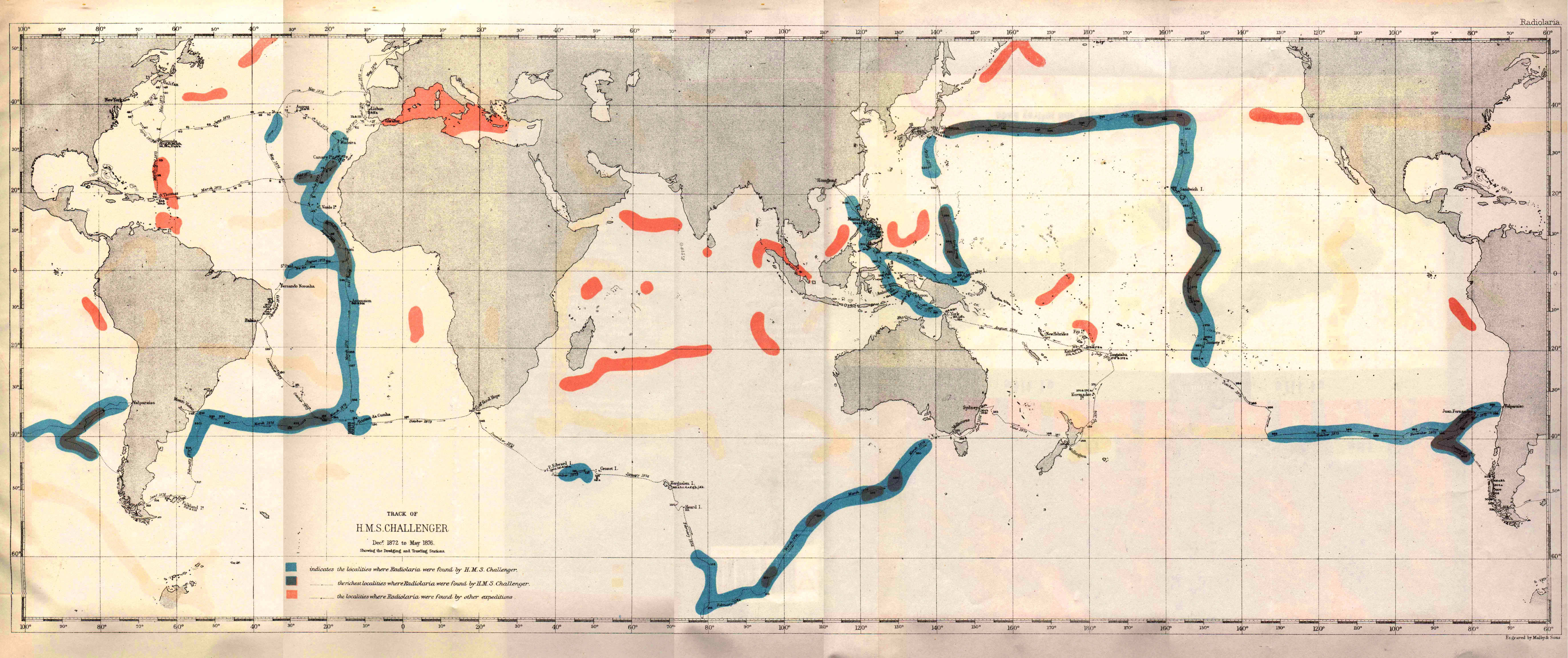
| MAP, showing the Geographical Distribution of the Radiolaria. | ||||
PLATE 1.
Legion SPUMELLARIA.
Order COLLOIDEA.
Family Thalassicollida.
| 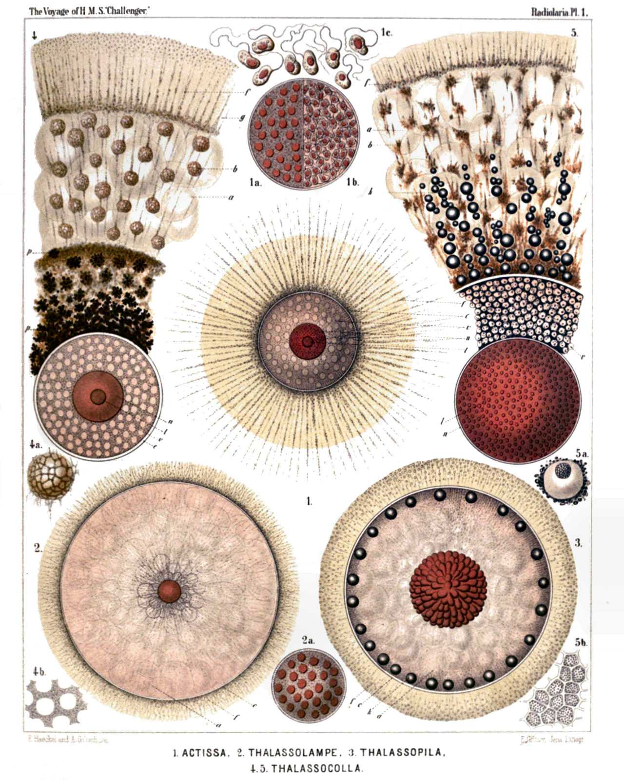 | |||||||||||||||||||||||||||||||||||||||||||||||||||||||||||||
| PLATE 1. | |||
| Thalassicollida. | |||
| Diam. | Page. | ||
Fig. 1. Actissa princeps, n. sp., | × | 300 | 13 |
| The entire living Spumellarium. c, The spherical central capsule containing finely granulated protoplasm, which is radially striated in the cortical zone; v, spherical vacuoles enclosed by the protoplasm; n, the spherical nucleus in the centre; l, the concentric nucleolus; f, the radial pseudopodia which pierce the calymma or the (yellowish) jelly-envelope of the central capsule and arise from the granular sarcomatrix. | |||
| Fig. 1a. Half of the central capsule of another specimen, in which the original central nucleus is cleft into numerous small nuclei, | × | 400 | |
| Fig. 1b. Half of the central capsule of another specimen, filled up by flagellate spores, | × | 400 | |
| Fig. 1c. Eight isolated flagellate spores, | × | 800 | |
| Fig. 2. Thalassolampe maxima, n. sp., | × | 8 | 17 |
| The entire living Spumellarium. c, The big spherical central capsule; a, the large alveoles filling the central capsule and surrounding a central nucleus; f, the pseudopodia piercing the extracapsular calymma. | |||
| Fig. 2a. The nucleus alone, with numerous nucleoli, | × | 30 | |
| Fig. 3. Thalassopila cladococcus, n. sp., | × | 20 | 17 |
| c, The big central capsule; a, numerous large alveoles contained in the central capsule; k, oil globules, many of which are placed in the radially striped cortical zone; the nucleus placed centrally, is covered with numerous radial apophyses or cæcal sacs. f, The radially striped calymma. | |||
| Fig. 4. Thalassicolla maculata, n. sp., | × | 100 | 21 |
| c, The central capsule; v, vacuoles filling this capsule; n, the central nucleus; l, the concentric nucleolus; g, the voluminous calymma, a small radial piece of which is only represented; a, the large alveoles; b, peculiar exoplasmatic bodies; p, black pigment in the inner zone; f, the retracted pseudopodia in the outer zone. | |||
| Fig. 4a. An exoplasmatic body, | × | 300 | |
| Fig. 4b, Vacuoles in the endoplasm, | × | 300 | |
| Fig. 5. Thalassicolla melacapsa, n. sp., | × | 300 | 21 |
| n, The large nucleus; l, numerous small nucleoli inside the nucleus; v, the vacuoles filling up the central capsule and separated by black pigment; a, large alveoles in the calymma; k, oil globules; b, exoplasmatic bodies; f, the retracted pseudopodia in the outer zone of the calymma. | |||
| Fig. 5a. An endoplasmatic vacuole, resembling a cell, | × | 600 | |
| Fig. 5b. A piece of the central capsule, | × | 600 | |
PLATE 2.
Legion SPUMELLARIA.
Order BELOIDEA.
Family Thalassosphærida.
| 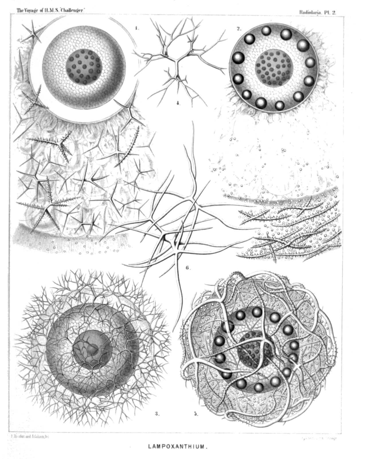 | ||||||||||||||||||||||||||||||||||||||||||
| PLATE 2. | |||
| Thalassosphærida. | |||
| Diam. | Page. | ||
Fig. 1. Lampoxanthium pandora, n. sp., | × | 120 | 38 |
| The central capsule exhibits distinct pore-canals in its membrane, and a clear interval between this and the coagulated and vacuolated protoplasm. The central nucleus contains numerous dark nucleoli. The spicula are scattered throughout the alveolate calymma. | |||
| Fig. 2. Thalassoplancta brevispicula, n. sp. (vel Lampoxanthium brevispiculum), | × | 120 | 36 |
| The central capsule contains numerous clear vacuoles, and in the cortical zone a layer of large oil-globules. The central nucleus includes numerous dark nucleoli. The calymma is alveolate. The spicula lie only in the cortical zone. | |||
| Fig. 3. Thalassoxanthium cervicorne, n. sp., | × | 300 | 33 |
| The central capsule is filled up by clear vacuoles and contains a large central nucleus, with a single nucleolus. The spicula surround the thin calymma. | |||
| Fig. 4. Thalassoxanthium cervicorne, n. sp., | × | 600 | 33 |
| A single spiculum. | |||
| Fig. 5. Thalassoxanthium medusinum, n. sp., | × | 120 | 32 |
| The central capsule is filled up by clear vacuoles and contains on its cortical zone a layer of large oil-globules. The central nucleus contains numerous dark nucleoli. The calymma is radially striped, contains numerous small xanthellæ, and is surrounded by the spicula. | |||
| Fig. 6. Thalassoxanthium octoceras, n. sp., | × | 400 | 34 |
| Three isolated spicula. | |||
PLATE 3.
Legion SPUMELLARIA.
Order COLLOIDEA.
Family Collozoida.
| 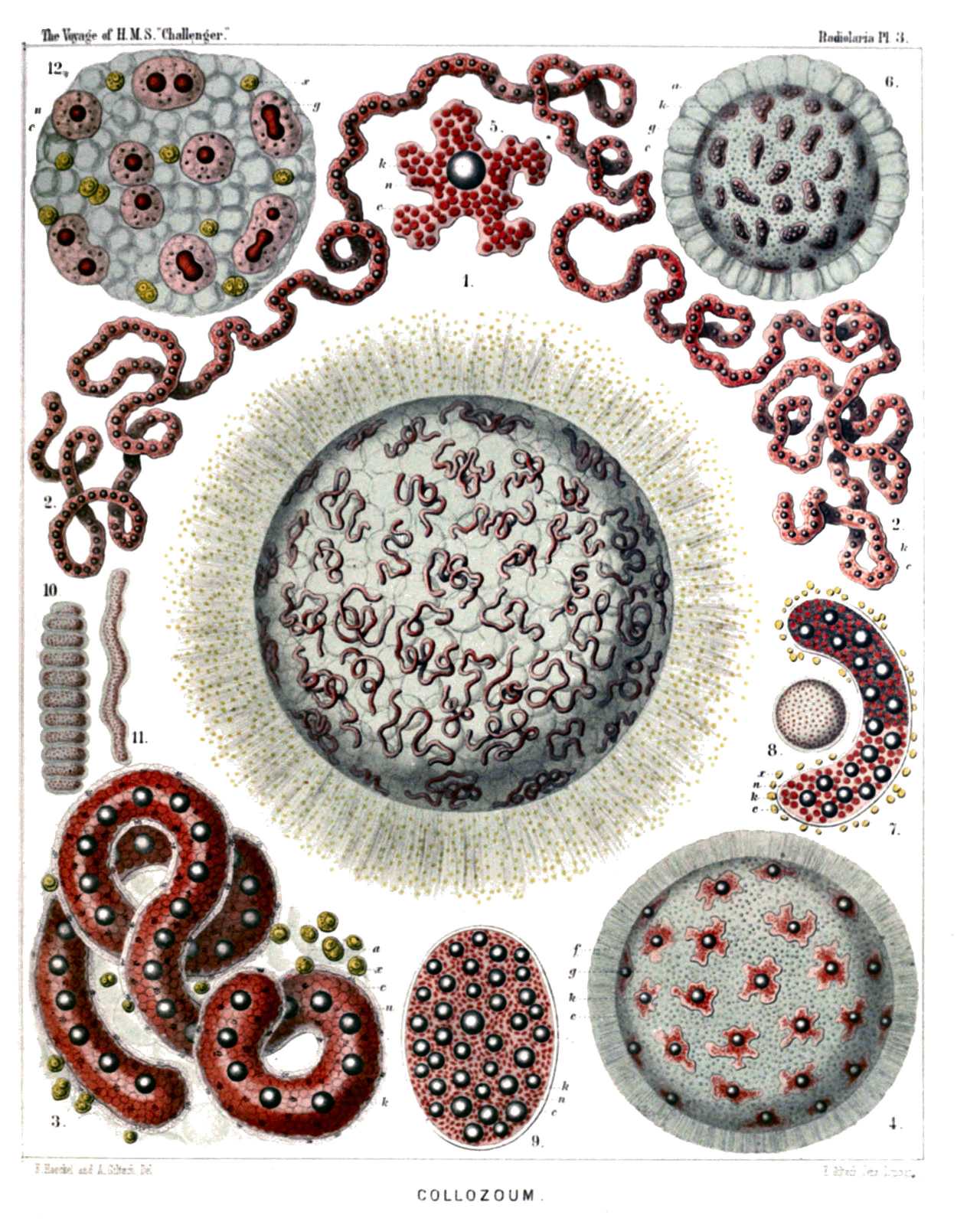 | ||||||||||||||||||||||||||||||||||||||||||||||||||||||||||||||||||||||||
| PLATE 3. | |||
| Collozoida. | |||
| Diam. | Page. | ||
Fig. 1. Collozoum serpentinum, n sp (vel Collophidium serpentinum, Hkl), | × | 10 | 26 |
| A living cœnobium, with expanded pseudopodia. The spherical calymma (or the common jelly-mass of the colony) is alveolate and contains numerous cylindrical, serpentine, central capsules. Numerous yellow cells or xanthellæ are scattered between the radial pseudopodia in the periphery. | |||
| Fig. 2. Collozoum serpentinum, n. sp., | × | 50 | 26 |
| An isolated, cylindrical, worm-shaped, central capsule, with an axial series of oil-globules; the red points are nuclei. | |||
| Fig. 3. Collozoum serpentinum, n. sp., | × | 150 | 26 |
| An isolated, cylindrical, serpentine, central capsule. k, Oil-globules forming an axial series; n, densely placed, red-coloured nuclei;, c, the capsule membrane under which are scattered small black pigment spots in the colourless cortical zone of the endoplasm; a, extracapsular alveoles; x, xanthellæ or "yellow cells." | |||
| Fig. 4. Collozoum amœboides, n. sp., | × | 100 | 28 |
| A spherical cœnobium or jelly-colony. Each amœboid central capsule contains an oil-globule; the small red points are nuclei. | |||
| Fig. 5. Collozoum amœboides, n. sp., | × | 400 | 28 |
| c, A single isolated central capsule; n, nuclei; k, oil-globule. | |||
| Fig. 6. Collozoum vermiforme, n. sp., | × | 30 | 27 |
| g, A spherical cœnobium or jelly-colony; a, large alveoles, forming a cortical zone; c, central capsules; k, oil-globules. | |||
| Fig. 7. Collozoum vermiforme, n. sp., | × | 100 | 27 |
| c, A single isolated central capsule; x, xanthellæ surrounding this central capsule; k, oil-globules; n, nuclei. | |||
| Fig. 8. Collozoum ellipsoides, n. sp., | × | 2 | 26 |
| A spherical colony; the red points are central capsules. | |||
| Fig. 9. Collozoum ellipsoides, n. sp., | × | 150 | 26 |
| c, A single isolated central capsule; k, oil-globules; n, nuclei. | |||
| Fig. 10. Collozoum inerme, Hkl., | × | 2 | 25 |
| An old, cylindrical, articulated cœnobium; the red points are centralcapsules. | |||
| Fig. 11. Collozoum inerme, Hkl., | × | 2 | 25 |
| A young cylindrical cœnobium; the red points are central capsules. | |||
| Fig. 12. Collozoum inerme, Hkl., | × | 400 | 25 |
| A piece of a young colony with eight small central capsules, without oil-globules. n, The central nucleus in different stages of division. Two capsules are also dividing. x, Xanthellæ in the jelly-like calymma (blue), which also contains numerous vacuoles. | |||
PLATE 4.
Legion SPUMELLARIA.
Orders BELOIDEA.
Families Sphærozoida.
| 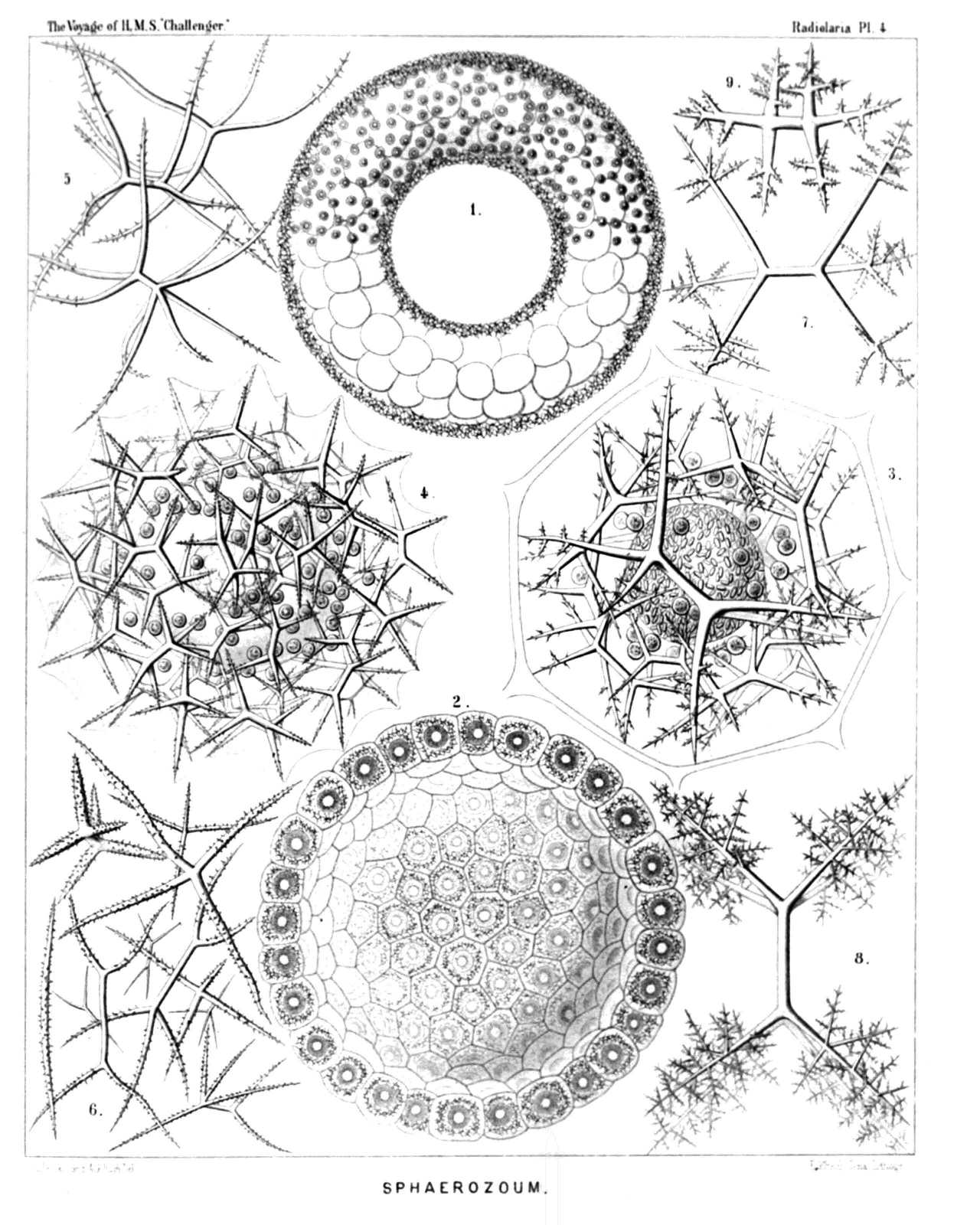 | |||||||||||||||||||||||||||||||||||||||||||||||||||||||||
| PLATE 4. | |||
| Sphærozoida | |||
| Diam. | Page. | ||
Fig. 1. Sphærozoum trigeminum, n. sp., | × | 50 | 43 |
| An annular colony. The main mass of the jelly-colony is filled up by large alveoles; the entire surface is densely covered with spicula, and beyond this skeleton-cover lie the spherical central capsules, each with an oil-globule. This species is by mistake not mentioned in the text. | |||
| Fig. 2. Sphærozoum alveolatum, n. sp., | × | 50 | 43 |
| Section through a spherical colony; displaying the inside of a hemisphere. All the central capsules lie in a single stratum on the surface of the jelly-sphere, each being surrounded by a thick-walled alveole. The spicula lie between the alveole and the capsule, which includes a central oil-globule. | |||
| Fig. 3. Sphærozoum alveolatum, n. sp., | × | 400 | 43 |
| A single central capsule, filled up by crystal-spores. Numerous geminato-radiate spicula and spherical xanthellæ lie between the capsule and the including thick-walled alveole. In the jelly-calymma, between the capsule and the alveole, numerous thin ramified pseudopodia are expanded. | |||
| Fig. 4. Sphærozoum geminatum, n. sp., | × | 400 | 45 |
| A single central capsule, with a central oil-globule, surrounded by numerous spicula and spherical xanthellæ. The jelly-substance of the calymma is expanded between the points of the spicula. | |||
| Fig. 5. Sphærozoum variabile, n. sp., | × | 300 | 45 |
| Three isolated spicula. | |||
| Fig. 6. Sphærozoum pandora, n. sp. (vel Rhaphidozoum pandora), | × | 300 | 49 |
| A group of various spicula. | |||
| Fig. 7. Sphærozoum verticillatum, n. sp., | × | 300 | 44 |
| A single spiculum. | |||
| Fig. 8. Sphærozoum arborescens, n. sp., | × | 300 | 44 |
| A single spiculum. | |||
| Fig. 9. Sphærozoum armatum, n. sp., | × | 300 | 43 |
| A single spiculum. | |||
PLATE 5.
Legion SPUMELLARIA.
Order SPHÆROIDEA.
Family Collosphærida.
| 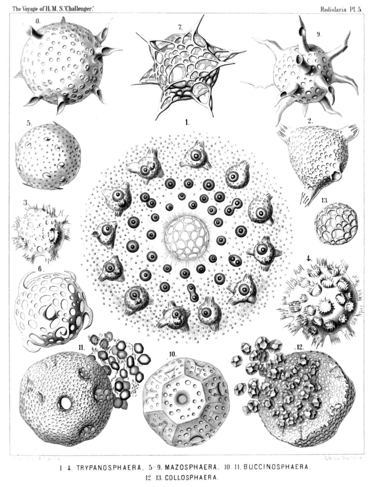 | ||||||||||||||||||||||||||||||||||||||||||||||||||||||||||||||||||||
| PLATE 5. | |||
| Collosphærida. | |||
| Diam. | Page. | ||
Fig. 1. Trypanosphæra transformata, n. sp., | × | 150 | 111 |
| A living colony. The centre of the spherical cœnobium contains a large alveole, surrounded by a net of sarcode. The entire calymma is filled up by smaller, thin-walled alveoles. Its inner part contains numerous small, young, central capsules (each with an oil-globule) without shells; in the cortical zone of the calymma lie larger capsules, each of which is enclosed by a fenestrated shell with from two to four or more dentated tubes. Between the radiant pseudopodia very numerous small yellow cells (xanthellæ), which are scattered everywhere. | |||
| Fig. 2. Trypanosphæra transformata, n. sp., | × | 300 | 111 |
| A single shell. | |||
| Fig. 3. Trypanosphæra coronata, n. sp., | × | 300 | 110 |
| Fig. 4. Trypanosphæra trepanata, n. sp., | × | 300 | 110 |
| Fig. 5. Odontosphæra monodon, n. sp., | × | 300 | 102 |
| Fig. 6. Odontosphæra cyrtodon, n. sp., | × | 300 | 102 |
| Fig. 7. Acrosphæra inflata, n. sp., | × | 300 | 101 |
| Fig. 8. Mazosphæra hippotis, n. sp., | × | 400 | 108 |
| Fig. 9. Mazosphæra lagotis, n. sp., | × | 300 | 108 |
| Fig. 10. Pharyngosphæra stomodæa, n. sp., | × | 400 | 98 |
| Fig. 11. Buccinosphæra invaginata. n. sp., | × | 500 | 99 |
| Each shell contains numerous larger and smaller crystals. | |||
| Fig. 12. Tribonosphæra centripetalis, n. sp., | × | 500 | 98 |
| Each shell contains numerous large crystals. | |||
| Fig. 13. Collosphæra polygona, n. sp., | × | 200 | 96 |
PLATE 6.
Legion SPUMELLARIA.
Order SPHÆROIDEA.
Family Collosphærida.
| 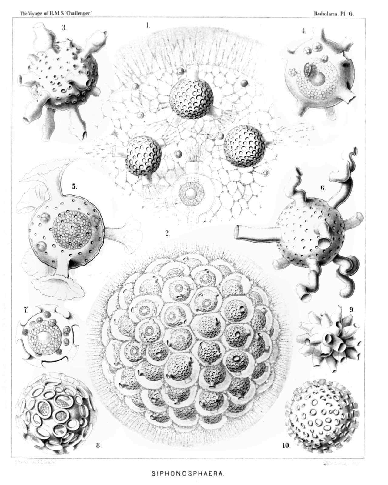 | ||||||||||||||||||||||||||||||||||||||||||||||||||||||||
| PLATE 6. | |||
| Collosphærida. | |||
| Diam. | Page. | ||
Fig. 1. Siphonosphæra socialis, n. sp., | × | 500 | 106 |
| A small piece of the surface of a living cœnobium, seen from the surface. Only four individuals are visible, the central capsule of which contains numerous small nuclei and a central oil-globule. The including spherical lattice-shell is provided with a few (one to four) larger apertures, which are prolonged into short cylindrical tubules. Through these latter radiate bundles of fine pseudopodia, branching and anastomosing, and forming a fine sarcode network between the alveoles of the calymma. On the surface of the alveolated jelly-sphere the pseudopodia form a dense radiating zone. Xanthella or yellow cells are everywhere scattered. | |||
| Fig. 2. Siphonosphæra socialis, n. sp., | × | 300 | 106 |
| A small cœnobium or colony in the state of alveolation, forming a jelly-sphere, composed of a great number of capsulated individuals, densely aggregated. Each central capsule contains an oil-globule, and is enclosed by a spherical lattice-shell, which bears a few (one to four) short cylindrical tubules. Each shell is again enveloped by a membranous polyhedral alveole and separated from it by structureless jelly. The thick cortical jelly-envelope, which surrounds the whole spherical colony, exhibits a fine radial striation, produced by radiating pseudopodia; many xanthellæ or yellow cells are scattered in the calymma. | |||
| Fig. 3. Siphonosphæra pipetta, n. sp., | × | 300 | 108 |
| Fig. 4. Siphonosphæra tubulosa, J. Müller, | × | 300 | 105 |
| The central capsule, enclosed in the cavity of the shell, has a central oil-globule, and is surrounded by a few xanthella. | |||
| Fig. 5. Siphonosphæra chonophora, n. sp., | × | 300 | 107 |
| Fig. 6. Siphonosphæra serpula, n. sp., | × | 300 | 107 |
| Fig. 7. Siphonosphæra patinaria, n sp., | × | 300 | 105 |
| The central capsule, enclosed in the cavity of the shell, contains a central oil-globule, and is surrounded by a few xanthella. | |||
| Fig. 8. Siphonosphæra patinaria, n. sp., | × | 300 | 105 |
| Fig. 9. Siphonosphæra conifera, n. sp., | × | 300 | 106 |
| Fig. 10. Siphonosphæra cyathina, n. sp., | × | 300 | 105 |
PLATE 7.
Legion SPUMELLARIA.
Order SPHÆROIDEA.
Family Collosphærida.
| 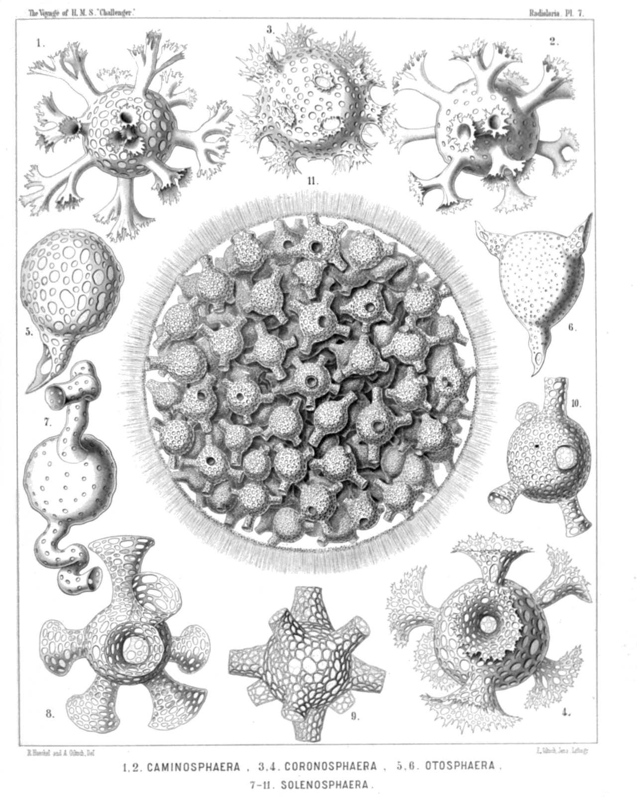 | |||||||||||||||||||||||||||||||||||||||||||||||||||||||||
| PLATE 7. | |||
| Collosphærida. | |||
| Diam. | Page. | ||
Fig. 1 Caminosphæra dendrophora, n. sp., | × | 300 | 112 |
| Fig. 2. Caminosphæra dichotoma, n. sp., | × | 300 | 112 |
| Fig. 3. Coronosphæra diadema, n. sp., | × | 300 | 117 |
| Fig. 4. Coronosphæra calycina, n. sp., | × | 300 | 117 |
| Fig. 5. Otosphæra auriculata, n. sp., | × | 300 | 116 |
| Fig. 6. Otosphæra polymorpha, n. sp., | × | 300 | 116 |
| Fig. 7. Solenosphæra serpentina, n. sp., | × | 300 | 114 |
| Fig. 8. Solenosphæra cornucopia, n. sp., | × | 300 | 115 |
| Fig. 9. Solenosphæra ascensionis, n. sp., | × | 300 | 115 |
| Fig. 10. Solenosphæra pandora, n. sp., | × | 300 | 113 |
| Fig. 11. Solenosphæra pandora, n. sp., | × | 100 | 113 |
| An entire spherical cœnobium. The shells of the colony bear a variable number of fenestrated radial tubes and are densely crowded in the jelly-sphere of the calymma, the cortical zone of which is radially striped. | |||
PLATE 8.
Legion SPUMELLARIA.
Order SPHÆROIDEA.
Family Collosphærida.
| 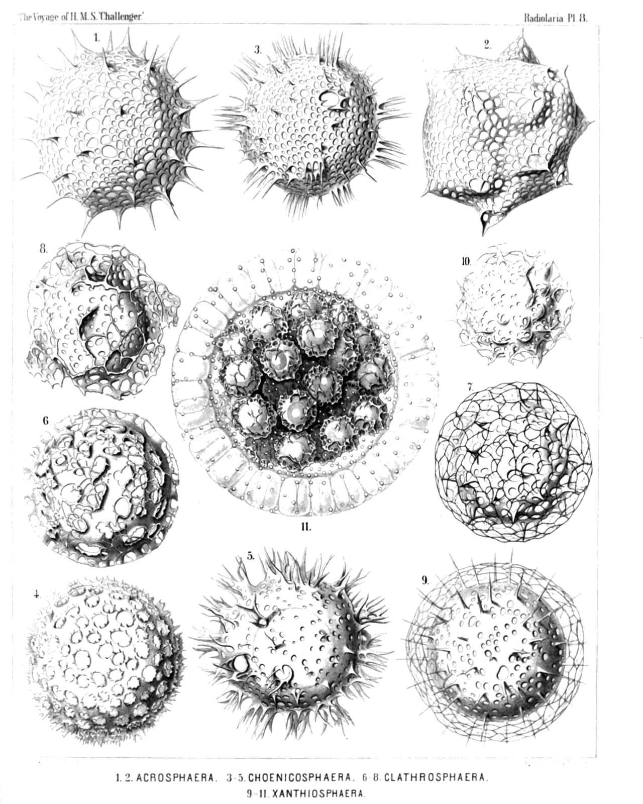 | |||||||||||||||||||||||||||||||||||||||||||||||||||||||||
| PLATE 8. | |||
| Collosphærida. | |||
| Diam. | Page. | ||
Fig. 1. Acrosphæra echinoides, n. sp., | × | 400 | 100 |
| Fig. 2. Acrosphæra collina, n. sp., | × | 300 | 101 |
| Fig. 3. Chænicosphæra nassiterna, n. sp., | × | 400 | 103 |
| Fig. 4. Chænicosphæra murrayana, n. sp., | × | 300 | 102 |
| Fig. 5. Chænicosphæra flammabunda, n. sp., | × | 300 | 103 |
| Fig. 6. Clathrosphæra circumtexta, n. sp., | × | 400 | 118 |
| Fig. 7. Clathrosphæra arachnoides, n. sp., | × | 300 | 119 |
| Fig. 8. Clathrosphæra lamellosa, n. sp., | × | 300 | 119 |
| Fig. 9. Xanthiosphæra erinacea, n. sp., | × | 400 | 120 |
| Fig. 10. Xanthiosphæra lappacea, n. sp., | × | 300 | 120 |
| Fig. 11. Xanthiosphæra lappacea, n. sp., | × | 100 | 120 |
| A complete spherical cœnobium. The associated central capsules (each with a double shell) are densely crowded in the central part of the calymma, whilst its peripheral part is occupied by a layer of large alveoles. Numerous xanthellæ or yellow cells an scattered in the calymma. | |||
PLATE 9.
Legion SPUMELLARIA.
Order LARCOIDEA.
Family Pylonida.
| 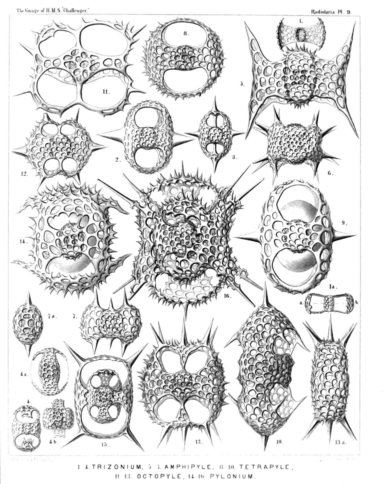 | |||||||||||||||||||||||||||||||||||||||||||||||||||||||||||||||||||||||||||||||||||||||||||||||
| PLATE 9. | |||
| Pylonida. | |||
| Diam. | Page. | ||
Fig. 1. Monozonium alatum, n. sp., | × | 300 | 633 |
| Dorsal view. | |||
| Fig. 1a. Apical view. | |||
| Fig. 2. Dizonium pleuracanthum, n. sp., | × | 400 | 636 |
| Fig. 3. Dizonium stauracanthum, n. sp., | × | 300 | 636 |
| Fig. 4. Trizonium tricinctum, n. sp., | × | 300 | 637 |
| Dorsal view. | |||
| Fig. 4a. Lateral view. | |||
| Fig. 4b. Apical view. | |||
| Fig. 5. Amphipyle tetraceros, n. sp., | × | 400 | 642 |
| Dorsal view. | |||
| Fig. 6. Amphipyle callizona, n. sp., | × | 300 | 644 |
| Dorsal view. | |||
| Fig. 7. Amphipyle amphiptera, n. sp., | × | 300 | 642 |
| Dorsal view. | |||
| Fig. 7a. Lateral view. | |||
| Fig. 8. Tetrapyle circularis, n. sp., | × | 300 | 645 |
| Dorsal view. | |||
| Fig. 9. Tetrapyle pleuracantha, n. sp., | × | 400 | 646 |
| Dorsal view. The lentelliptical central capsule is visible between medullary and cortical shell. | |||
| Fig. 10. Tetrapyle turrita, n. sp., | × | 400 | 649 |
| Oblique view, half dorsal, half lateral. | |||
| Fig. 11. Octopyle stenozona, n. sp., | × | 400 | 652 |
| Dorsal view. | |||
| Fig. 12. Octopyle sexangulata, n. sp., | × | 300 | 653 |
| Dorsal view. | |||
| Fig. 13. Octopyle decastyle, n. sp., | × | 300 | 654 |
| Dorsal view. | |||
| Fig. 13a. Lateral view. | |||
| Fig. 14. Pylonium quadricorne, n. sp., | × | 400 | 655 |
| Dorsal view. | |||
| Fig. 15. Tetrapylonium quadrangulare, n. sp., | × | 300 | 658 |
| Dorsal view. | |||
| Fig. 16. Pylozonium octacanthum, n. sp., | × | 300 | 660 |
| Dorsal view. | |||
PLATE 10.
Legion SPUMELLARIA.
Order LARCOIDEA.
Family Tholonida.
| 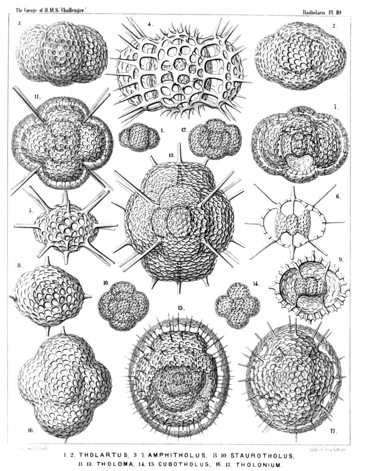 | |||||||||||||||||||||||||||||||||||||||||||||||||||||||||||||||||||||||||||||||||
| PLATE 10. | |||
| Tholonida. | |||
| Diam. | Page. | ||
Fig. 1. Tholartus tricolus, n. sp., | × | 200 | 664 |
| Fig. 2. Tholodes cupula, n. sp., | × | 500 | 665 |
| Fig. 3. Amphitholus artiscus, n. sp., | × | 400 | 666 |
| Fig. 4. Amphitholus panicium, n. sp., | × | 500 | 668 |
| Fig. 5. Amphitholus acanthometra, n. sp., | × | 300 | 667 |
| Fig. 6. Amphitholus acanthometra, n. sp., | × | 300 | 667 |
| Frontal section of the shell. | |||
| Fig. 7. Amphitholonium tricolonium, n. sp., | × | 300 | 669 |
| Fig. 8. Staurotholus tetrastylus, n. sp., | × | 300 | 673 |
| Fig. 9. Staurotholus dodecastylus, n. sp., | × | 400 | 674 |
| Fig. 10. Tholoma quadrigeminum, n. sp., | × | 200 | 672 |
| Fig. 11. Staurotholonium octodoronium, n. sp., | × | 300 | 676 |
| Fig. 12. Tholocubus tessellatus, n. sp., | × | 200 | 677 |
| Fig. 13. Tholoma metallasson, n. sp., | × | 300 | 672 |
| Fig. 14. Cubotholus regularis, n. sp., | × | 200 | 680 |
| Fig. 15. Cubotholonium ellipsoides, n. sp., | × | 300 | 682 |
| Fig. 16. Tholocubus tesseralis, n. sp., | × | 400 | 678 |
| Fig. 17. Tholonium hexonium, | × | 400 | 679 |
PLATE 11.
Legion SPUMELLARIA.
Order SPHÆROIDEA.
Family Astrosphærida.
| 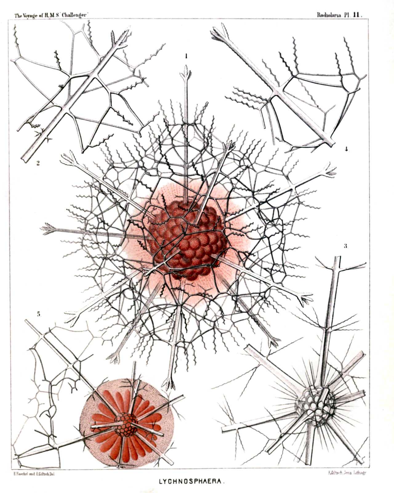 | |||||||||||||||||||||||||||||||||||||
| PLATE 11. | |||
| Astrosphærida. | |||
| Diam. | Page. | ||
Fig. 1. Lychnosphæra regina, n. sp., | × | 200 | 277 |
| The entire shell and the central capsule. Numerous club-shaped radial apophyses or cœcal sacs arise from the pink central capsule and are protruded through the pores of the medullary shell, which is completely hidden by them. The sarcomatrix in the calymma, surrounding the central capsule, exhibits a fine radial striation. Numerous retracted pseudopodia, bearing red granules, arise from the sarcomatrix and pierce the calymma radially. The interval between the two concentric shells is filled up by the hyaline calymma. | |||
| Fig. 2. Lychnosphæra regina, n. sp., | × | 400 | 277 |
| A part of the cortical shell, with a radial spine. | |||
| Fig. 3. Lychnosphæra regina, n. sp., | × | 400 | 277 |
| The medullary shell and the basal parts of the radial spines arising from it. | |||
| Fig. 4. Lychnosphæra regina, n. sp., | × | 400 | 277 |
| Distal end of a radial spine. | |||
| Fig. 5. Rhizoplegma lychnosphæra, n. sp., | × | 200 | 276 |
| The central capsule and the enclosed parts of the skeleton. The protoplasm is radially striped. The central nucleus (red) sends out numerous radial apophyses, which are protruded through the pores of the medullary shell. | |||
PLATE 12.
Legion SPUMELLARIA.
Orders PHÆOSPHÆRIA et SPHÆROIDEA.
Families Orosphærida, Astrosphærida et Liosphærida.
| 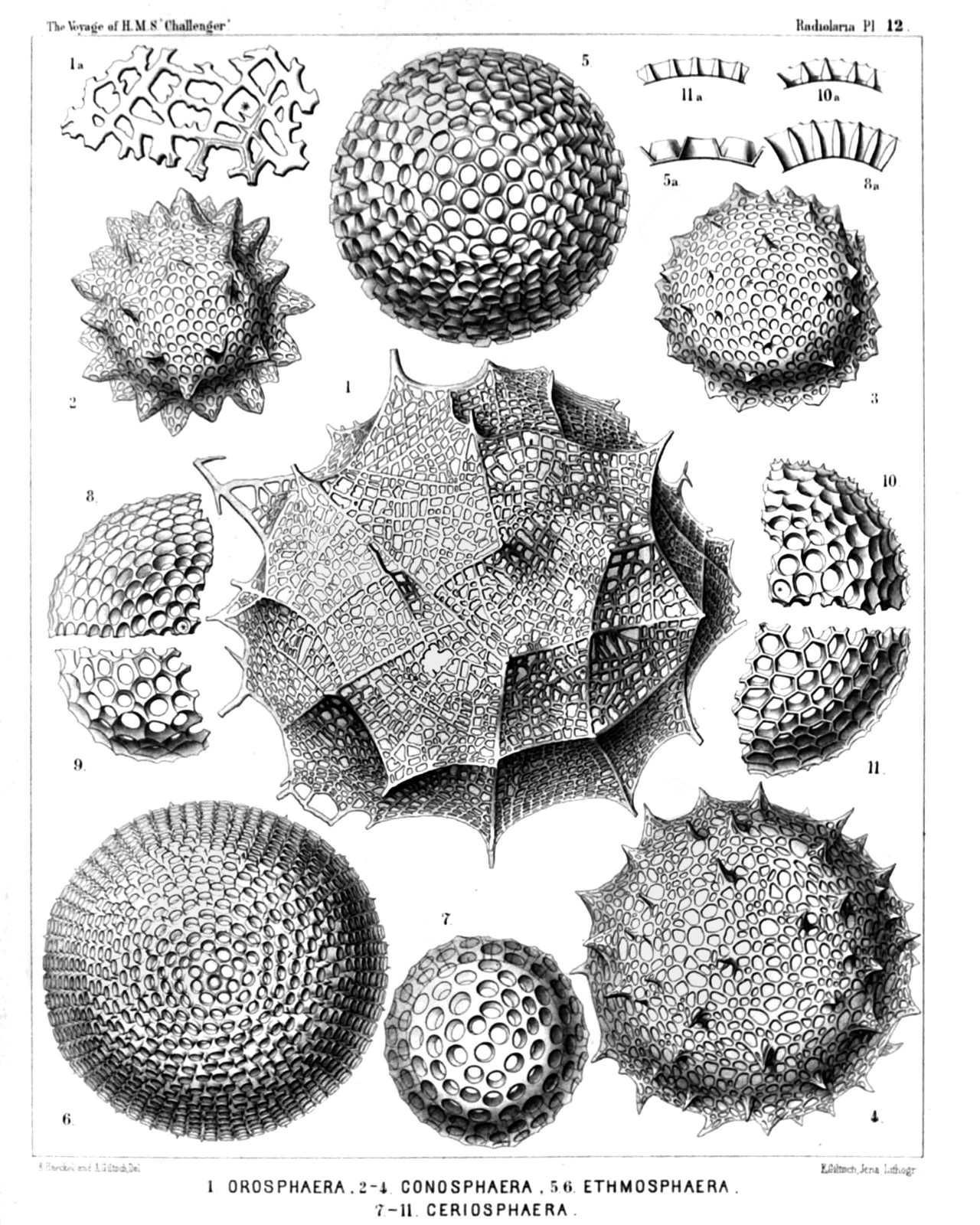 | ||||||||||||||||||||||||||||||||||||||||||||||||||||||||||||||||
| PLATE 12. | |||
| Orosphærida, Astrosphærida et Liosphærida. | |||
| Diam. | Page. | ||
Fig. 1. Orosphæra huxleyii, n. sp. (vel Oroscena huxleyii), | × | 50 | 1599 |
| Fig. 1a. A piece of the network, the bars of which contain partly an axial canal, | × | 200 | 1599 |
| Fig. 2. Conosphæra orthoconus, n. sp., | × | 200 | 221 |
| Fig. 3. Conosphæra platyconus, n. sp., | × | 300 | 221 |
| Fig. 4. Conosphæra plagioconus, n. sp., | × | 300 | 222 |
| Fig. 5. Ethmosphæra conosiphonia, n. sp., | × | 400 | 69 |
| Fig. 5a. Vertical section through the wall. | |||
| Fig. 6. Ethmosphæra polysiphonia, n. sp., | × | 400 | 70 |
| Fig. 7. Cenosphæra compacta, n. sp., | × | 300 | 65 |
| Fig. 8. Cenosphæra elysia, n. sp., | × | 300 | 64 |
| Fig. 8a. Vertical section through the wall. | |||
| Fig. 9. Cenosphæra mellifica, n. sp., | × | 300 | 62 |
| Fig. 10. Cenosphæra favosa, n. sp., | × | 300 | 62 |
| Fig. 10a. Vertical section through the wall. | |||
| Fig. 11. Cenosphæra vesparia, n. sp., | × | 300 | 62 |
| Fig. 11a. Vertical section through the wall. | |||
PLATE 13.
Legion SPUMELLARIA.
Orders SPHÆROIDEA et PRUNOIDEA.
Families Stylosphærida et Ellipsida.
| 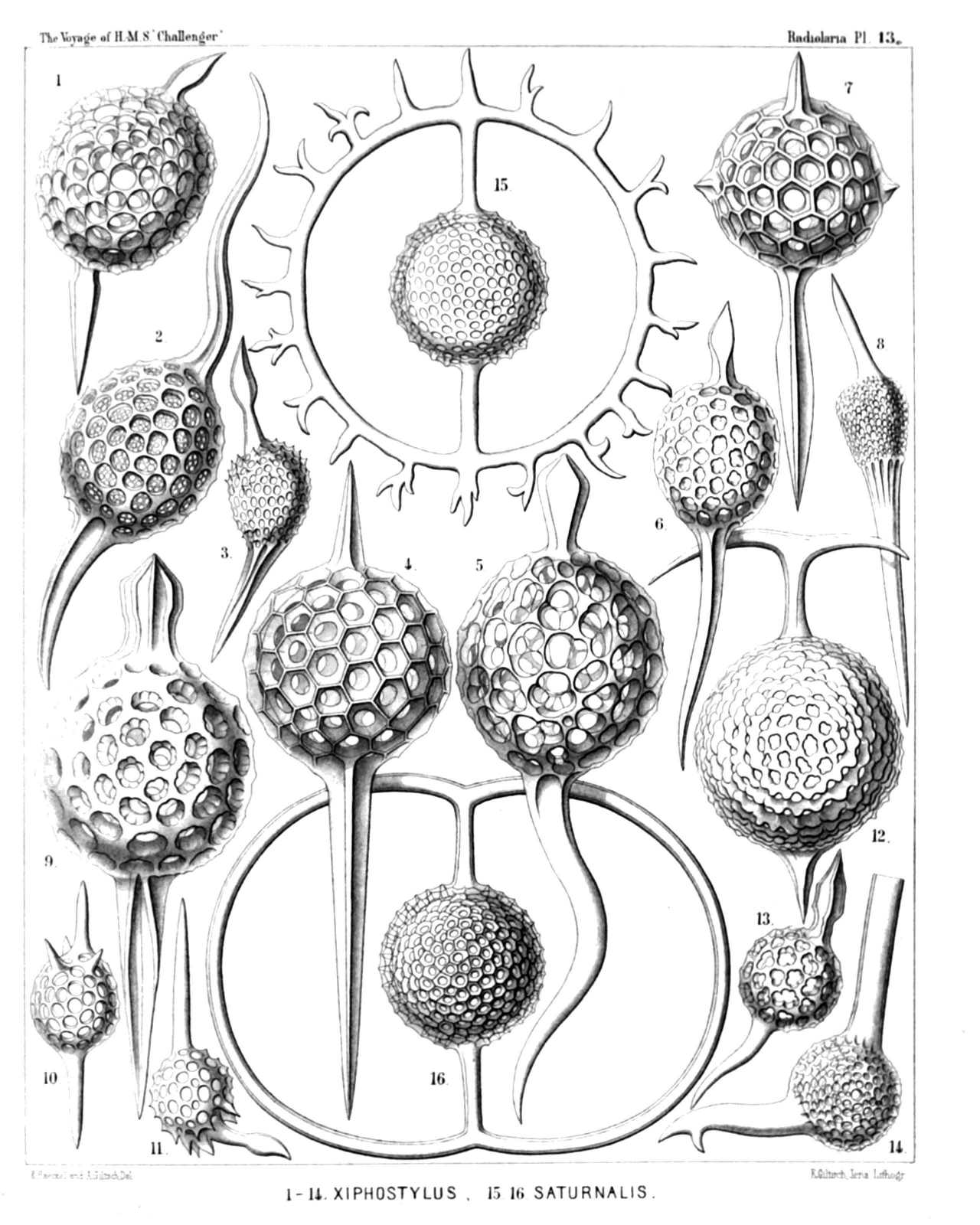 | |||||||||||||||||||||||||||||||||||||||||||||||||||||||||||||||||||||||||||||
| PLATE 13. | |||
| Stylosphærida et Ellipsida. | |||
| Diam. | Page. | ||
Fig. 1. Ellipsostylus aquila, n. sp., | × | 300 | 300 |
| Fig. 2. Ellipsostylus hirundo, n. sp., | × | 300 | 301 |
| Fig. 3. Ellipsostylus columba, n. sp., | × | 300 | 300 |
| Fig. 4. Xiphostylus alcedo, n. sp., | × | 400 | 127 |
| Fig. 5. Xiphostylus edolius, n. sp., | × | 400 | 130 |
| Fig. 6. Ellipsostylus psittacus, n. sp., | × | 400 | 300 |
| Fig. 7. Stylostaurus caudatus, n. sp., | × | 400 | 157 |
| Fig. 8. Ellipsostylus ciconia, n. sp., | × | 300 | 300 |
| Fig. 9. Xiphostylus phasianus, n. sp., | × | 400 | 127 |
| Fig. 10. Xiphostylus trochilus, n. sp., | × | 300 | 129 |
| Fig. 11. Xiphostylus emberiza, n. sp., | × | 300 | 131 |
| Fig. 12. Saturnalis circoideus, n. sp., | × | 400 | 132 |
| Not fully developed. | |||
| Fig. 13. Xiphostylus alca, n. sp., | × | 300 | 130 |
| Fig. 14. Xiphostylus falco, n. sp., | × | 300 | 130 |
| Fig. 15. Saturnalis rotula, n. sp., | × | 400 | 133 |
| Fig. 16. Saturnalis annularis, n. sp., | × | 400 | 132 |
PLATE 14.
Legion SPUMELLARIA.
Orders SPHÆROIDEA et PRUNOIDEA.
Families Stylosphærida et Ellipsida.
| 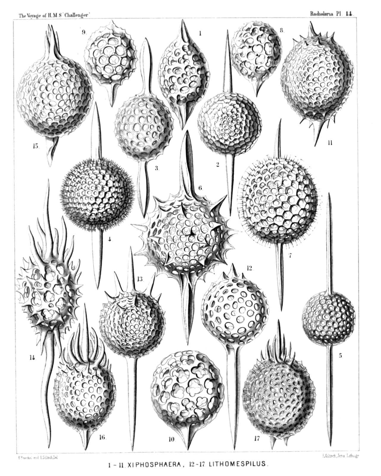 | ||||||||||||||||||||||||||||||||||||||||||||||||||||||||||||||||||||||||||||||||
| PLATE 14. | |||
| Stylosphærida et Ellipsida. | |||
| Diam. | Page. | ||
Fig. 1. Ellipsoxiphus atractus, n. sp., | × | 300 | 298 |
| Fig. 2. Xiphosphæra venus, n. sp., | × | 300 | 123 |
| Fig. 3. Ellipsoxiphus claviger, n. sp., | × | 300 | 297 |
| Fig. 4. Xiphosphæra pallas, n. sp., | × | 400 | 124 |
| Fig. 5. Xiphosphæra gæa, n. sp., | × | 400 | 123 |
| Fig. 6. Xiphosphæra vesta, n. sp., | × | 300 | 126 |
| Fig. 7. Ellipsoxiphus elegans, n. sp., var. palliatus, | × | 400 | 296 |
| Fig. 8. Lithapium halicapsa, n. sp., | × | 300 | 303 |
| Fig. 9. Lithapium pyriforme, n. sp., | × | 300 | 303 |
| Fig. 10. Lithapium monocyrtis, n. sp., | × | 300 | 304 |
| Fig. 11. Ellipsoxiphus bipolaris, n. sp., | × | 600 | 297 |
| Fig. 12. Xiphostylus trogon, n. sp., | × | 400 | 129 |
| Fig. 13. Xiphostylus picus, n. sp., | × | 300 | 129 |
| Fig. 14. Lithomespilus flammabundus, n. sp., | × | 400 | 303 |
| Fig. 15. Xiphostylus alauda, n. sp., | × | 400 | 128 |
| Fig. 16. Lithomespilus phloginus, n. sp., | × | 600 | 302 |
| Fig. 17. Lithomespilus phlogoides, n. sp., | × | 600 | 302 |
PLATE 15.
Legion SPUMELLARIA.
Orders SPHÆROIDEA et PRUNOIDEA.
Families Staurosphærida et Druppulida.
| 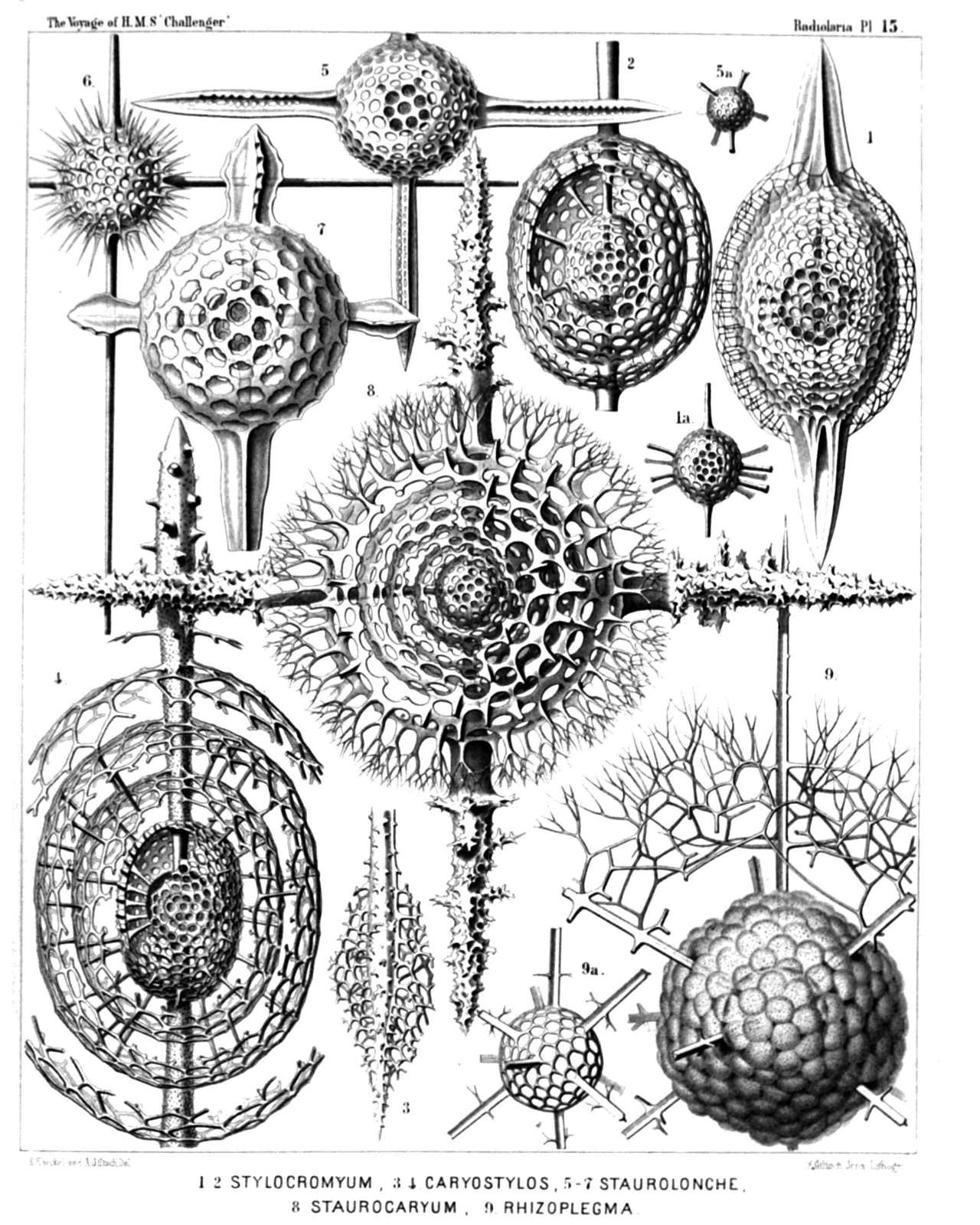 | ||||||||||||||||||||||||||||||||||||||||||||||||||||
| PLATE 15. | |||
| Staurosphærida et Druppulida. | |||
| Diam. | Page. | ||
Fig. 1. Cromyatractus tetracelyphus, n. sp., | × | 300 | 335 |
| Fig. 1a. The two inner medullary shells. | |||
| Fig. 2. Cromyatractus tetraphractus, n. sp., | × | 300 | 335 |
| Fig. 3. Cromyatractus cepicius, n. sp., | × | 300 | 336 |
| The spongy distal part of a polar spine. | |||
| Fig. 4. Cromyatractus ceparius, n. sp. (vel Caryostylus ceparius), | × | 300 | 336 |
| Fig. 5. Staurolonche pertusa, n. sp., | × | 300 | 159 |
| Fig. 5a. Its medullary shell. | |||
| Fig. 6. Staurosphæra philippi, n. sp., | × | 300 | 154 |
| Fig. 7. Stauroxiphus gladius, n. sp., | × | 400 | 163 |
| Fig. 8. Staurocaryum arborescens, n. sp., | × | 300 | 167 |
| Fig. 9. Rhizoplegma radicatum, n. sp., | × | 200 | 276 |
| Fig. 9a. The medullary shell, which is completely hidden in fig. 9 by the numerous club-shaped apophyses of the central capsule. | |||
PLATE 16.
Legion SPUMELLARIA.
Orders SPHÆROIDEA et PRUNOIDEA.
Families STYLOSPHÆRIDA et Druppulida.
| 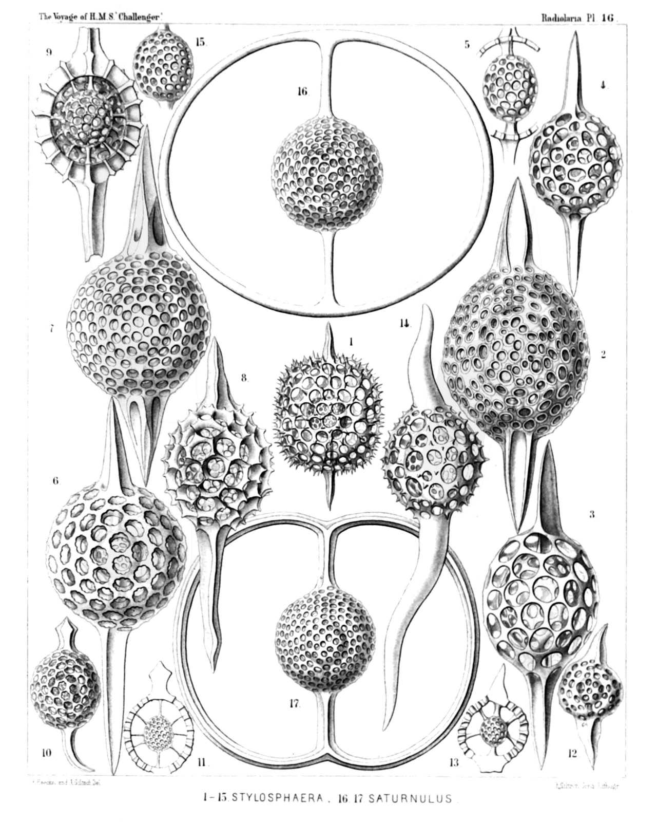 | |||||||||||||||||||||||||||||||||||||||||||||||||||||||||||||||||||||||||||||||||||||||||
| PLATE 16. | |||
| STYLOSPHÆRIDA et Druppulida. | |||
| Diam. | Page. | ||
Fig. 1. Stylosphæra melpomene, n. sp., | × | 300 | 135 |
| Fig. 2. Lithatractus jugatus, n. sp., (vel Stylosphæra jugata), | × | 400 | 323 |
| Fig. 3. Lithatractus fragilis, n. sp. (vel Stylosphæra fragilis), | × | 400 | 319 |
| Fig. 4. Stylosphæra lithatractus, n. sp., | × | 300 | |
| The entire shell. | |||
| Fig. 5. Stylosphæra lithatractus, n. sp., | × | 300 | |
| The greater part of the cortical shell and the two spines taken off. | |||
| The description of Stylosphæra lithatractus (intermediate between Stylosphæra jugata and Stylosphæra terpsichore, p. 137) is by mistake not given in the text. | |||
| Fig. 6. Stylosphæra calliope, n. sp., | × | 400 | 134 |
| Fig. 7. Stylosphæra clio, n. sp., | × | 400 | 134 |
| Fig. 8. Druppatractus ostracion, n. sp., | × | 300 | 326 |
| The entire shell. | |||
| Fig. 9. Druppatractus ostracion, n. sp., | × | 300 | 326 |
| The anterior half of the cortical shell has been removed. | |||
| Fig. 10. Druppatractus hippocampus, n. sp., | × | 300 | 324 |
| The entire shell. | |||
| Fig. 11. Druppatractus hippocampus, n. sp., | × | 300 | 324 |
| The greater part of the cortical shell has been removed. | |||
| Fig. 12. Stylosphæra nana, n. sp., | × | 300 | 136 |
| The entire shell | |||
| Fig. 13. Stylosphæra nana, n. sp., | × | 300 | 136 |
| The greater part of the cortical shell taken off. | |||
| Fig. 14. Sphærostylus ophidium, n. sp., | × | 300 | 140 |
| The entire shell. | |||
| Fig. 15. Sphærostylus ophidium, n. sp., | × | 300 | 140 |
| The medullary shell alone. | |||
| Fig. 16. Saturnulus ellipticus, n. sp., | × | 400 | 141 |
| Fig. 17. Saturnulus planetes, n. sp., | × | 400 | 142 |
PLATE 17.
Legion SPUMELLARIA.
Orders SPHÆROIDEA et PRUNOIDEA.
Families Stylosphærida, Druppulida et Spongurida.
| 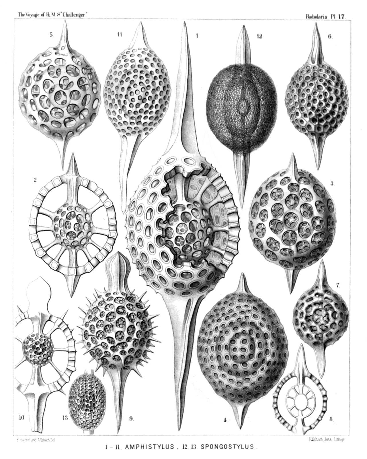 | ||||||||||||||||||||||||||||||||||||||||||||||||||||||||||||||||||||||||
| PLATE 17. | |||
| Stylosphærida, Druppulida et Spongurida. | |||
| Diam. | Page. | ||
Fig. 1. Stylatractus giganteus, n. sp. (vel Amphistylus giganteus), | × | 300 | 329 |
| Fig. 2. Stylatractus sethoporus, n. sp., | × | 400 | 330 |
| The greater part of the cortical shell taken off. | |||
| Fig. 3. Stylatractus sethoporus, n. sp., | × | 400 | 330 |
| The entire cortical shell. | |||
| Fig. 4. Stylatractus compactus, n. sp., | × | 400 | 329 |
| Fig. 5. Amphisphæra cronos, n. sp. (vel Amphistylus cronos), | × | 400 | 144 |
| Fig. 6. Stylatractus neptunus, n. sp. (vel Amphisphæra neptunus), | × | 300 | 328 |
| Fig. 7. Amphisphæra pluto, n. sp., | × | 300 | 144 |
| The entire cortical shell. | |||
| Fig. 8. Amphisphæra pluto, n. sp., | × | 300 | 144 |
| Meridional section through the three concentric shells. | |||
| Fig. 9. Xiphatractus glyptodon, n. sp., | × | 400 | 334 |
| The entire cortical shell. | |||
| Fig. 10. Xiphatractus glyptodon, n. sp., | × | 400 | 334 |
| The greater part of the cortical shell taken off. | |||
| Fig. 11. Xiphatractus armadillo, n. sp., | × | 400 | 332 |
| Fig. 12. Spongoxiphus prunococcus, n. sp., | × | 300 | 354 |
| The spongy cortical shell. | |||
| Fig. 13. Spongoxiphus prunococcus, n. sp., | × | 600 | 354 |
| The two concentric latticed medullary shells. | |||
PLATE 18.
Legion SPUMELLARIA.
Order SPHÆROIDEA.
Families Liosphærida et Astrosphærida.
| 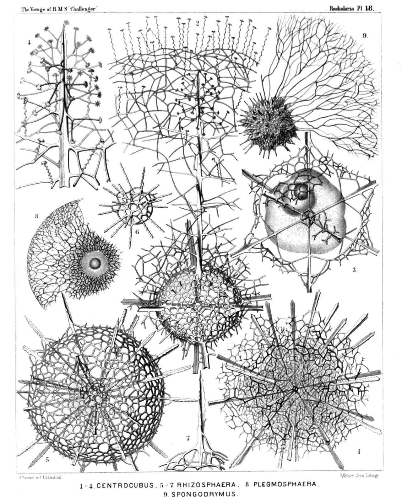 | |||||||||||||||||||||||||||||||||||||||||||||||||||||||
| PLATE 18. | |||
| Liosphærida et Astrosphærida. | |||
| Diam. | Page. | ||
Fig. 1. Centrocubus cladostylus, n. sp., | × | 100 | 278 |
| Fig. 2. Octodendron spathillatum, n. sp., | × | 300 | 280 |
| The entire inner shell, but a small part only of the outer shell is represented. | |||
| Fig. 3. Octodendron cubocentron, n. sp., | × | 400 | 279 |
| The central capsule (somewhat irregular by compression?) exhibits a large excentric nucleus (probably dislocated artificially?). | |||
| Fig. 4. Octodendron spathillatum, n. sp., | × | 800 | 280 |
| Free distal end of a radial spine, with the spathillæ on the end of the branches. | |||
| Fig. 5. Rhizosphæra serrata, n. sp., | × | 300 | 284 |
| Fig. 6. Rhizosphæra serrata, n. sp., | × | 300 | 284 |
| Medullary shell. | |||
| Fig. 7. Rhizosphæra serrata, n. sp., | × | 600 | 284 |
| A single radial spine. | |||
| Fig. 8. Plegmosphæra exodictyon, n. sp., | × | 200 | 89 |
| The central shell-cavity encloses the spherical central capsule and the concentric nucleus. | |||
| Fig. 9. Spongodrymus elaphococcus, n. sp., | × | 150 | 272 |
| The entire inner shell, but only a small part of the outer spongy envelope is represented. | |||
PLATE 19.
Legion SPUMELLARIA.
Order SPHÆROIDEA.
Family Astrosphærida.
| 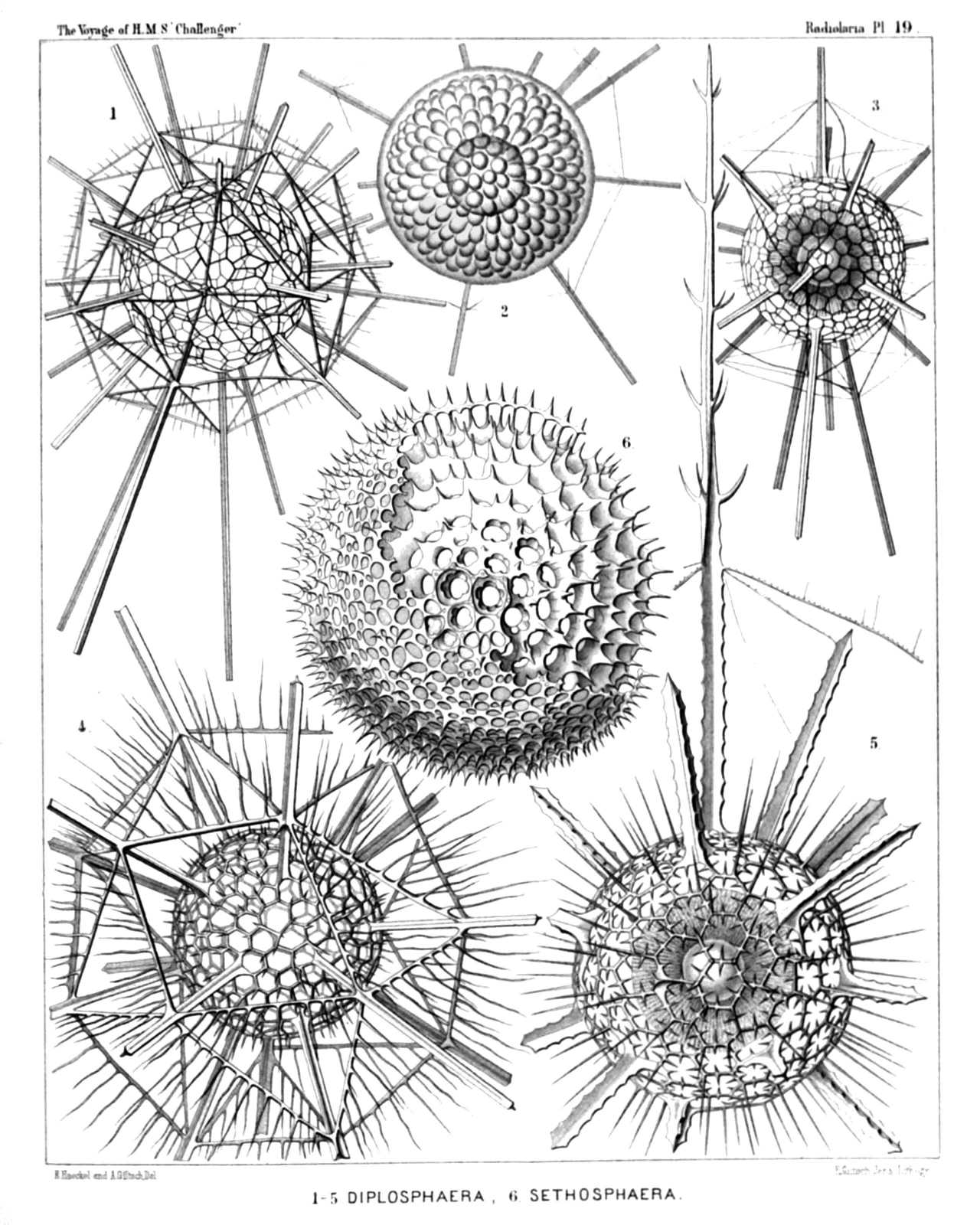 | ||||||||||||||||||||||||||||||||||||||||
| PLATE 19. | |||
| Astrosphærida. | |||
| Diam. | Page. | ||
Fig. 1. Drymosphæra polygonalis, n. sp., | × | 200 | 249 |
| Fig. 2. Leptosphæra hexagonalis, n. sp., | × | 200 | 244 |
| Showing the central capsule (forming numerous club-shaped protuberances) and the simple spherical nucleus in its centre. The skeleton is nearly the same as in Diplosphæra hexagonalis (fig. 3). | |||
| Fig. 3. Diplosphæra hexagonalis, n. sp., | × | 200 | 246 |
| The spherical central capsule, with radially striped protoplasm, is enclosed in the inner shell, and exhibits in its centre the clear spherical nucleus. | |||
| Fig. 4. Astrosphæra hexagonalis, n. sp., | × | 300 | 250 |
| Fig. 5. Astrosphæra stellata, n. sp., | × | 300 | 251 |
| The central capsule, enclosed in the inner shell, exhibits a distinct radial striation of the protoplasm, and in the centre a clear spherical nucleus. | |||
| Fig. 6. Haliomma rhodococcus, n. sp. (vel Sethosphæra rhodococcus), | × | 400 | 237 |
| The greater part of the outer shell is removed. | |||
PLATE 20.
Legion SPUMELLARIA.
Order SPHÆROIDEA.
Families Liosphærida et Astrosphærida.
| 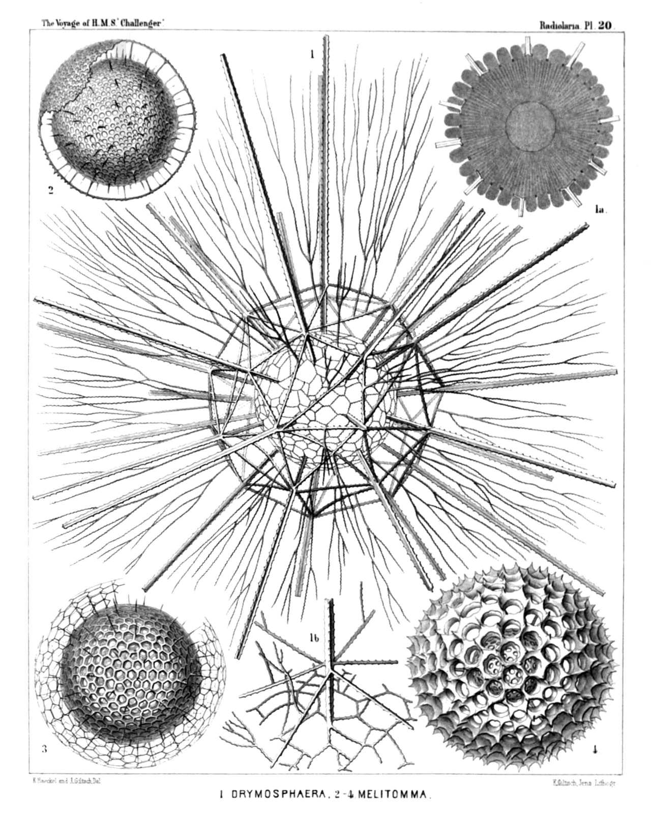 | |||||||||||||||||||||||||||||||||||
| PLATE 20. | |||
| Liosphærida et Astrosphærida. | |||
| Diam. | Page. | ||
Fig. 1. Drymosphæra dendrophora, n. sp., | × | 300 | 249 |
| Fig. 1a. Meridional section through the central capsule. In the centre the large spherical nucleus is visible. The protoplasm around it is distinctly radiate. From the central capsule arise numerous club-shaped apophyses or cæcal sacs, which are protruded through the meshes of the inner shell, | × | 300 | |
| Fig. 1b. Basal part of a single radial spine, and its connection with the network of the two shells, | × | 400 | |
| Fig. 2. Liosphæra polypora, n. sp., | × | 300 | 78 |
| The greater part of the outer shell is removed. | |||
| Fig. 3. Liosphæra hexagonia, n. sp., | × | 400 | 76 |
| Fig. 4. Carposphæra melitomma, n. sp. (vel Melitomma melittosphæra), | × | 400 | 73 |
PLATE 21.
Legion SPUMELLARIA.
Order SPHÆROIDEA.
Family Cubosphærida.
| 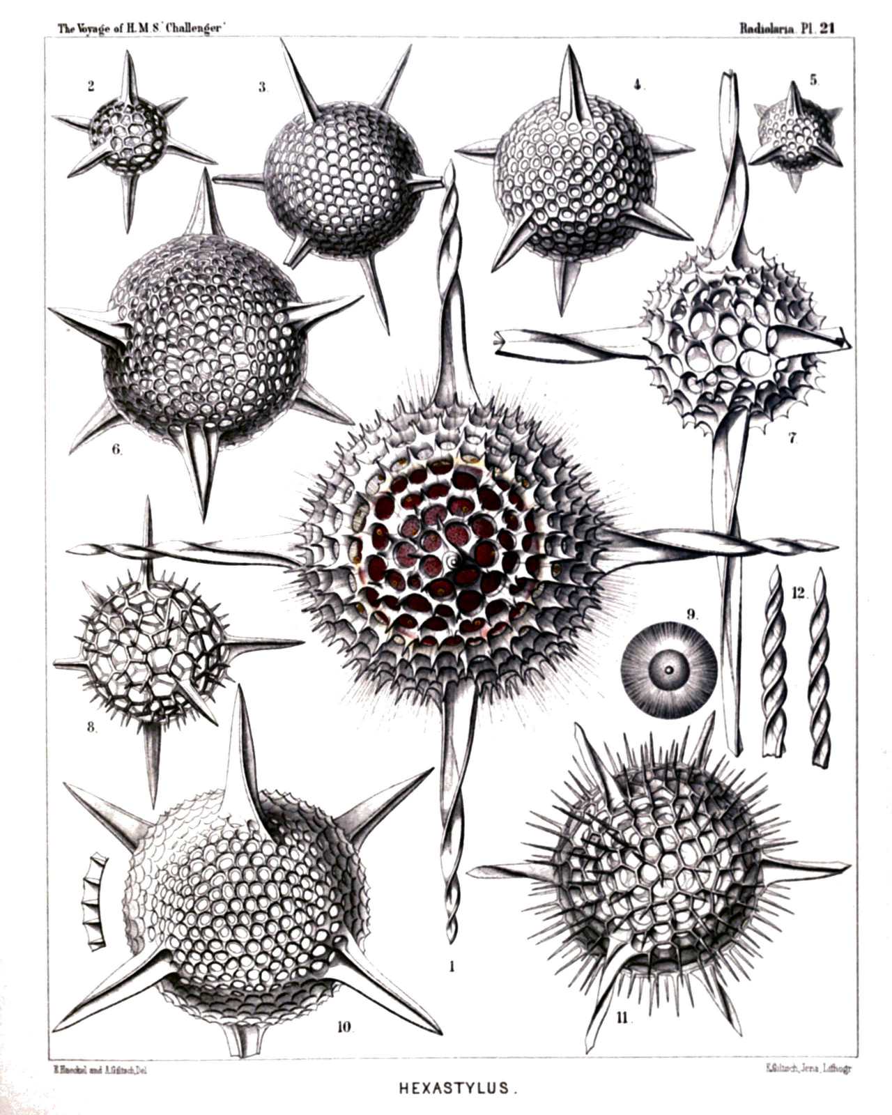 | |||||||||||||||||||||||||||||||||||||||||||||||||||||||||||||||
| PLATE 21. | |||
| Cubosphærida. | |||
| Diam. | Page. | ||
Fig. 1. Hexastylus cochleatus, n. sp., | × | 400 | 174 |
| From the central capsule, enclosed in the shell, numerous delicate radial pseudopodia arise, which are protruded through the pores of the shell. | |||
| Fig. 2. Hexastylus triaxonius, n. sp., | × | 400 | 175 |
| Fig. 3. Hexastylus phænaxonius, n. sp., | × | 300 | 171 |
| Fig. 4. Hexastylus thaletis, n. sp., | × | 400 | 172 |
| Fig. 5. Hexastylus minimus, n. sp., | × | 400 | 172 |
| Fig. 6. Hexastylus dimensivus, n. sp., | × | 400 | 175 |
| Fig. 7. Hexastylus spiralis, n. sp., | × | 400 | 177 |
| Fig. 8. Hexastylus dictyotus, n. sp., | × | 400 | 176 |
| Fig. 9. Hexastylus dictyotus, n. sp., | × | 400 | 176 |
| Central capsule with concentric nucleus and nucleolus; the protoplasm is radially striped. | |||
| Fig. 10. Hexastylus marginatus, n. sp., | × | 400 | 176 |
| Fig. 10a. Radial section through the shell-wall. | |||
| Fig. 11. Hexastylus solonis, n. sp., | × | 400 | 173 |
| Fig. 12. Hexastylus contortus, n. sp., | × | 300 | 177 |
PLATE 22.
Legion SPUMELLARIA.
Order SPHÆROIDEA.
Family Cubosphærida.
| 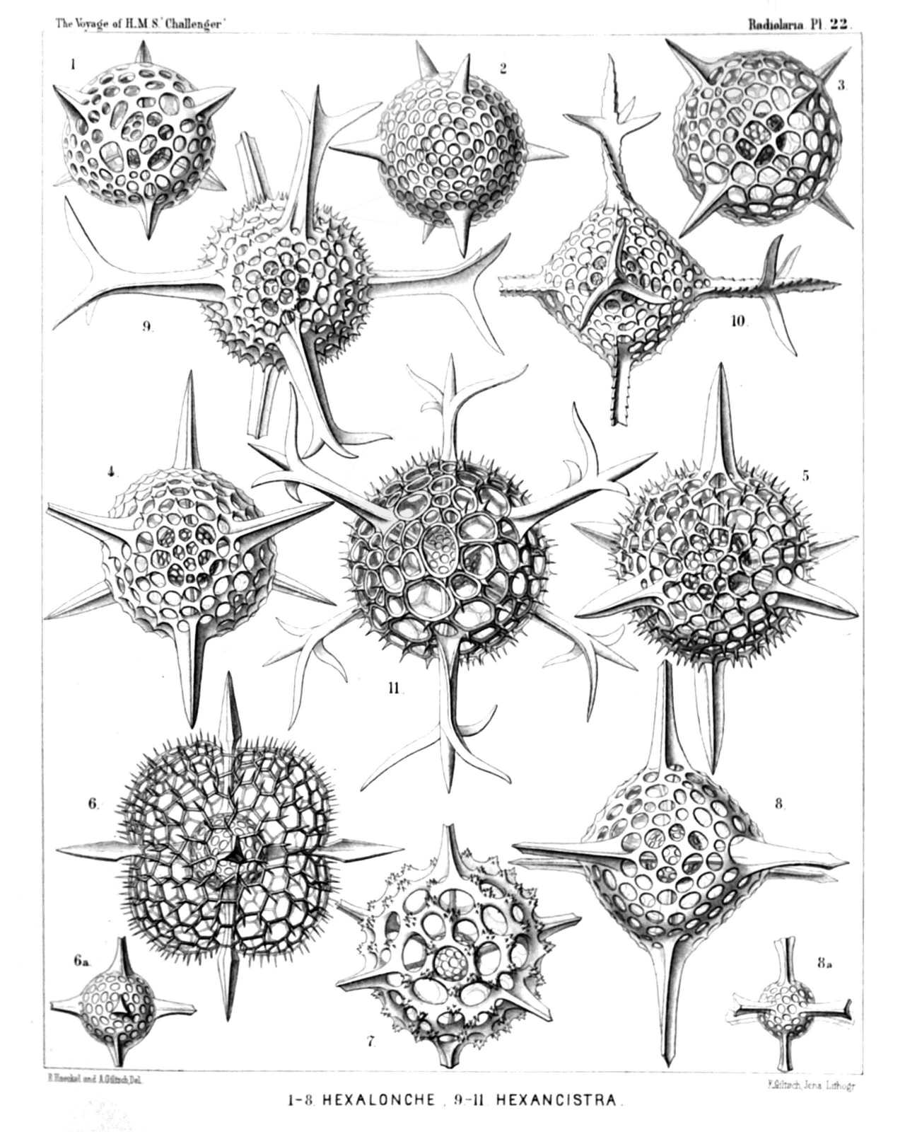 | ||||||||||||||||||||||||||||||||||||||||||||||||||||||||||
| PLATE 22. | |||
| Cubosphærida. | |||
| Diam. | Page. | ||
Fig. 1. Hexalonche pythagoræa, n. sp., | × | 300 | 185 |
| Fig. 2. Hexalonche conicornis, n. sp., | × | 300 | 181 |
| Fig. 3. Hexalonche aristarchi, n. sp., | × | 400 | 185 |
| Fig. 4. Hexalonche philosophica, n. sp., | × | 400 | 186 |
| Fig. 5. Hexalonche anaximandri, n. sp., | × | 400 | 182 |
| Fig. 6. Hexalonche octocolpa, n. sp., | × | 300 | 183 |
| Fig. 6a. The inner shell alone. | |||
| Fig. 7. Hexalonche heracliti, n. sp., | × | 300 | 187 |
| Fig. 8. Hexalonche octahedra, n. sp., | × | 400 | 181 |
| Fig. 8a. The inner shell alone. | |||
| Fig. 9. Hexancistra tricuspis, n. sp., | × | 300 | 188 |
| Fig. 10. Hexancistra triserrata, n. sp., | × | 300 | 188 |
| Fig. 11. Hexancistra quadricuspis, n. sp., | × | 300 | 189 |
PLATE 23.
Legion SPUMELLARIA.
Order SPHÆROIDEA.
Family Cubosphærida.
| 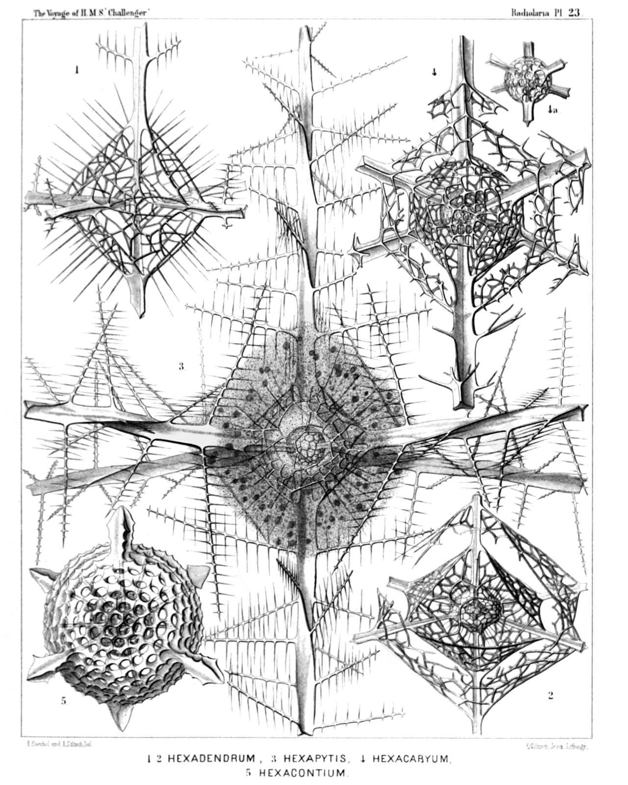 | |||||||||||||||||||||||||||||||||
| PLATE 23. | |||
| Cubosphærida. | |||
| Diam. | Page. | ||
Fig. 1. Hexadendron bipinnatum, n. sp., | × | 400 | 200 |
| Fig. 2. Hexacromyum octahedrum, n. sp., | × | 400 | 202 |
| Fig. 3. Hexancistra mirabilis, n. sp. (= Hexapitys mirabilis), | × | 400 | 189 |
| The spherical central capsule encloses the concentric spherical inner shell (which is filled up by the nucleus), and is surrounded by the octahedral outer shell. The latter is enveloped by the octahedral calymma, which is radially striated and contains numerous xanthellæ. | |||
| Fig. 4. Hexacaryum arborescens, n. sp., | × | 400 | 203 |
| Fig. 5. Hexacontium clavigerum, n. sp., | × | 300 | 19 |
PLATE 24.
Legion SPUMELLARIA.
Order SPHÆROIDEA.
Family Cubosphærida.
| 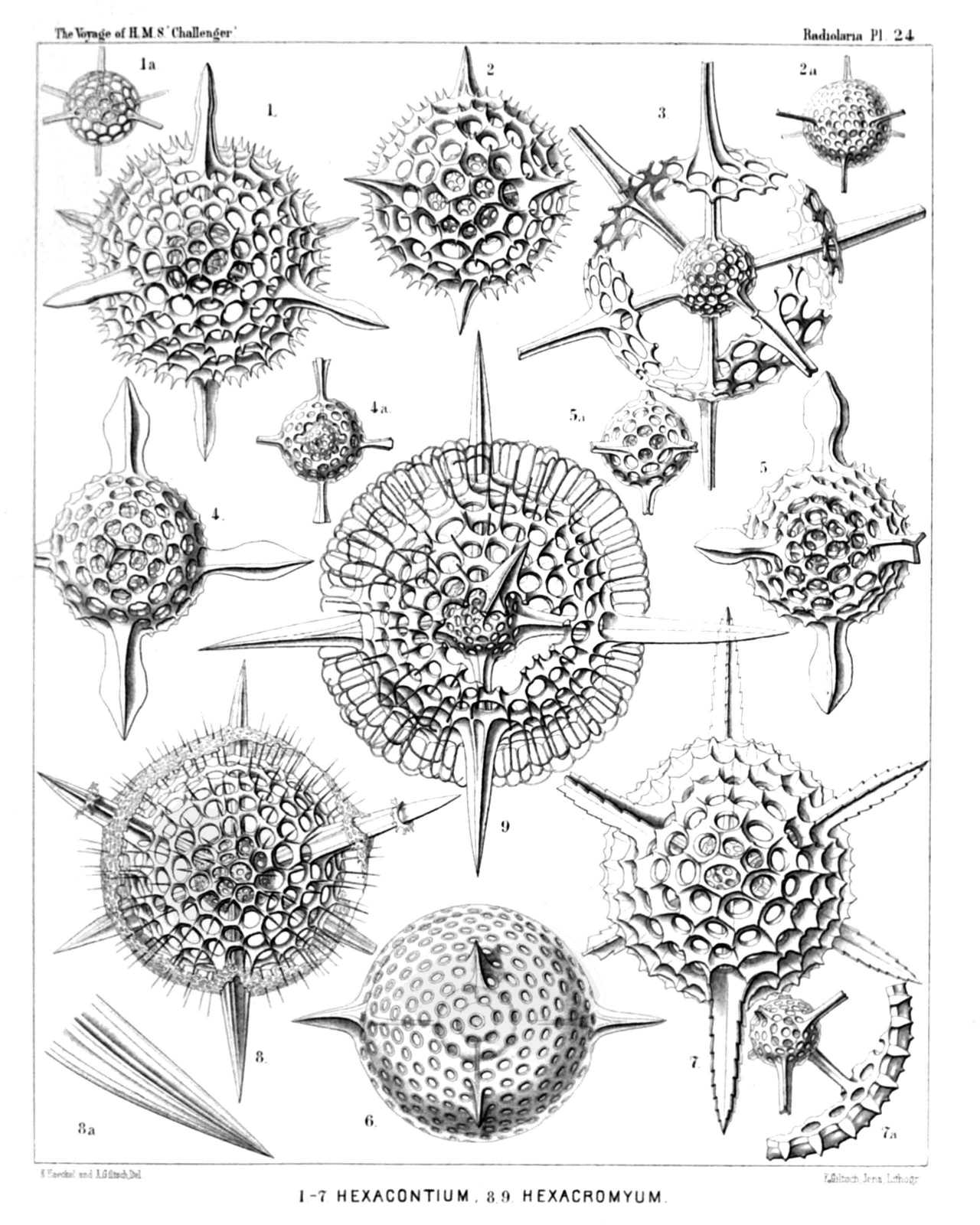 | |||||||||||||||||||||||||||||||||||||||||||||||||||||||||
| PLATE 24. | |||
| Cubosphærida. | |||
| Diam. | Page. | ||
Fig. 1. Hexacontium sceptrum, n. sp., | × | 400 | 194 |
| Fig. 1a. The two medullary shells. | |||
| Fig. 2. Hexacontium favosum, n. sp., | × | 400 | 194 |
| Fig. 2a. The two medullary shells. | |||
| Fig. 3. Hexacontium axotrias, n. sp., | × | 300 | 192 |
| The six lattice-plates, which form the cortical shell, are not yet fully developed. | |||
| Fig. 4. Hexacontium floridum, n. sp., | × | 300 | 195 |
| Fig. 4a. The two medullary shells. | |||
| Fig. 5. Hexacontium papillosum, n. sp., | × | 400 | 197 |
| Fig. 5a. The two medullary shells. | |||
| Fig. 6. Hexacontium lævigatum, n. sp., | × | 400 | 193 |
| The contours of the two medullary shells are visible in the centre. | |||
| Fig. 7. Hexacontium prionacanthum, n. sp., | × | 400 | 195 |
| Fig. 7a. The two medullary shells, connected with a fragment of the cortical shell. | |||
| Fig. 8. Cubosphæra cubaxonia, n. sp., | × | 400 | 203 |
| Fig. 8a. A single radial spine. | |||
| Fig. 9. Hexacromyum elegans, n. sp., | × | 400 | 201 |
| A part of the two cortical shells is broken off. | |||
PLATE 25.
Legion SPUMELLARIA.
Order SPHÆROIDEA.
Family Cubosphærida.
| 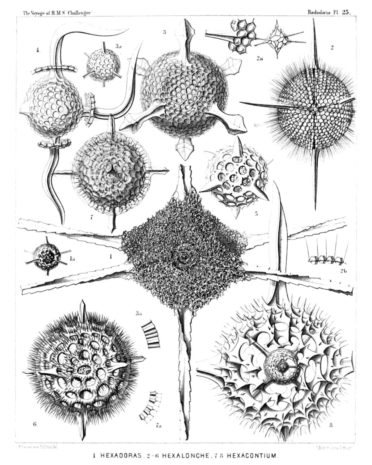 | ||||||||||||||||||||||||||||||||||||||||||||||||||||
| PLATE 25. | |||
| Cubosphærida. | |||
| Diam. | Page. | ||
Fig. 1. Hexadoridium streptacanthum, n. sp., | × | 400 | 206 |
| Fig. 1a. The two concentric medullary shells. | |||
| Fig. 2. Hexalonche amphisiphon, n. sp., | × | 300 | 182 |
| Fig. 2a. Medullary shell connected with a fragment of the cortical shell. | |||
| Fig. 2b. Vertical section through the wall of the cortical shell. (Below the centre of the Plate, also lettered 3a by mistake.) | |||
| Fig. 3. Hexalonche rosetta, n. sp., | × | 400 | 180 |
| Fig. 3a. Medullary shell. | |||
| Fig. 3b. Vertical section through the wall of the cortical shell. | |||
| Fig. 4. Hexalonche curvicornis, n. sp., | × | 300 | 181 |
| Outer shell not yet complete, or partly broken off (?). | |||
| Fig. 5. Hexalonche anaximenis, n. sp., | × | 400 | 183 |
| Fig. 6. Hexalonche hystricina, n. sp., | × | 300 | 187 |
| Fig. 7. Hexacontium circumtextum, n. sp., | × | 400 | 193 |
| Fig. 7a. Vertical section through the double wall of the cortical shell. | |||
| Fig. 8. Hexacontium gladiatum, n. sp., | × | 400 | 198 |
| A part of the two outer shells and of the radial spines is broken off. | |||
PLATE 26.
Legion SPUMELLARIA.
Order SPHÆROIDEA.
Families Liosphærida et Astrosphærida.
| 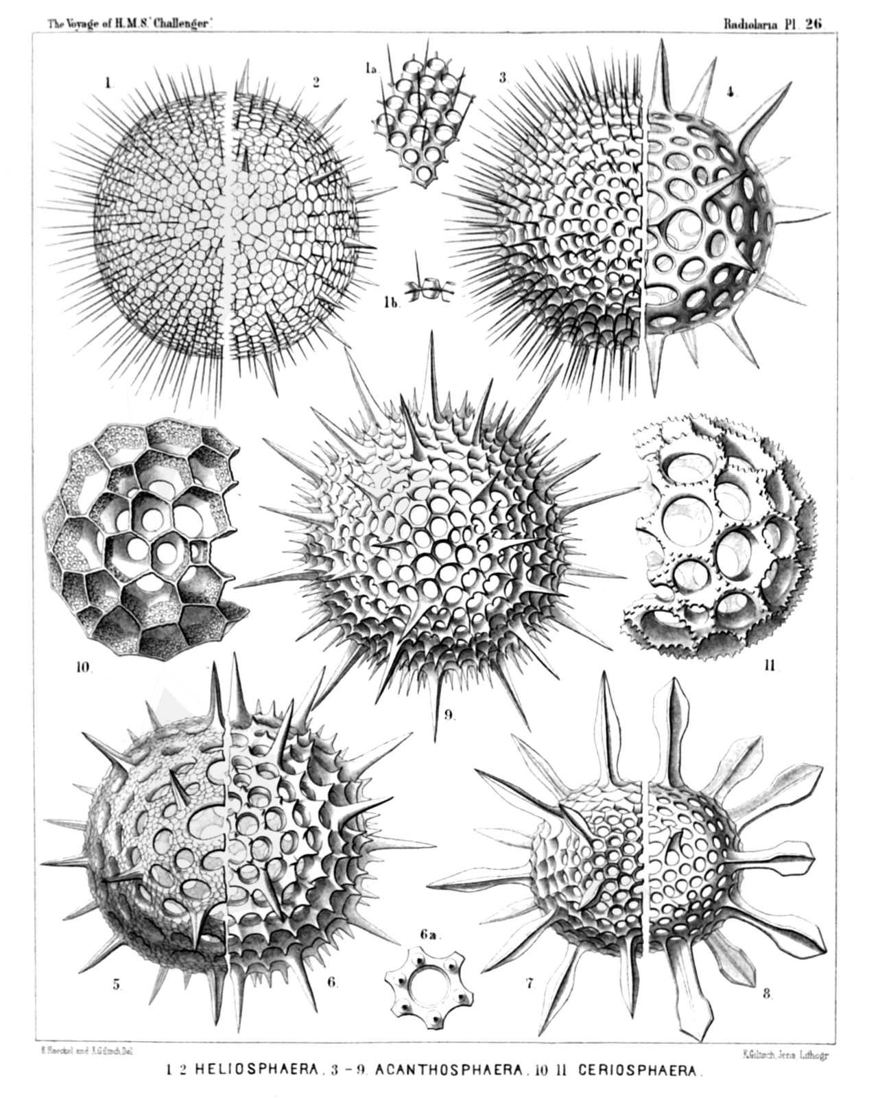 | |||||||||||||||||||||||||||||||||||||||||||||||||||||||||||||||||
| PLATE 26. | |||
| Liosphærida et Astrosphærida. | |||
| Diam. | Page. | ||
Fig. 1. Coscinomma amphisiphon, n. sp., | × | 300 | 222 |
| Fig. 1a. A piece of the lattice-shell, | × | 600 | |
| Fig. 1b. Vertical section through the shell-wall, | × | 600 | |
| Fig. 2. Heliosphæra hexagonaria, n. sp., | × | 300 | 217 |
| Fig. 3. Acanthosphæra castanea, n. sp., | × | 400 | 211 |
| Fig. 4. Acanthosphæra angulata, n. sp., | × | 300 | 216 |
| Fig. 5. Acanthosphæra reticulata, n. sp., | × | 300 | 217 |
| Fig. 6. Heliosphæra coronata, n. sp., | × | 400 | 219 |
| Fig. 6a. A single pore with its coronal, | × | 300 | |
| Fig. 7. Acanthosphæra mucronata, n. sp., | × | 400 | 212 |
| Fig. 8. Acanthosphæra clavata, n. sp., | × | 400 | 212 |
| Fig. 9. Heliosphæra pectinata, n. sp., | × | 400 | 218 |
| Fig. 10. Cenosphæra perforata, n. sp., | × | 400 | 66 |
| Fig. 11. Cenosphæra coronata, n. sp., | × | 400 | 67 |
PLATE 27.
Legion SPUMELLARIA.
Order SPHÆROIDEA.
Family Astrosphærida.
| 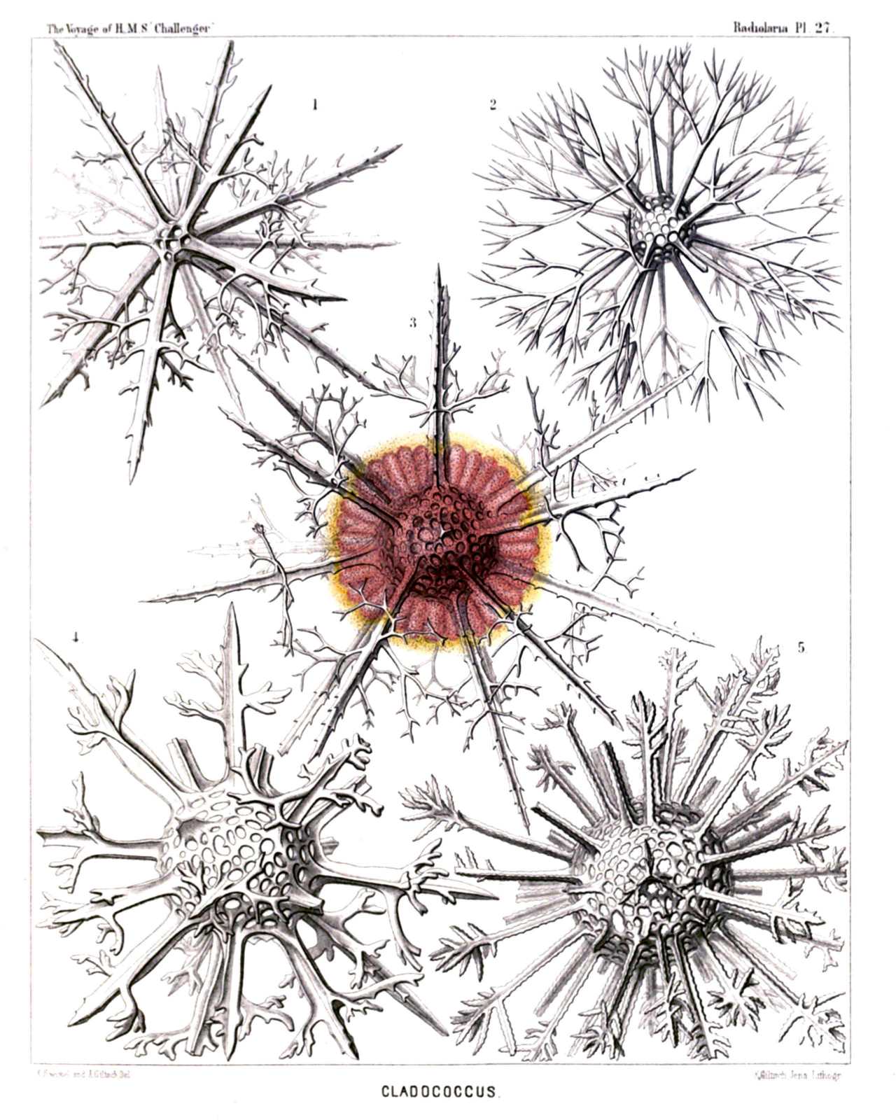 | |||||||||||||||||||||||||||||||||
| PLATE 27. | |||
| Astrosphærida. | |||
| Diam. | Page. | ||
Fig. 1. Cladococcus pinetum, n. sp., | × | 300 | 226 |
| Fig. 2. Cladococcus scoparius, n. sp., | × | 300 | 225 |
| Fig. 3. Cladococcus abietinus, n. sp., | × | 300 | 226 |
| The central capsule, enclosed originally in the shell, sends out numerous club-shaped apophyses through the pores of the lattice-sphere. The central spherical nucleus fills up half the shell-cavity. | |||
| Fig. 4. Cladococcus stalactites, n. sp., | × | 300 | 227 |
| Fig. 5. Cladococcus dendrites, n. sp., | × | 200 | 227 |
PLATE 28.
Legion SPUMELLARIA.
Order SPHÆROIDEA.
Families Liosphærida et Astrosphærida.
| 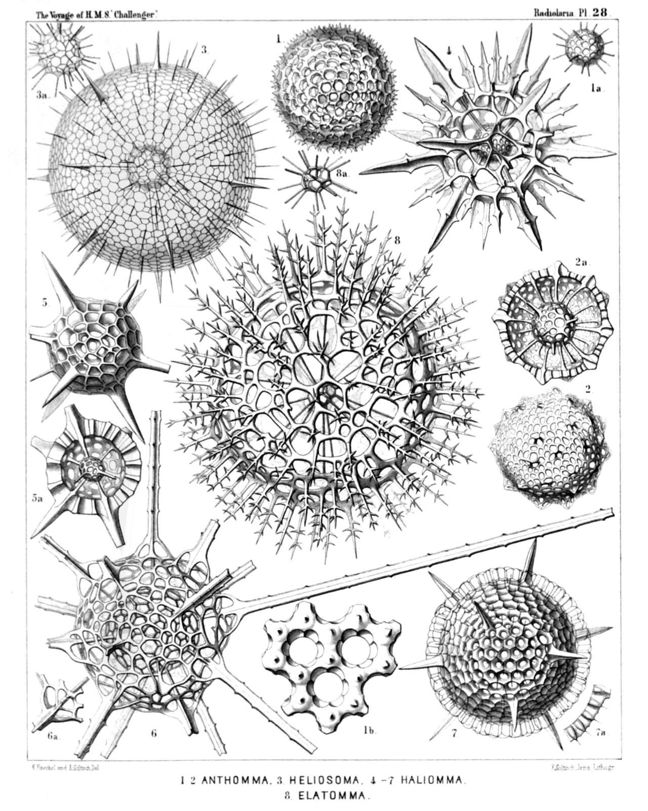 | ||||||||||||||||||||||||||||||||||||||||||||||||||||||||||||||
| PLATE 28. | |||
| Liosphærida et Astrosphærida. | |||
| Diam. | Page. | ||
Fig. 1. Haliomma lirianthus, n. sp., | × | 300 | 232 |
| Fig. 1a. Medullary shell, | × | 300 | |
| Fig. 1b. Three pores of the cortical shell, | × | 900 | |
| Fig. 2. Carposphæra nodosa, n. sp., | × | 300 | 76 |
| Fig. 2a. The medullary shell is visible, the upper half of the cortical shell being taken off, | × | 300 | |
| Fig. 3. Heliosoma radians, n. sp., | × | 300 | 240 |
| Fig. 3a. Medullary shell, | × | 300 | |
| Fig. 4. Heliosoma hastatum, n. sp., | × | 400 | 241 |
| Fig. 5. Haliomma compactum, n. sp., | × | 400 | 239 |
| Fig. 5a. The upper half of the cortical shell is removed, | × | 300 | |
| Fig. 6. Haliomma macrodoras, n. sp., | × | 400 | 238 |
| Fig. 7. Haliomma circumtextum, n sp., | × | 400 | 233 |
| Fig. 8. Elatomma juniperinum, n. sp., | × | 400 | 243 |
| Fig. 8a. Medullary shell, | × | 400 | |
PLATE 29.
Legion SPUMELLARIA.
Order SPHÆROIDEA.
Family Astrosphærida.
| 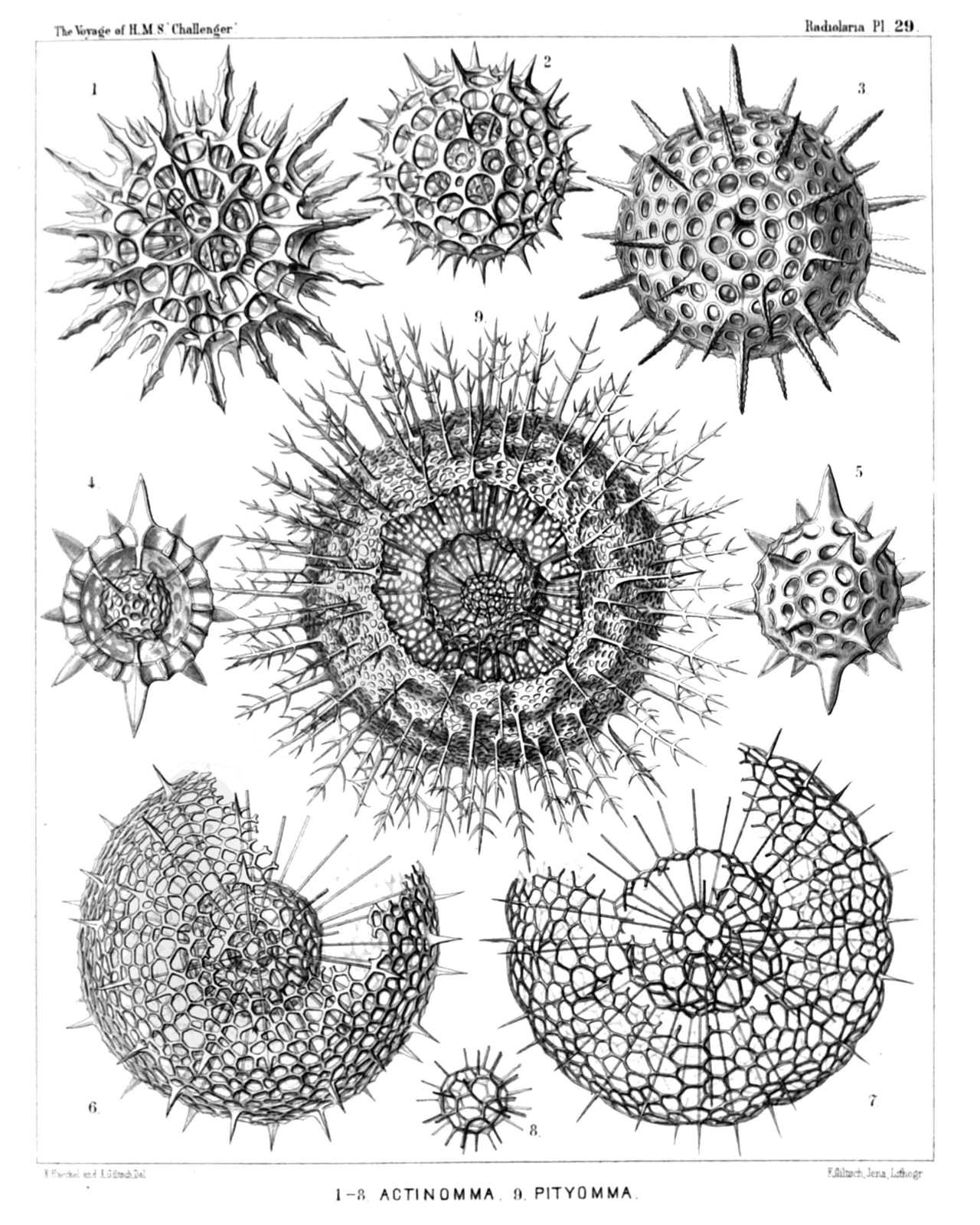 | ||||||||||||||||||||||||||||||||||||||||||||||||||||
| PLATE 29. | |||
| Astrosphærida. | |||
| Diam. | Page. | ||
Fig. 1. Echinomma toxopneustes, n. sp., | × | 400 | 259 |
| Fig. 2. Echinomma sphærechinus, n. sp., | × | 400 | 258 |
| Fig. 3. Actinomma denticulatum, n. sp., | × | 400 | 254 |
| Fig. 4. Actinomma pachyderma, n. sp., | × | 400 | 254 |
| The half of the cortical shell is removed. | |||
| Fig. 5. Actinomma pachyderma, n. sp., | × | 400 | 254 |
| Fig. 6. Actinomma capillaceum, n. sp., | × | 300 | 255 |
| Fig. 7. Actinomma arcadophorum, n. sp., | × | 400 | 255 |
| A part of the two outer shells is removed. | |||
| Fig. 8. Actinomma arcadophorum, n. sp., | × | 400 | 255 |
| Inner medullary shell. | |||
| Fig. 9. Pityomma drymodes, n. sp., | × | 300 | 260 |
| A part of the two outer shells is removed. | |||
PLATE 30.
Legion SPUMELLARIA.
Order SPHÆROIDEA.
Families Liosphærida et Astrosphærida.
| 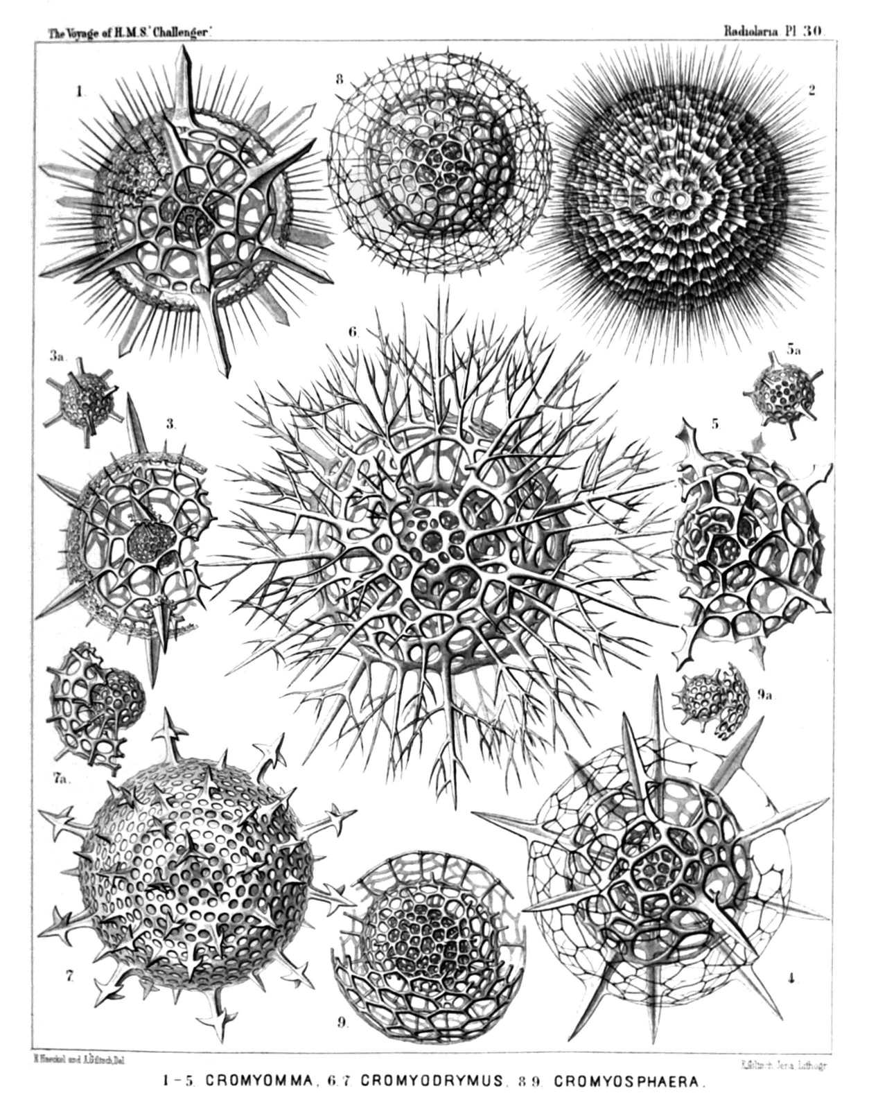 | ||||||||||||||||||||||||||||||||||||||||||||||||||||
| PLATE 30. | |||
| Liosphærida et Astrosphærida. | |||
| Diam. | Page. | ||
Fig. 1. Cromyechinus icosacanthus, n. sp., | × | 300 | 263 |
| Fig. 2. Cromyomma villosum, n. sp., | × | 300 | 261 |
| Fig. 3. Cromyechinus dodecacanthus, n. sp., | × | 400 | 264 |
| Fig. 3a. The innermost shells. | |||
| Fig. 4. Cromyomma circumtextum, n. sp., | × | 300 | 262 |
| Fig. 5. Cromyomma mucronatum, n. sp., | × | 200 | 263 |
| Fig. 5a. The innermost shells. | |||
| Fig. 6. Cromyodrymus abietinus, n. sp., | × | 300 | 265 |
| Fig. 7. Cromyodrymus quadricuspis, n. sp., | × | 400 | 264 |
| Fig. 7a. The inner concentric shells. | |||
| Fig. 8. Cromyomma perspicuum, n. sp., | × | 300 | 262 |
| Fig. 9. Cromyosphæra quadruplex, n. sp., | × | 300 | 84 |
| Fig. 9a. The innermost shells. | |||
PLATE 31.
Legion SPUMELLARIA.
Order DISCOIDEA.
Families Cenodiscida et Phacodiscida.
| 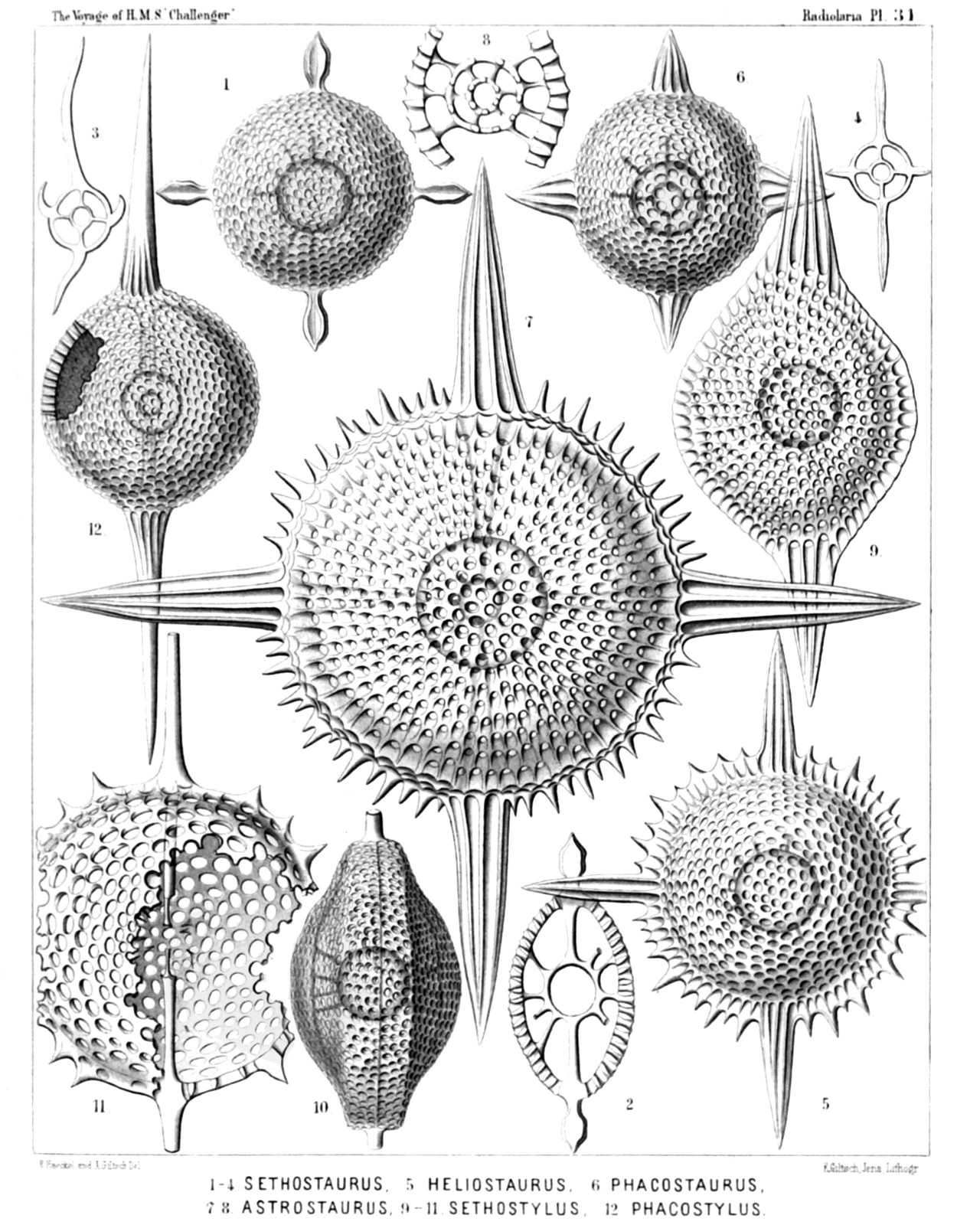 | |||||||||||||||||||||||||||||||||||||||||||||||||||||||||||||||||
| PLATE 31. | |||
| Cenodiscida et Phacodiscida. | |||
| Diam. | Page. | ||
Fig. 1. Sethostaurus orthostaurus, n. sp., | × | 300 | 433 |
| Fig. 2. Sethostaurus orthostaurus, n. sp., | × | 300 | 433 |
| Vertical section through the centrum. | |||
| Fig. 3. Sethostaurus recurvatus, n. sp., | × | 100 | 434 |
| Optical section through the equatorial plane. | |||
| Fig. 4. Sethostaurus rhombostaurus, n. sp., | × | 100 | 434 |
| Optical section through the equatorial plane. | |||
| Fig. 5. Sethostaurus cruciatus, n. sp. (vel Heliostaurus cruciatus), | × | 300 | 434 |
| Fig. 6. Phacostaurus oceanidum, n. sp., | × | 300 | 435 |
| Fig. 7. Phacostaurus magnificus, n. sp., | × | 400 | 436 |
| Fig. 8. Phacostaurus magnificus, n. sp., | × | 200 | 436 |
| Vertical section through the centrum. | |||
| Fig. 9. Sethostylus distyliscus, n. sp., | × | 400 | 428 |
| Fig. 10. Sethostylus dicylindrus, n. sp., | × | 300 | 428 |
| Marginal view. | |||
| Fig. 11. Stylodiscus endostylus, n. sp. (vel Sethostylus endostylus), | × | 300 | 413 |
| Fig. 12. Phacostylus amphistylus, n. sp., | × | 300 | 430 |
PLATE 32.
Legion SPUMELLARIA.
Order DISCOIDEA.
Family Phacodiscida.
| 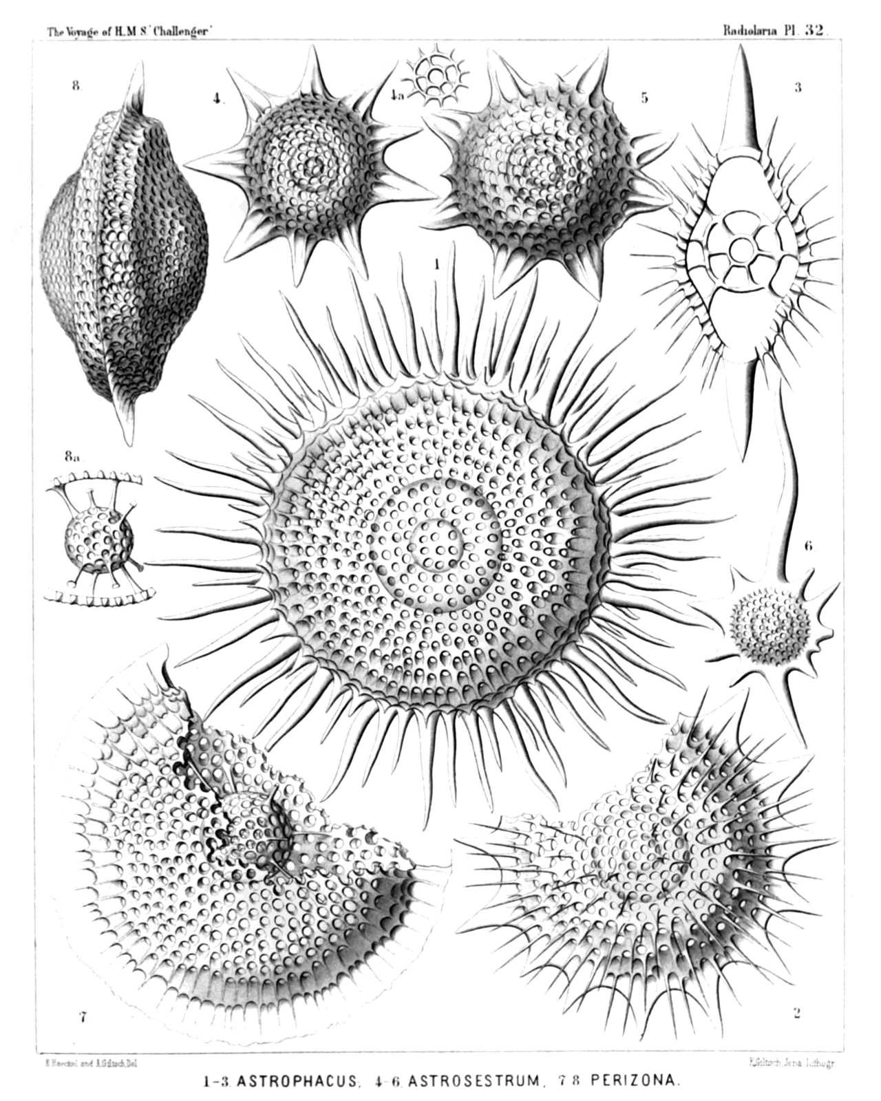 | |||||||||||||||||||||||||||||||||||||||||||||||||||||
| PLATE 32. | |||
| Phacodiscida. | |||
| Diam. | Page. | ||
Fig. 1. Astrophacus solaris, n. sp., | × | 300 | 453 |
| Fig. 2. Astrophacus apollinis, n. sp., | × | 300 | 455 |
| Fig. 3. Astrophacus phacodiscus, n. sp., | × | 300 | 454 |
| Vertical section through the centrum. | |||
| Fig. 4. Astrosestrum ephyra, n. sp., | × | 300 | 442 |
| Fig. 4a. Transverse section through the double medullary shell, | × | 300 | 442 |
| Fig. 5. Astrosestrum nauphanta, n. sp., | × | 300 | 442 |
| Fig. 6. Phacostylus caudatus, n. sp. (vel Astrosestrum caudatum), | × | 200 | 431 |
| Fig. 7. Perizona scutella, n. sp., | × | 400 | 427 |
| Fig. 8. Perizona pterygota, n. sp., | × | 400 | 427 |
| Fig. 8a. Medullary shells and radial beams connecting them with the disk, | × | 300 | 427 |
PLATE 33.
Legion SPUMELLARIA.
Order DISCOIDEA.
Family Phacodiscida.
| 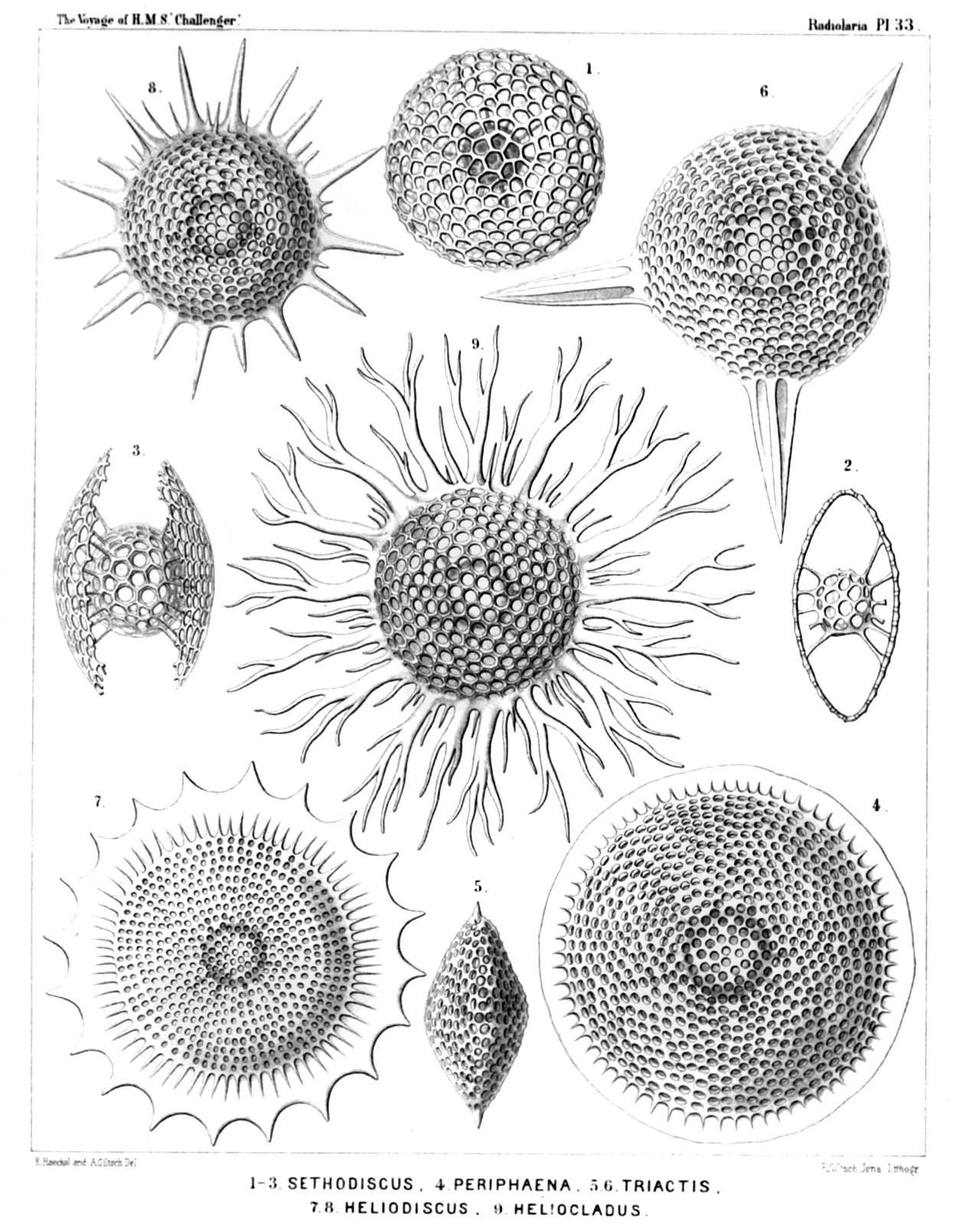 | |||||||||||||||||||||||||||||||||||||||||||||||||||
| PLATE 33. | |||
| Phacodiscida. | |||
| Diam. | Page. | ||
Fig. 1. Sethodiscus lenticula, n. sp., | × | 300 | 423 |
| Fig. 2. Sethodiscus lenticula, n. sp., | × | 300 | 423 |
| Vertical section. | |||
| Fig. 3. Sethodiscus macrococcus, n. sp., | × | 300 | 423 |
| Young shell, not yet closed, seen from the margin. | |||
| Fig. 4. Periphæna cincta, n. sp., | × | 400 | 426 |
| Fig. 5. Triactiscus tricuspis, n. sp., | × | 300 | 432 |
| Marginal view. | |||
| Fig. 6. Triactiscus tripyramis, n. sp., | × | 400 | 432 |
| Fig. 7. Heliodiscus cingillum, n. sp., | × | 300 | 448 |
| Fig. 8. Heliodiscus asteriscus, n. sp., | × | 300 | 445 |
| Fig. 9. Heliodrymus dendrocyclus, n. sp.(vel Heliocladus dendrocyclus), | × | 300 | 451 |
PLATE 34.
Legion SPUMELLARIA.
Order DISCOIDEA.
Family Phacodiscida.
| 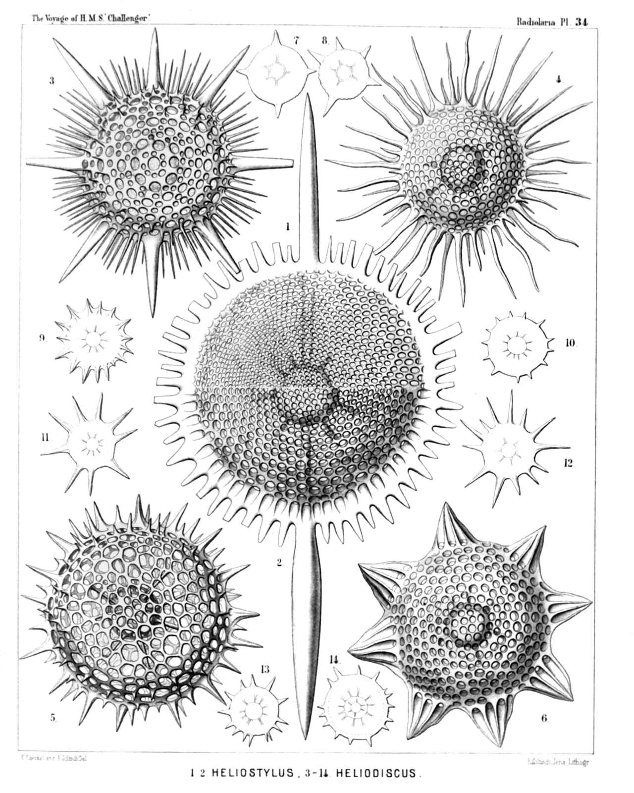 | ||||||||||||||||||||||||||||||||||||||||||||||||||||||||||||||||||||||||
| PLATE 34. | |||
| Phacodiscida. | |||
| Diam. | Page. | ||
Fig. 1. Sethostylus dentatus, n. sp. (vel Heliostylus dentatus), | × | 300 | 429 |
| Upper half of the disk. | |||
| Fig. 2. Sethostylus serratus, n. sp. (vel Heliostylus serratus), | × | 300 | 429 |
| Lower half of the disk. | |||
| Fig. 3. Heliosestrum octonum, n. sp., | × | 300 | 440 |
| Fig. 4. Heliodiscus solaster, n. sp., | × | 300 | 447 |
| Fig. 5. Heliodiscus echiniscus, n. sp., | × | 400 | 448 |
| Fig. 6. Heliosestrum medusinum, n. sp., | × | 300 | 438 |
| Fig. 7. Sethostaurus conostaurus, n. sp., | × | 100 | 433 |
| Normal form with four regular spines. | |||
| Fig. 8. Sethostaurus conostaurus, n. sp., | × | 100 | 433 |
| Abnormal form with five spines. | |||
| Fig. 9. Heliodiscus marginatus, n. sp., | × | 100 | 449 |
| Fig. 10. Heliodiscus trochiscus, n. sp., | × | 100 | 445 |
| Fig. 11. Heliodiscus polymorphus, n. sp., | × | 100 | 447 |
| Fig. 12. Heliodiscus polymorphus, n. sp., | × | 100 | 447 |
| Fig. 13. Heliodiscus trochiscus, n. sp., | × | 100 | 445 |
| Fig. 14. Astrophacus trochiscus, n. sp., | × | 100 | 453 |
PLATE 35.
Legion SPUMELLARIA.
Order DISCOIDEA.
Family Phacodiscida.
| 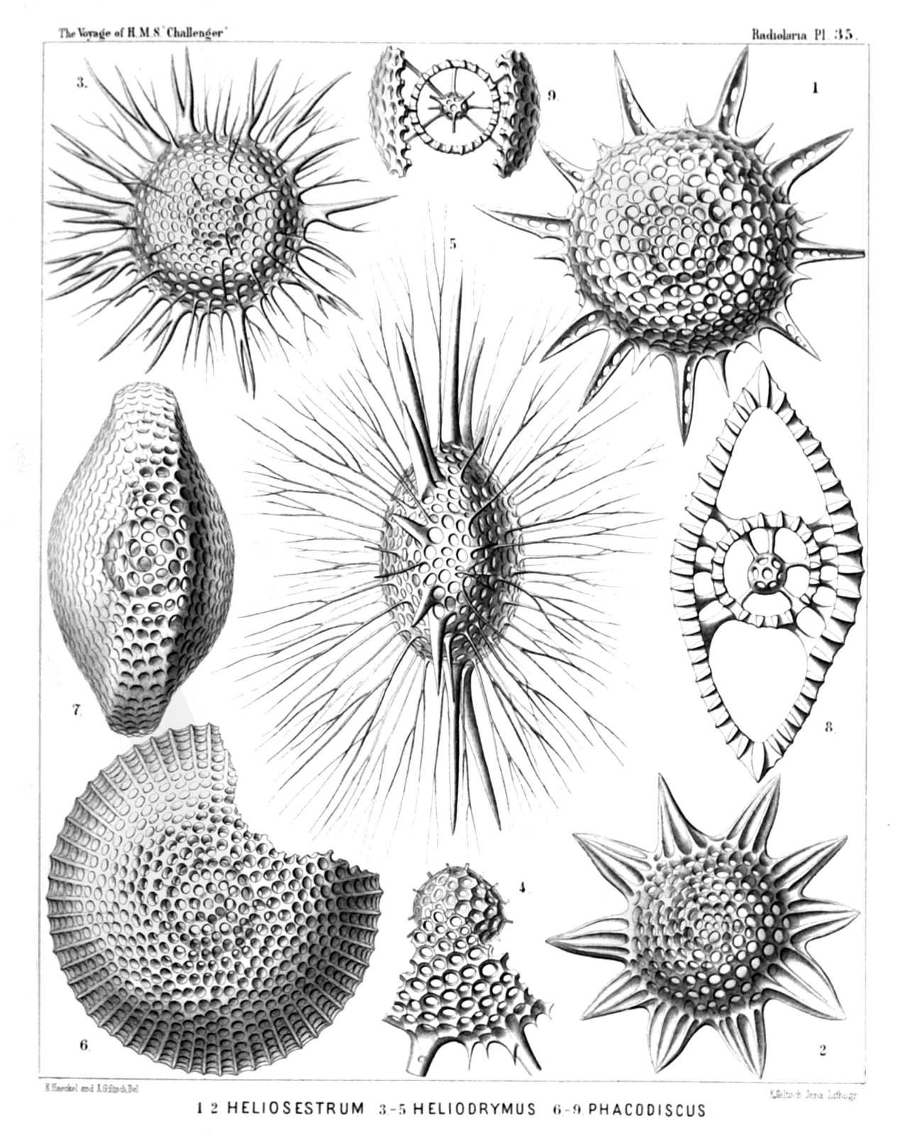 | ||||||||||||||||||||||||||||||||||||||||||||||||||||||
| PLATE 35. | |||
| Phacodiscida. | |||
| Diam. | Page. | ||
Fig. 1. Heliodiscus pertusus, n. sp. (vel Heliosestrum pertusum), | × | 400 | 448 |
| Irregular form with ten (instead of eight) larger latticed spines. | |||
| Fig. 2. Heliodiscus glyphodon, n. sp. (vel Heliosestrum glyphodon), | × | 300 | 446 |
| Fig. 3. Heliodrymus ramosus, n. sp., | × | 300 | 452 |
| Fig. 4. Heliodrymus ramosus, n. sp., | × | 500 | 452 |
| Medullary shell and a segment of the disk. | |||
| Fig. 5. Heliodrymus viminalis, n. sp., | × | 400 | 452 |
| Marginal view. | |||
| Fig. 6. Phacodiscus clypeus, n. sp., | × | 400 | 425 |
| Fig. 7. Phacodiscus rotula, n. sp., | × | 400 | 424 |
| Marginal view. | |||
| Fig. 8. Phacodiscus lentiformis, n. sp., | × | 400 | 425 |
| Vertical section nearly through the centre. | |||
| Fig. 9. Phacodiscus clypeus, n. sp., | × | 400 | 425 |
| Vertical section nearly through the centre. | |||
PLATE 36.
Legion SPUMELLARIA.
Order DISCOIDEA.
Family Coccodiscida.
| 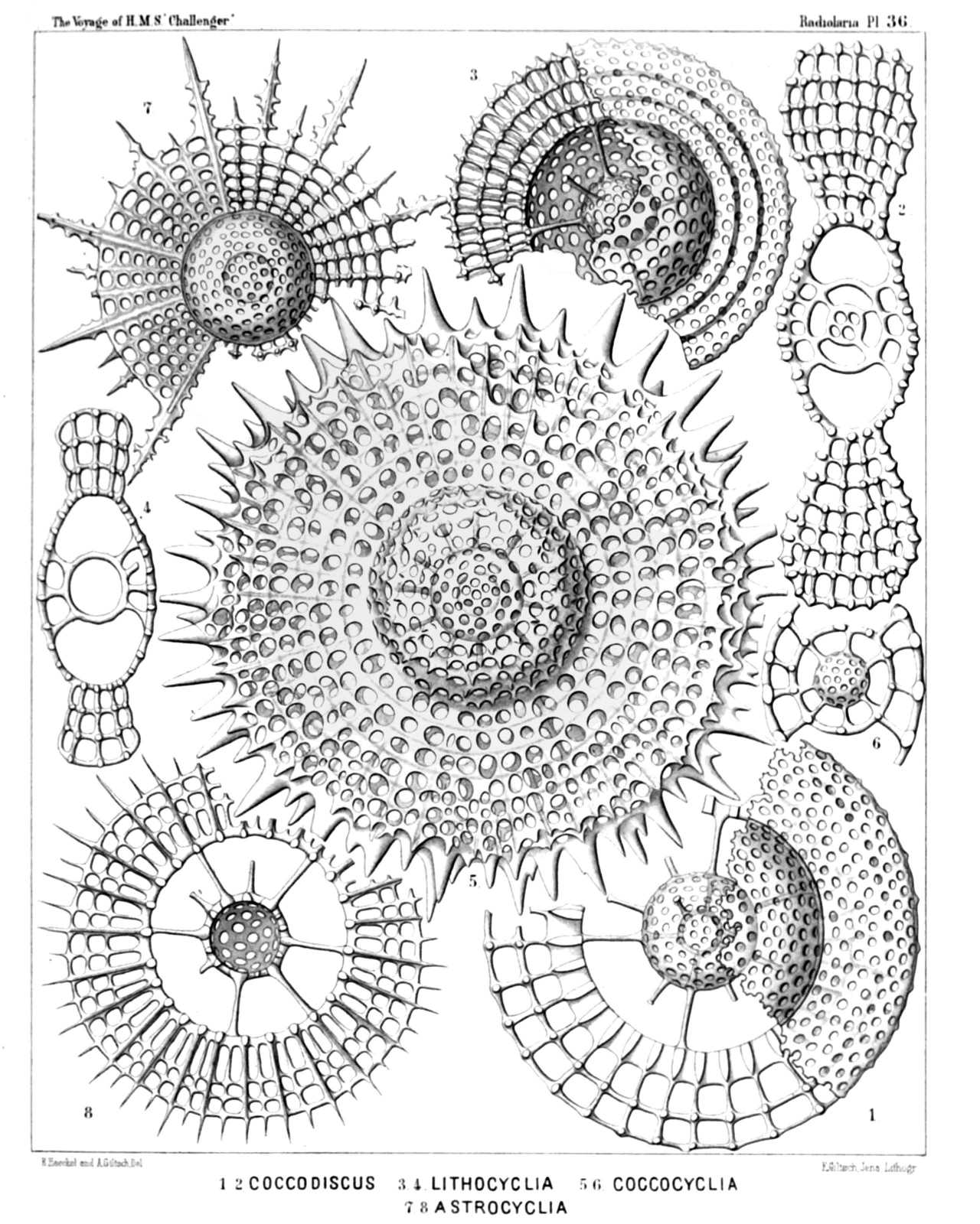 | |||||||||||||||||||||||||||||||||||||||||||||||||
| PLATE 36. | |||
| Coccodiscida. | |||
| Diam. | Page. | ||
Fig. 1. Coccodiscus lamarckii, n. sp., | × | 500 | 459 |
| The left half of the figure represents a horizontal section through the peripheral shell, the right half a view of the surface. | |||
| Fig. 2. Coccodiscus gœthei, n. sp., | × | 500 | 461 |
| Vertical section nearly through the centre. | |||
| Fig. 3. Lithocyclia lenticula, n. sp., | × | 400 | 459 |
| Fig. 4. Lithocyclia lenticula, n. sp., | × | 400 | 459 |
| Vertical section through the centre. | |||
| Fig. 5. Coccocyclia helianthus, n. sp., | × | 400 | 468 |
| Fig. 6. Coccocyclia helianthus, n. sp., | × | 500 | 468 |
| Vertical section through the outer medullary shell, showing the inner. | |||
| Fig. 7. Astrocyclia solaster, n. sp., | × | 300 | 466 |
| Fig. 8. Astrocyclia heterocycla, n. sp., | × | 400 | 468 |
| Horizontal section through the equatorial plane. | |||
PLATE 37.
Legion SPUMELLARIA.
Order DISCOIDEA.
Family Coccodiscida.
| 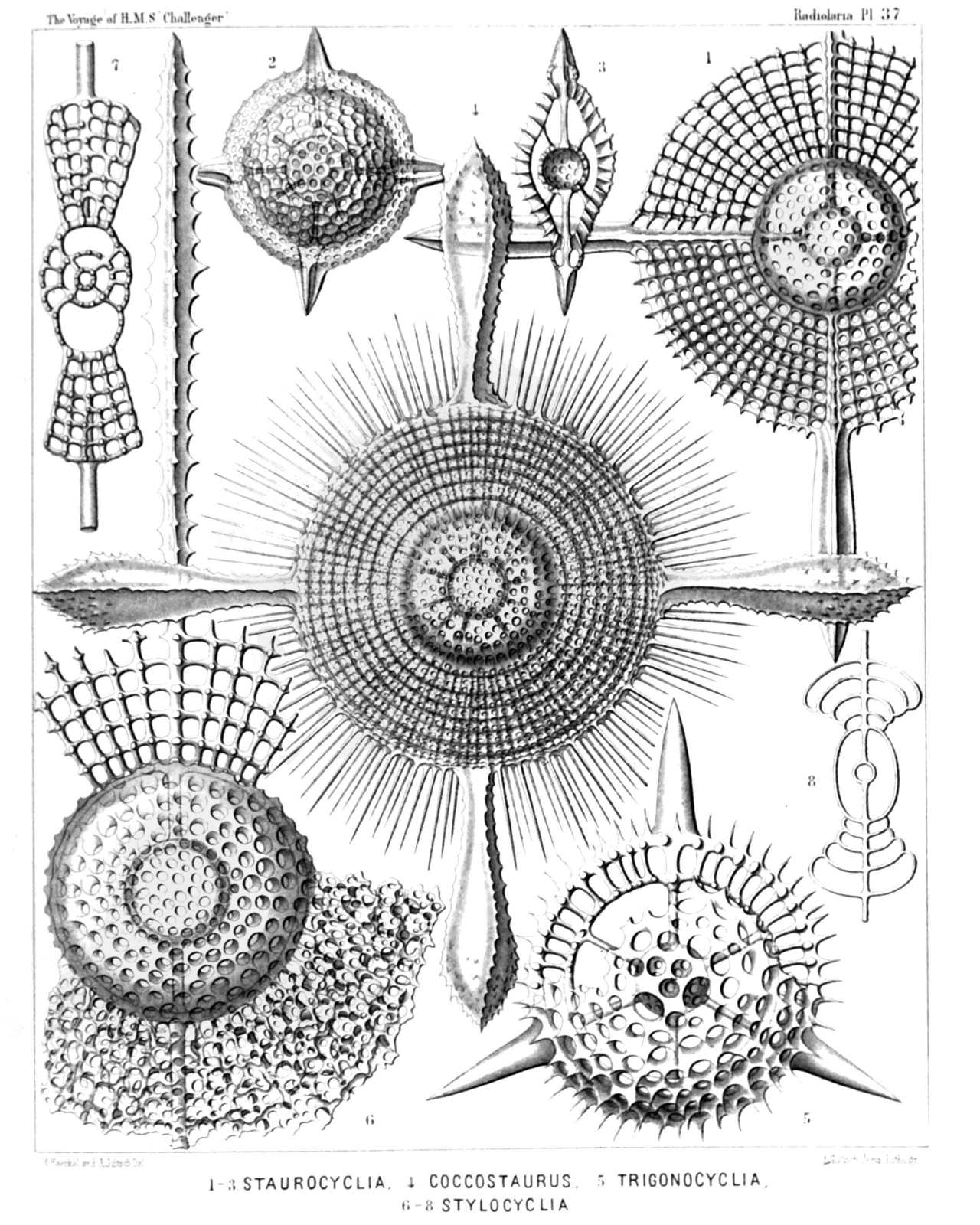 | ||||||||||||||||||||||||||||||||||||||||||||||||
| PLATE 37. | |||
| Coccodiscida. | |||
| Diam. | Page. | ||
Fig. 1. Staurocyclia cruciata, n. sp., | × | 400 | 465 |
| Fig. 2. Staurocyclia phacostaurus, n. sp., | × | 300 | 465 |
| Fig. 3. Staurocyclia phacostaurus, n. sp., | × | 300 | 465 |
| Vertical section through the centre. | |||
| Fig. 4. Staurocyclia magniducis, n. sp. (Coccostaurus magniducis), | × | 300 | 466 |
| Fig. 5. Trigonocyclia triangularis, n. sp., | × | 400 | 464 |
| Fig. 6. Stylocyclia prionacantha, n. sp., | × | 500 | 462 |
| A great part of the peripheral shell is removed. | |||
| Fig. 7. Amphicyclia amphistyla, n. sp., | × | 300 | 464 |
| Vertical section through the centre. | |||
| Fig. 8. Stylocyclia excavata, n. sp., | × | 200 | 463 |
| Vertical section through the centre. | |||
PLATE 38.
Legion SPUMELLARIA.
Order DISCOIDEA.
Family Coccodiscida.
| 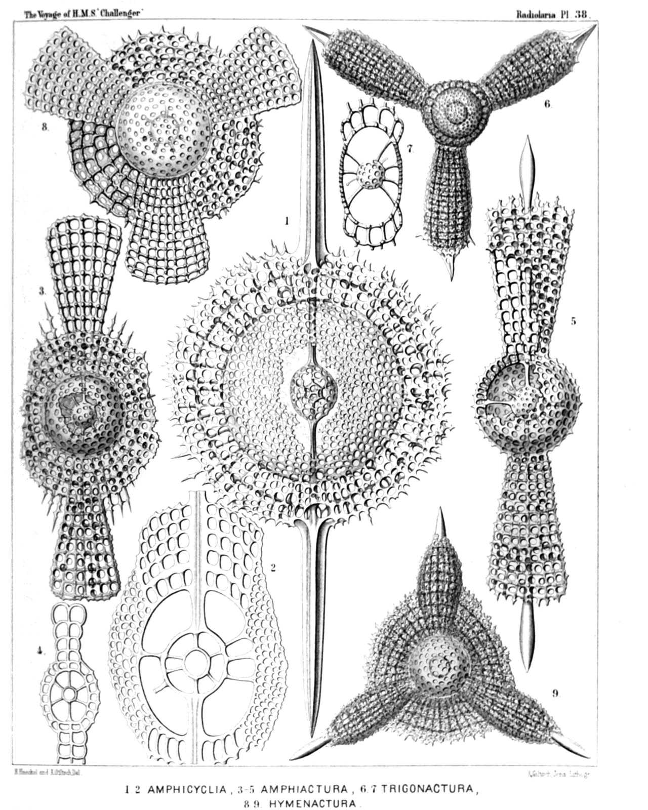 | |||||||||||||||||||||||||||||||||||||||||||||||||||
| PLATE 38. | |||
| Coccodiscida. | |||
| Diam. | Page. | ||
Fig. 1. Amphicyclia chronometra, n. sp., | × | 400 | 463 |
| Fig. 2. Amphicyclia pachydiscus, n. sp., | × | 500 | 464 |
| Vertical section through the centre. | |||
| Fig. 3. Amphiactura amphibrachia, n. sp., | × | 300 | 470 |
| Fig. 4. Amphiactura amphibrachia, n. sp., | × | 150 | 470 |
| Vertical section through the centre. | |||
| Fig. 5. Diplactura diploconus, n. sp., | × | 300 | 470 |
| Fig. 6. Trigonactura triacantha, n. sp., | × | 200 | 472 |
| Fig. 7. Trigonactura triacantha, n. sp., | × | 400 | 472 |
| Vertical section nearly through the centre. | |||
| Fig. 8. Hymenactura archimedis, n. sp., | × | 300 | 473 |
| Fig. 9. Hymenactura copernici, n. sp., | × | 200 | 475 |
PLATE 39.
Legion SPUMELLARIA.
Order PRUNOIDEA.
Families Ellipsida, Druppulida, Artiscida et Cyphinida.
| 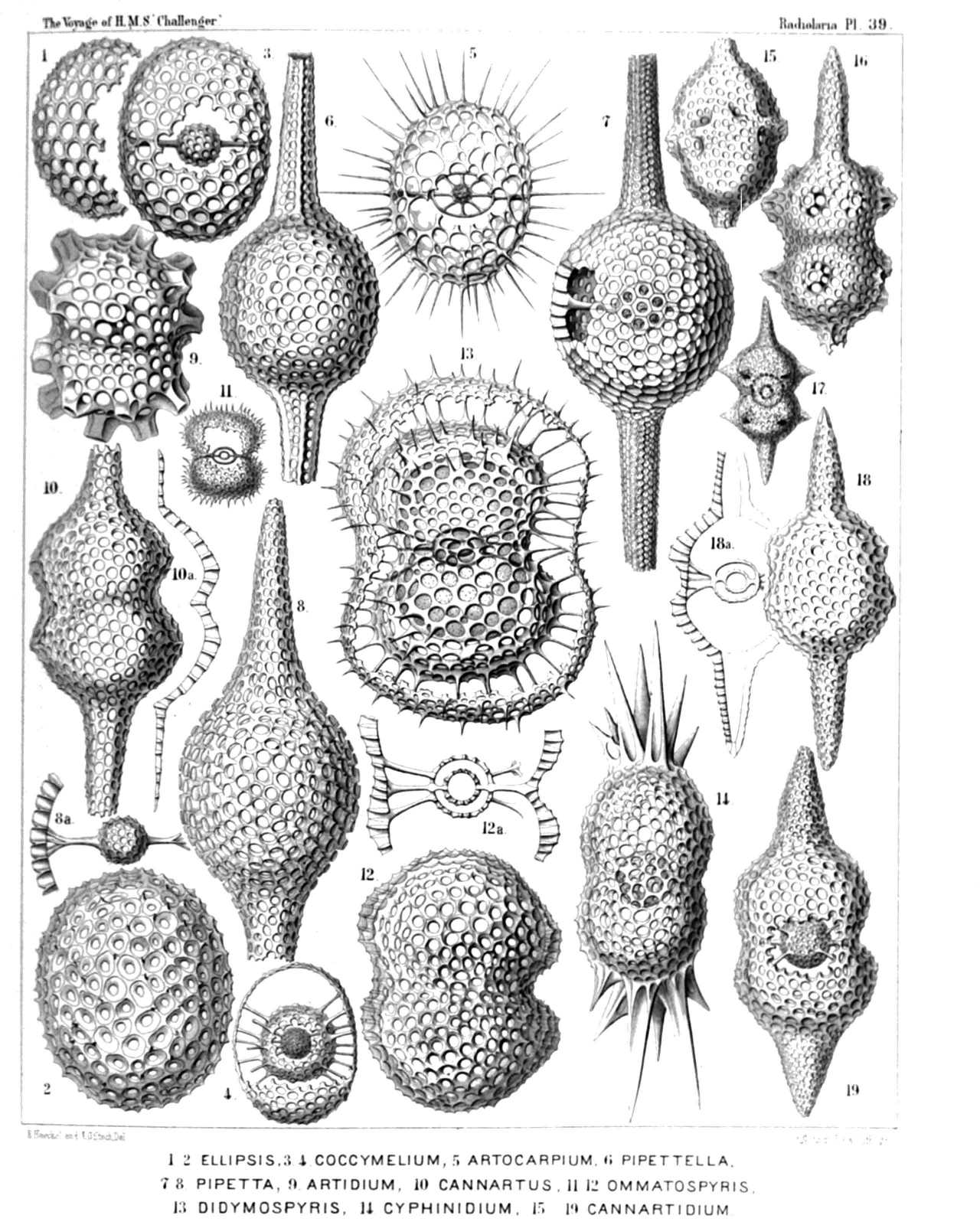 | ||||||||||||||||||||||||||||||||||||||||||||||||||||||||||||||||||||||||||||||||||||||||||||
| PLATE 39. | |||
| Ellipsida, Druppulida, Artiscida et Cyphinida. | |||
| Diam. | Page. | ||
Fig. 1. Cenellipsis faceta, n. sp. (vel Ellipsis faceta), | × | 300 | 291 |
| Fig. 2. Cenellipsis infundibulum, n. sp. (vel Ellipsis infundibulum), | × | 300 | 292 |
| Fig. 3. Druppula pandanus, n. sp. (vel Coccymelium pandanus), | × | 300 | 308 |
| Fig. 4. Prunulum coccymelium, n. sp. (vel Coccymelium prunulum), | × | 300 | 313 |
| Fig. 5. Prunocarpus artocarpium, n. sp. (vel Artocarpium indicum), | × | 300 | 316 |
| Fig. 6. Pipettella prismatica, n. sp., | × | 300 | 305 |
| Fig. 7. Pipetta tuba, n. sp., | × | 300 | 337 |
| Fig. 8. Pipetta fusus, n. sp., | × | 300 | 337 |
| Fig. 8a. The enclosed medullary shell. | |||
| Fig. 9. Artiscus nodosus, n. sp. (vel Artidium nodosum), | × | 400 | 356 |
| Fig. 10. Cannartus violina, n. sp., | × | 300 | 358 |
| Fig. 11. Cyphonium cribellum, n. sp., | × | 200 | 365 |
| Fig. 12. Cyphonium virgineum, n. sp. (vel Ommatospyris virginea), | × | 400 | 363 |
| Fig. 12a. Vertical section through the double medullary shell. | |||
| Fig. 13. Cypassis puella, n. sp. (vel Didymospyris puella), | × | 400 | 367 |
| The enclosed central capsule is visible. | |||
| Fig. 14. Cyphinus amphilophus, N. sp., | × | 300 | 370 |
| Fig. 15. Pipettaria tubaria, n. sp., | × | 300 | 339 |
| Fig. 16. Cannartidium mammiferum, n. sp., | × | 300 | 375 |
| Fig. 17. Cannartidium mastophorum, n. sp., | × | 150 | 375 |
| Fig. 18. Cannartidium bicinctum, n. sp., | × | 300 | 374 |
| Fig. 18a. Vertical section through the main axis. | |||
| Fig. 19. Cannartiscus amphiconiscus, n. sp., | × | 300 | 372 |
PLATE 40.
Legion SPUMELLARIA.
Order PRUNOIDEA.
Families Panartida et Zygartida.
| 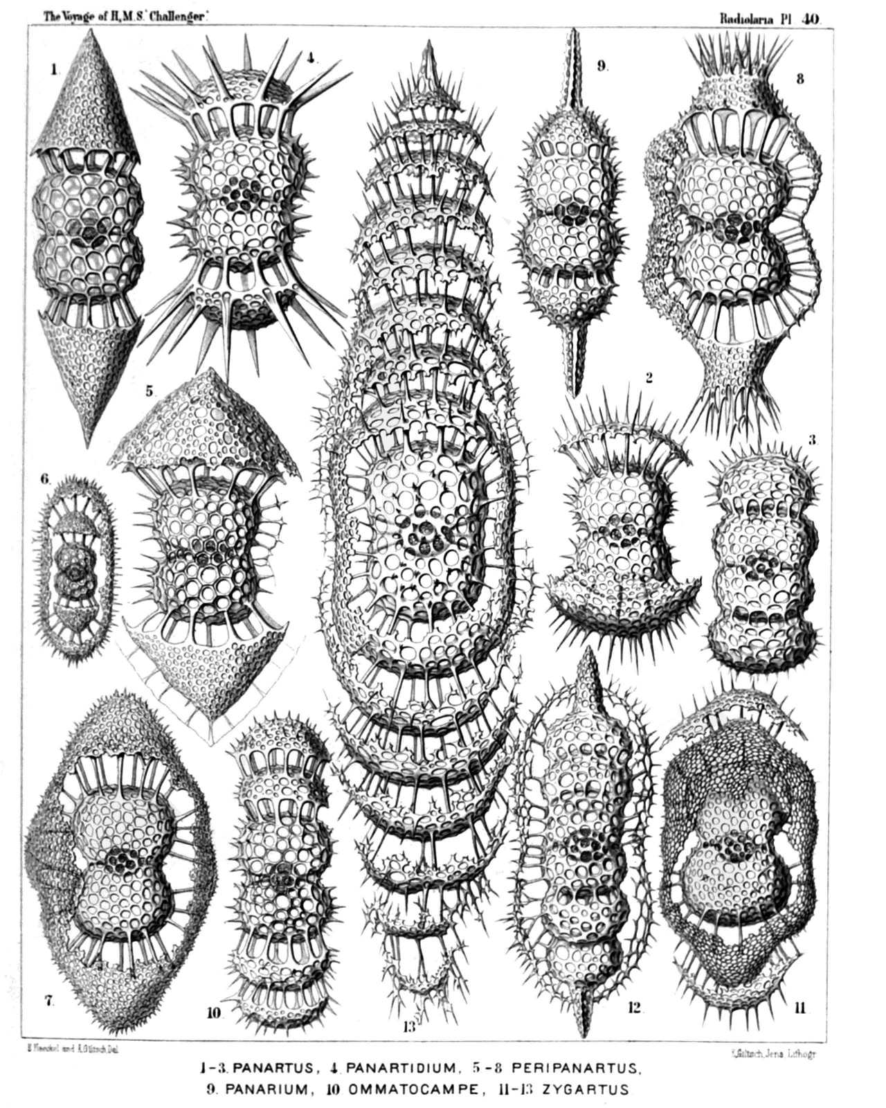 | ||||||||||||||||||||||||||||||||||||||||||||||||||||||||||||||||
| PLATE 40. | |||
| Panartida et Zygartida. | |||
| Diam. | Page. | ||
Fig. 1. Panartus diploconus, n. sp., | × | 300 | 379 |
| Fig. 2. Panartus pluteus, n. sp., | × | 300 | 382 |
| Fig. 3. Panartus tetrathalamus, n. sp., | × | 300 | 378 |
| Fig. 4. Panicium coronatum, n. sp. (vel Panartidium coronatum), | × | 300 | 386 |
| Fig. 5. Peripanartus amphiconus, n. sp., | × | 300 | 383 |
| Fig. 6. Peripanartus cylindrus, n. sp., | × | 150 | 384 |
| Fig. 7. Peripanartus atractus, n. sp., | × | 300 | 384 |
| Fig. 8. Peripanicium amphicorona, n. sp., | × | 300 | 387 |
| Fig. 9. Panarium tubularium, n. sp., | × | 300 | 390 |
| Fig. 10. Ommatocampe nereides, n. sp., | × | 300 | 394 |
| Fig. 11. Cyphocolpus virginis, n. sp. (vel Zygartus virginis) | × | 300 | 369 |
| Fig. 12. Desmartus larvalis, n. sp. (vel Zygartus larvalis), | × | 300 | 398 |
| Fig. 13. Zygartus chrysalis, n. sp. (vel Zygocampe chrysalis), | × | 400 | 401 |
PLATE 41.
Legion SPUMELLARIA.
Order DISCOIDEA.
Families Porodiscida et Spongodiscida.
| 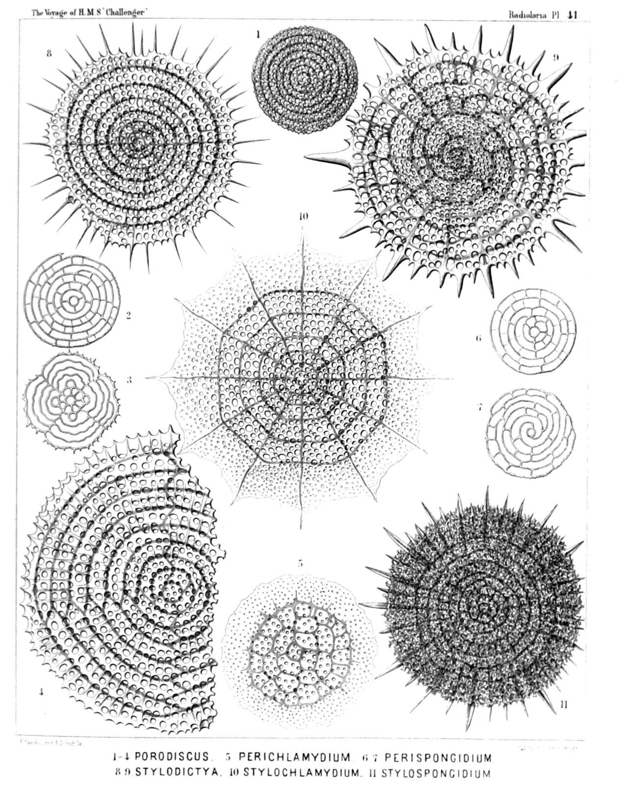 | ||||||||||||||||||||||||||||||||||||||||||||||||||||||||||||
| PLATE 41. | |||
| Porodiscida et Spongodiscida. | |||
| Diam. | Page. | ||
Fig. 1. Porodiscus flustrella, n. sp., | × | 300 | 493 |
| Fig. 2. Porodiscus perispira, n. sp., | × | 200 | 495 |
| The rings alone (equatorial section). | |||
| Fig. 3. Porodiscus quadrigatus, n. sp., | × | 200 | 494 |
| The rings alone (equatorial section). | |||
| Fig. 4. Porodiscus semispiralis, n. sp., | × | 500 | 497 |
| Fig. 5. Perichlamydium saturnus, n. sp., | × | 300 | 499 |
| Fig. 6. Porodiscus centrospira, n. sp. (vel Perispongidium centrospira), | × | 200 | 495 |
| The rings alone (equatorial section). | |||
| Fig. 7. Porodiscus irregularis, n. sp. (vel Perispongidium irregulare), | × | 200 | 498 |
| The rings alone (equatorial section). | |||
| Fig. 8. Stylodictya heliospira, n. sp., | × | 400 | 512 |
| Fig. 9. Stylodictya centrospira, n. sp., | × | 400 | 512 |
| Fig. 10. Stylochlamydium asteriscus, n. sp., | × | 400 | 514 |
| Fig. 11. Stylotrochus geddesii, n. sp., | × | 300 | 585 |
PLATE 42.
Legion SPUMELLARIA.
Order DISCOIDEA.
Family Porodiscida.
| 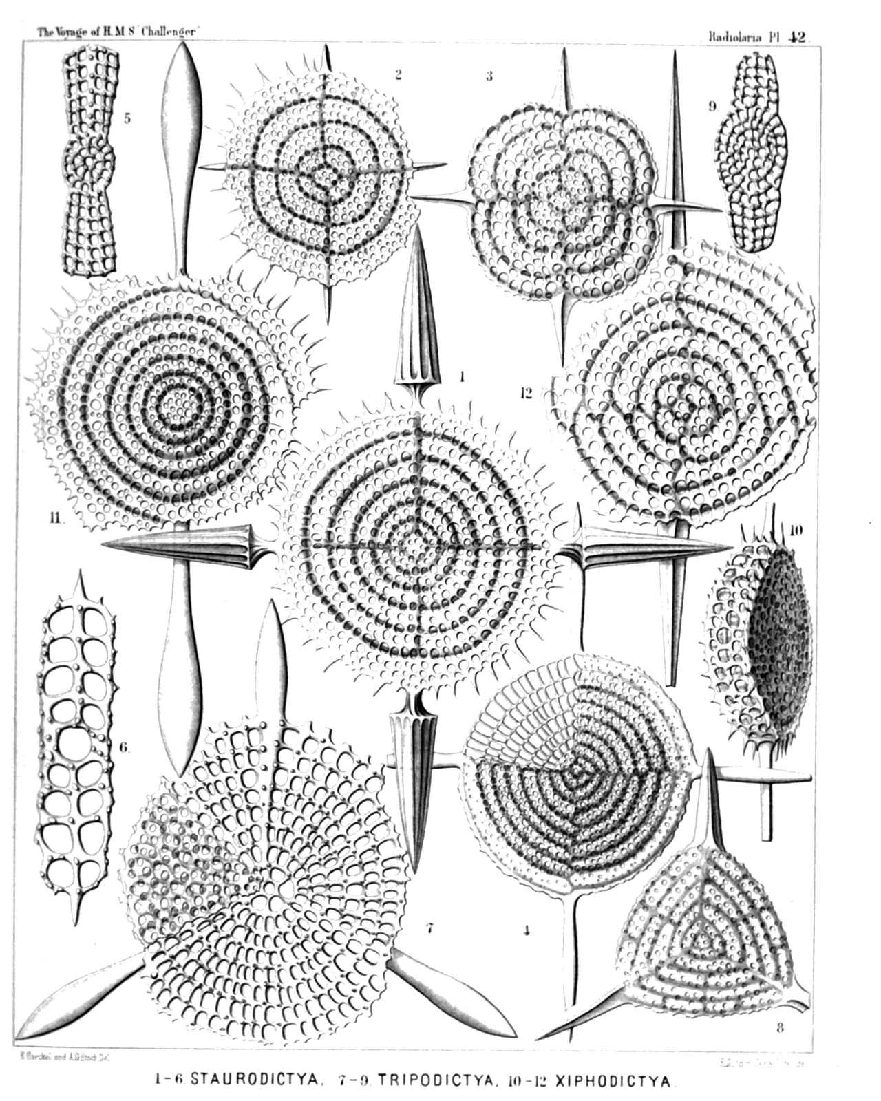 | ||||||||||||||||||||||||||||||||||||||||||||||||||||||||||||||||
| PLATE 42. | |||
| Porodiscida. | |||
| Diam. | Page. | ||
Fig. 1. Staurodictya elegans, n. sp., | × | 500 | 507 |
| Fig. 2. Staurodictya ciliata, n. sp., | × | 400 | 506 |
| Fig. 3. Staurodictya medusa, n. sp., | × | 400 | 506 |
| Fig. 4. Staurodictya cruciata, n. sp., | × | 300 | 507 |
| Fig. 5. Staurodictya cruciata, n. sp., | × | 300 | 507 |
| Vertical section through the disk. | |||
| Fig. 6. Staurodictya grandis, n. sp., | × | 300 | 508 |
| Vertical section through the disk. | |||
| Fig. 7. Tripodictya triacantha, n. sp., | × | 400 | 505 |
| Fig. 8. Tripodictya trigonaria, n. sp., | × | 400 | 505 |
| Fig. 9. Tripodictya tribelonia, n. sp., | × | 400 | 505 |
| Vertical section through the disk. | |||
| Fig. 10. Xiphodictya amphibelonia, n. sp., | × | 300 | 503 |
| Marginal view. | |||
| Fig. 11. Xiphodictya amphirrhopalia, n. sp., | × | 400 | 504 |
| Fig. 12. Xiphodictya staurospira, n. sp., | × | 500 | 504 |
PLATE 43.
Legion SPUMELLARIA.
Order DISCOIDEA.
Family Porodiscida.
| 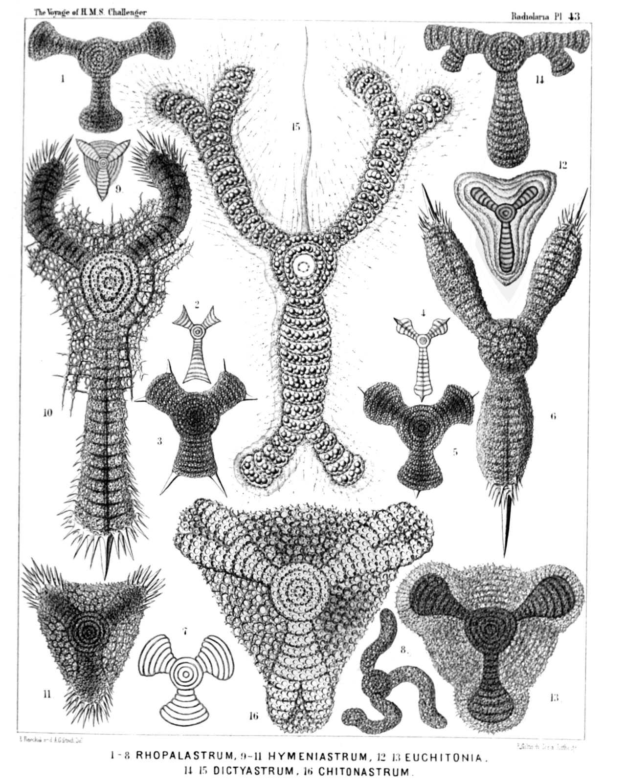 | |||||||||||||||||||||||||||||||||||||||||||||||||||||||||||||||||||||||||||||
| PLATE 43. | |||
| Porodiscida. | |||
| Diam. | Page. | ||
Fig. 1. Rhopalastrum malleus, n. sp., | × | 100 | 527 |
| Fig. 2. Rhopalastrum ypsilinum, n. sp., | × | 50 | 528 |
| Fig. 3. Rhopalastrum hexaceros, n. sp., | × | 100 | 529 |
| Fig. 4. Rhopalastrum triceros, n. sp., | × | 50 | 529 |
| Fig. 5. Rhopalastrum trispinosum, n. sp. (vel Dictyastrum trispinosum), | × | 150 | 525 |
| Fig. 6. Rhopalastrum arcticum, n. sp., | × | 300 | 539 |
| Fig. 7. Rhopalastrum hexagonum, n. sp. (vel Dictyastrum hexagonum), | × | 100 | 525 |
| Fig. 8. Rhopalastrum irregulare, n. sp., | × | 100 | 528 |
| Fig. 9. Euchitonia lanceolata, n. sp., | × | 80 | 534 |
| Fig. 10. Euchitonia carcinus, n. sp., | × | 300 | 535 |
| Fig. 11. Euchitonia echinata, n. sp., | × | 120 | 536 |
| Fig. 12. Euchitonia stohrii, n. sp., | × | 100 | 534 |
| Fig. 13. Hymeniastrum euclidis, n. sp., | × | 200 | 531 |
| Fig. 14. Chitonastrum jugatum, n. sp., | × | 200 | 537 |
| Fig. 15. Chitonastrum lyra, n. sp., | × | 500 | 538 |
| A living specimen observed. The entire shell is enveloped by the calymma and surrounded by radiating pseudopodia (drawn much too short). Between the two paired arms arises a large "sarcode-flagellum." The central chamber and the first enveloping ring are filled by the clear nucleus; the other rings and all the chambers of the arms contain numerous pink oil-globules. | |||
| Fig. 16. Trigonastrum regulare, n. sp. (vel Chitonastrum regulare), | × | 200 | 539 |
PLATE 44.
Legion SPUMELLARIA.
Order DISCOIDEA.
Family Porodiscida.
| 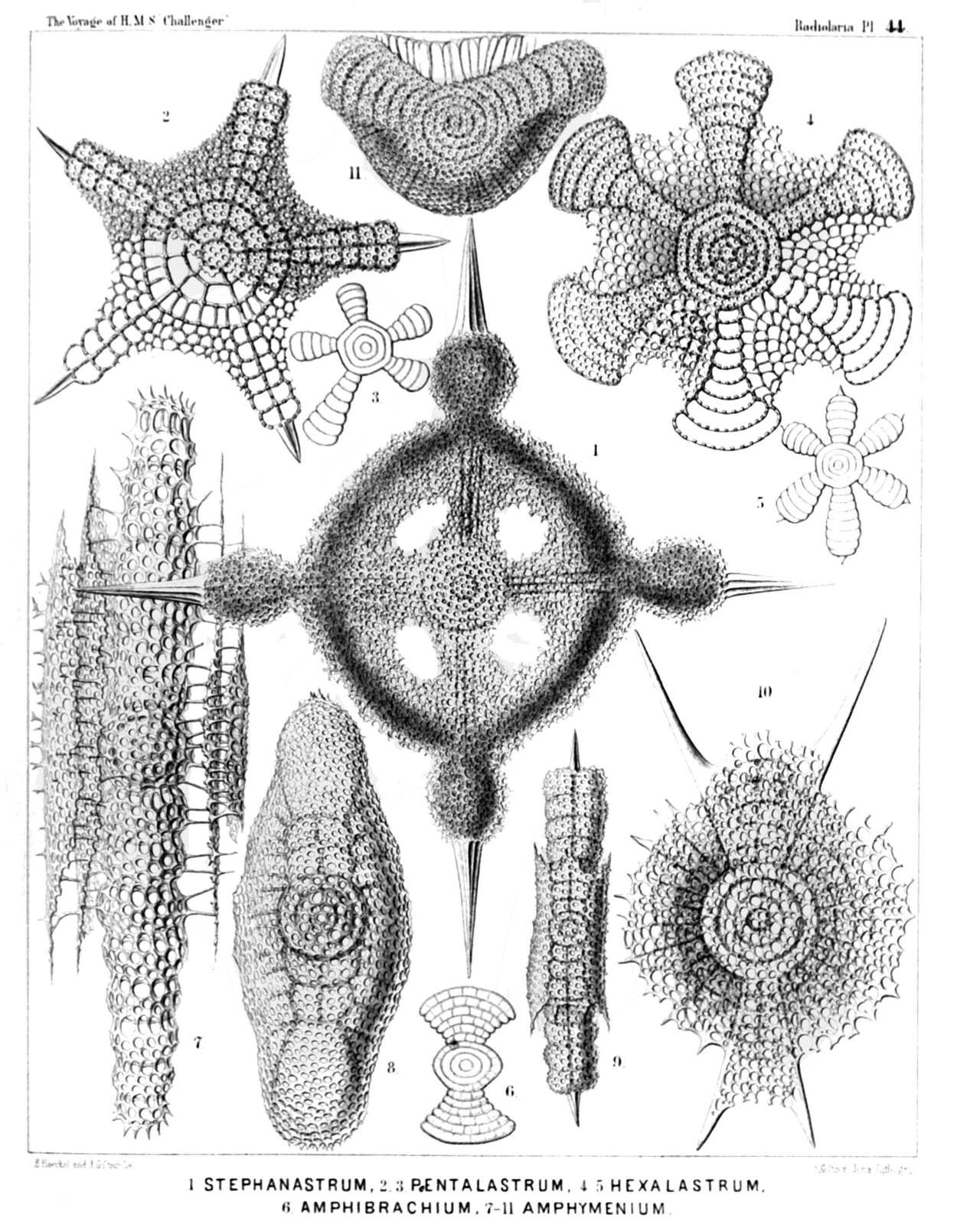 | ||||||||||||||||||||||||||||||||||||||||||||||||||||||||
| PLATE 44. | |||
| Porodiscida. | |||
| Diam. | Page. | ||
Fig. 1. Stephanastrum capitatum, n. sp., | × | 200 | 549 |
| Fig. 2. Pentinastrum asteriscus, n. sp., | × | 300 | 557 |
| Fig. 3. Pentalastrum ophidiaster, n. sp., | × | 100 | 557 |
| Fig. 4. Hexinastrum geryonidum, n. sp., | × | 300 | 560 |
| Fig. 5. Hexalastrum orchidaceum, n. sp., | × | 50 | 560 |
| Fig. 6. Amphibrachium dilatatum, n. sp., | × | 50 | 517 |
| Fig. 7. Amphymenium zygartus, n. sp., | × | 400 | 520 |
| Fig. 8. Amphymenium pupula, n. sp., | × | 300 | 519 |
| Fig. 9. Amphymenium amphistylium, n. sp., | × | 200 | 520 |
| Fig. 10. Amphicraspedum murrayanum, n. sp., | × | 300 | 523 |
| Fig. 11. Amphymenium monstrosum, n. sp., | × | 300 | 520 |
PLATE 45.
Legion SPUMELLARIA.
Order DISCOIDEA.
Family Porodiscida.
| 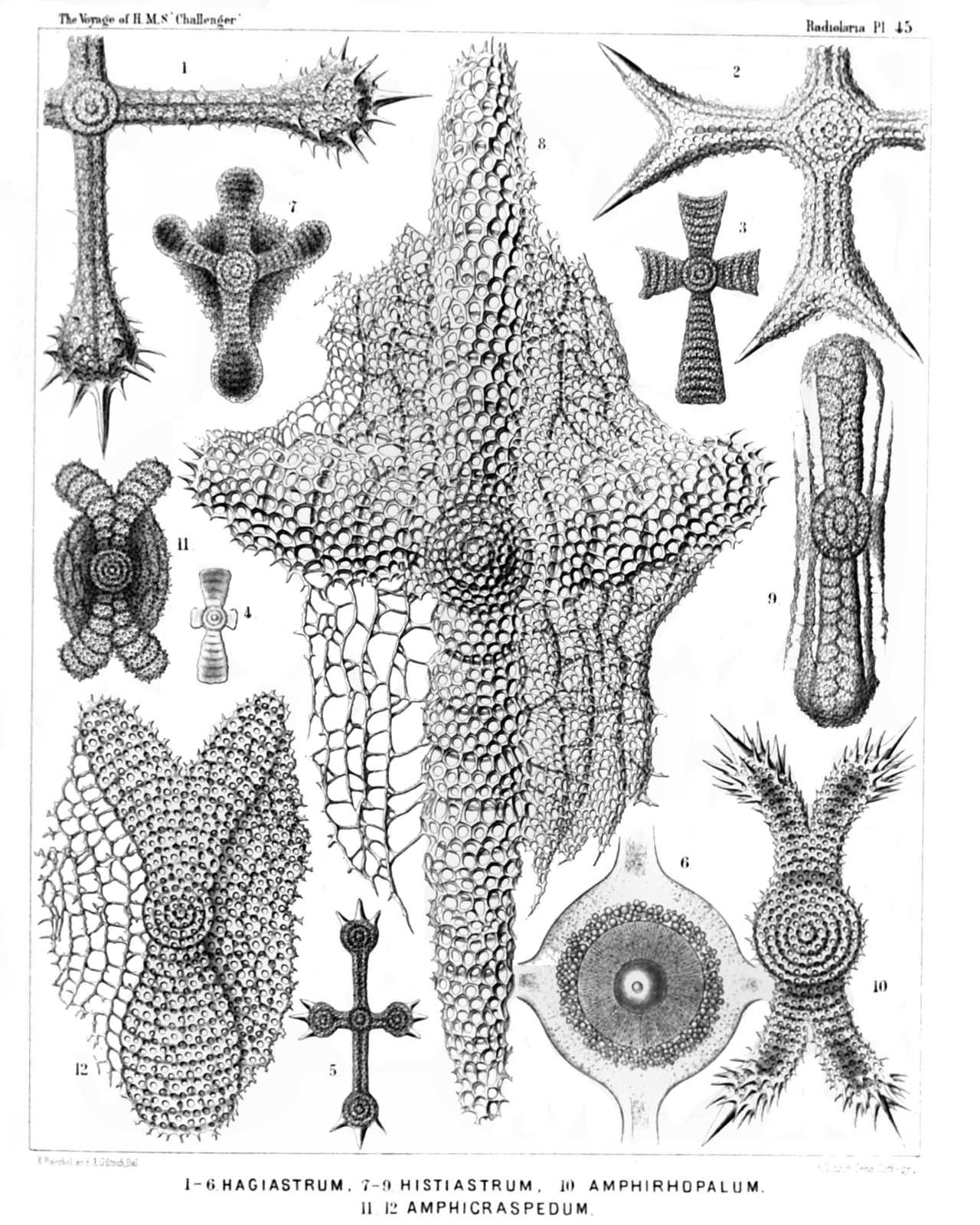 | |||||||||||||||||||||||||||||||||||||||||||||||||||||||||||||||
| PLATE 45. | |||
| Porodiscida. | |||
| Diam. | Page. | ||
Fig. 1. Stauralastrum rhopalophorum, n. sp., | × | 200 | 541 |
| Fig. 2. Dicranastrum cornutum, n. sp., | × | 200 | 551 |
| Fig. 3. Hagiastrum mosis, n. sp., | × | 100 | 543 |
| Fig. 4. Hagiastrum mosis, n. sp., | × | 50 | 543 |
| Lateral view, from the edge. | |||
| Fig. 5. Hagiastrum buddhæ, n. sp., | × | 50 | 542 |
| Fig. 6. Stauralastrum cruciforme, n. sp. (in glycerine), | × | 500 | 540 |
| The central capsule contains a large central nucleus with nucleolus, and is surrounded by the jelly calymma and numerous small zooxanthellæ. The endoplasm is radially striped. | |||
| Fig. 7. Tesserastrum democriti, n. sp., | × | 100 | 548 |
| Fig. 8. Tesserastrum straussii, n. sp., | × | 500 | 547 |
| Fig. 9. Tesserastrum brunonis, n. sp., | × | 200 | 548 |
| Disk seen from the edge. | |||
| Fig. 10. Amphirhopalum echinatum, n. sp., | × | 300 | 522 |
| Fig. 11. Amphicraspedum maclaganium, n. sp., | × | 100 | 523 |
| Fig. 12. Amphicraspedum wyvilleanum, n. sp., | × | 300 | 523 |
PLATE 46.
Legion SPUMELLARIA.
Order DISCOIDEA.
Family Porodiscida.
| 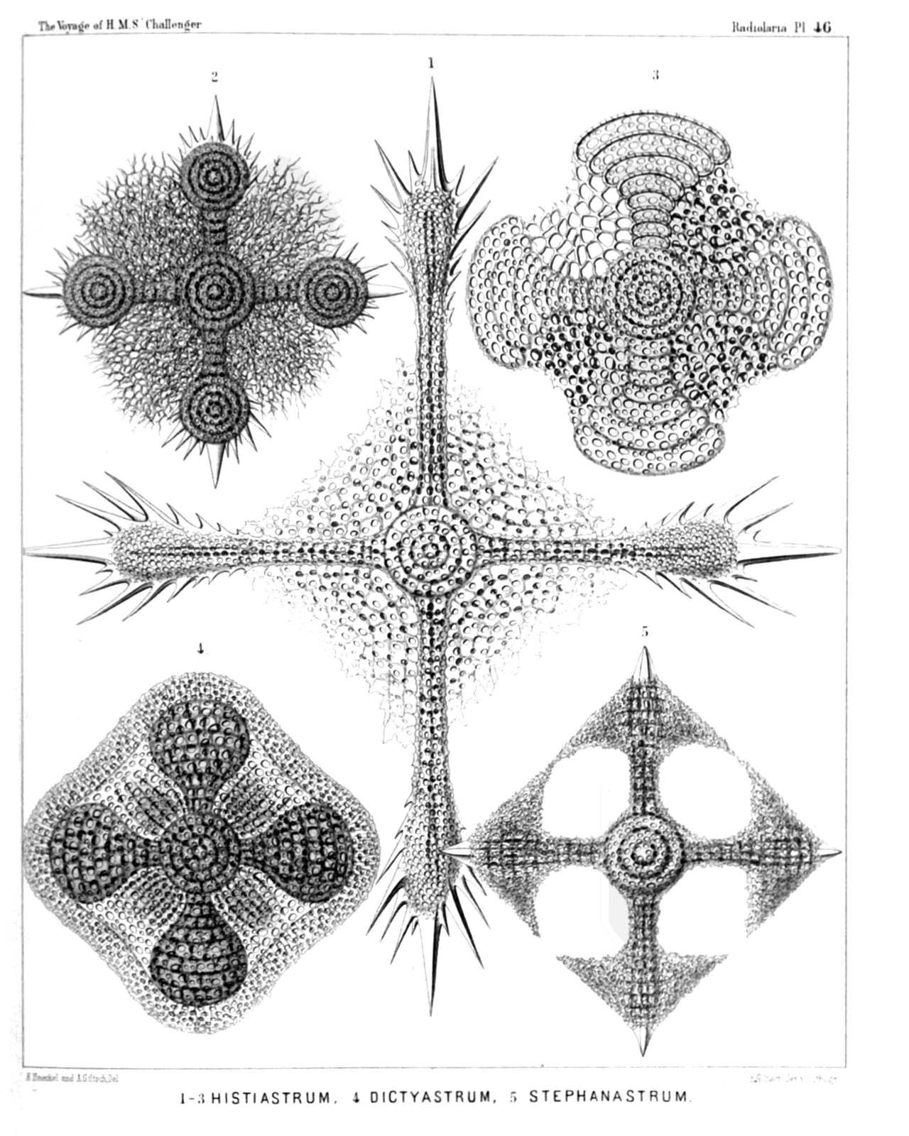 | ||||||||||||||||||||||||||||||||
| PLATE 46. | |||
| Porodiscida. | |||
| Diam. | Page. | ||
Fig. 1. Histiastrum boseanum, n. sp., | × | 400 | 546 |
| Fig. 2. Histiastrum pentadiscus, n. sp., | × | 200 | 546 |
| Fig. 3. Histiastrum quadrigatum, n. sp., | × | 300 | 544 |
| Fig. 4. Histiastrum velatum, n. sp., | × | 200 | 545 |
| Fig. 5. Stephanastrum quadratum, n. sp., | × | 200 | 549 |
PLATE 47.
Legion SPUMELLARIA.
Order DISCOIDEA.
Family Porodiscida.
| 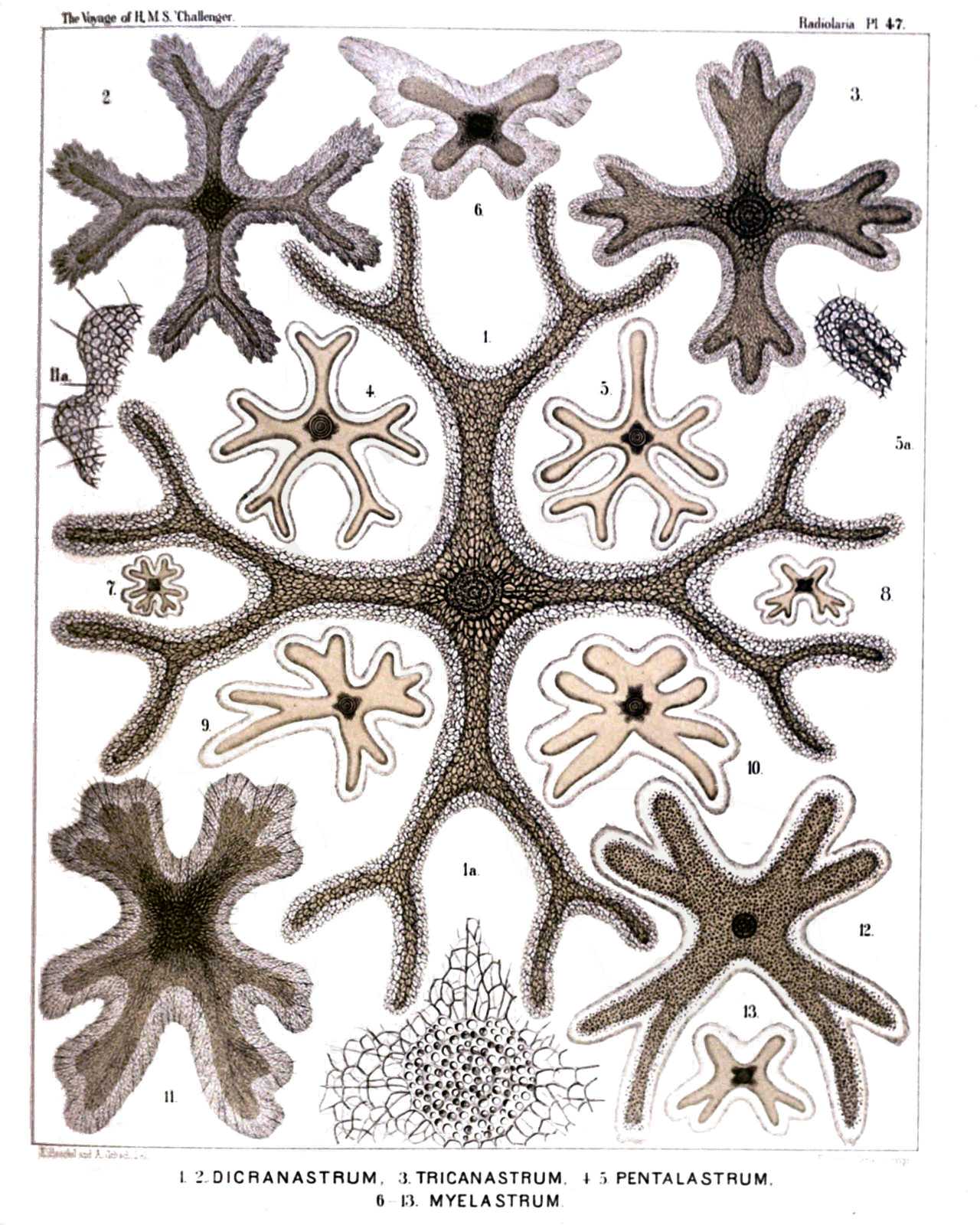 | |||||||||||||||||||||||||||||||||||||||||||||||||||||||||||||||||||
| PLATE 47. | |||
| Porodiscida. | |||
| Diam. | Page. | ||
Fig. 1. Dicranastrum bifurcatum, n. sp., | × | 200 | 552 |
| Fig. 1a. Central disc of the same, | × | 600 | |
| Fig. 2. Dicranastrum furcatum, n. sp., | × | 100 | 550 |
| Fig. 3. Dicranastrum wyvillei, n. sp., | × | 100 | 551 |
| Fig. 4. Pentophiastrum forcipatum, n. sp., | × | 50 | 559 |
| Fig. 5. Pentophiastrum caudatum, n. sp., | × | 50 | 559 |
| Fig. 6. Myelastrum papilio, n. sp., | × | 50 | 554 |
| Fig. 7. Myelastrum decaceros, n. sp., | × | 20 | 554 |
| Fig. 8. Myelastrum heteropterum, n. sp., | × | 20 | 553 |
| Fig. 9. Myelastrum anomalum, n. sp., | × | 50 | 556 |
| Fig. 10. Myelastrum farfalla, n. sp., | × | 50 | 554 |
| Fig. 11. Myelastrum dodecaceros, n. sp., | × | 100 | 554 |
| Fig. 12. Myelastrum octocorne, n. sp., | × | 90 | 553 |
| Fig. 13. Myelastrum medullare, n. sp., | × | 50 | 553 |
PLATE 48.
Legion SPUMELLARIA.
Orders PRUNOIDEA et DISCOIDEA.
Families Ellipsida, Artiscida, Spongurida, Cenodiscida, Porodiscida et Pylodiscida.
| 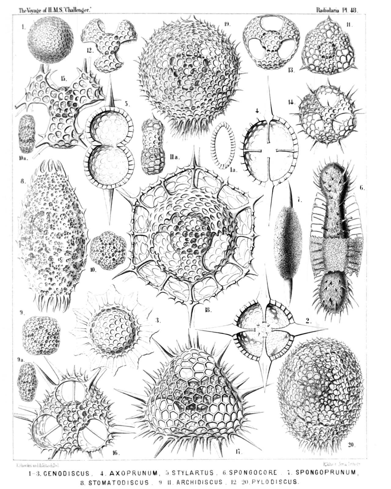 | |||||||||||||||||||||||||||||||||||||||||||||||||||||||||||||||||||||||||||||||||||||||||||||||||||
| PLATE 48. | |||
| Ellipsida, Artiscida, Spongurida, Cenodiscida, Porodiscida et Pylodiscida. | |||
| Diam. | Page. | ||
Fig. 1. Cenodiscus phacoides, n. sp., | × | 100 | 411 |
| Fig. 1a. Vertical section. | |||
| Fig. 2. Crucidiscus endostaurus, n. sp., | × | 200 | 416 |
| Equatorial section. | |||
| Fig. 3. Trochodiscus stellaris, n. sp., | × | 200 | 418 |
| Fig. 4. Axoprunum stauraxonium, n. sp., | × | 300 | 298 |
| Equatorial section. | |||
| Fig. 5. Stylartus bipolaris, n. sp., | × | 200 | 357 |
| Vertical section. | |||
| Fig. 6. Spongocore puella, n. sp., | × | 300 | 347 |
| Fig. 7. Spongoprunum amphilonche, n. sp., | × | 300 | 347 |
| Fig. 8. Stomatodiscus osculatus, n. sp., | × | 600 | 503 |
| Fig. 9. Archidiscus stauroniscus, n. sp., | × | 400 | 487 |
| Fig. 9a. Marginal view. | |||
| Fig. 10. Archidiscus hexoniscus, n. sp., | × | 400 | 488 |
| Fig. 10a. Marginal view. | |||
| Fig. 11. Archidiscus pyloniscus, n. sp., | × | 400 | 488 |
| Fig. 11a. Marginal view. | |||
| Fig. 12. Triolena primordialis, n. sp., | × | 800 | 564 |
| Fig. 13. Triopyle hexagona, n. sp., | × | 600 | 565 |
| Fig. 14. Triodiscus spinosus, n. sp., | × | 600 | 565 |
| Fig. 15. Pylolena armata, n. sp., | × | 300 | 568 |
| Fig. 16. Hexapyle dodecantha, n. sp., | × | 300 | 569 |
| Fig. 17. Pylodiscus triangularis, n. sp., | × | 400 | 570 |
| Fig. 18. Discozonium hexagonium, n. sp., | × | 400 | 572 |
| Fig. 19. Discopyle osculata, n. sp., | × | 400 | 573 |
| Fig. 20. Discopyle elliptica, n. sp., | × | 400 | 573 |
PLATE 49.
Legion SPUMELLARIA.
Order LARCOIDEA.
Families Lithelida, Streblonida, Phorticida et Soreumida.
| 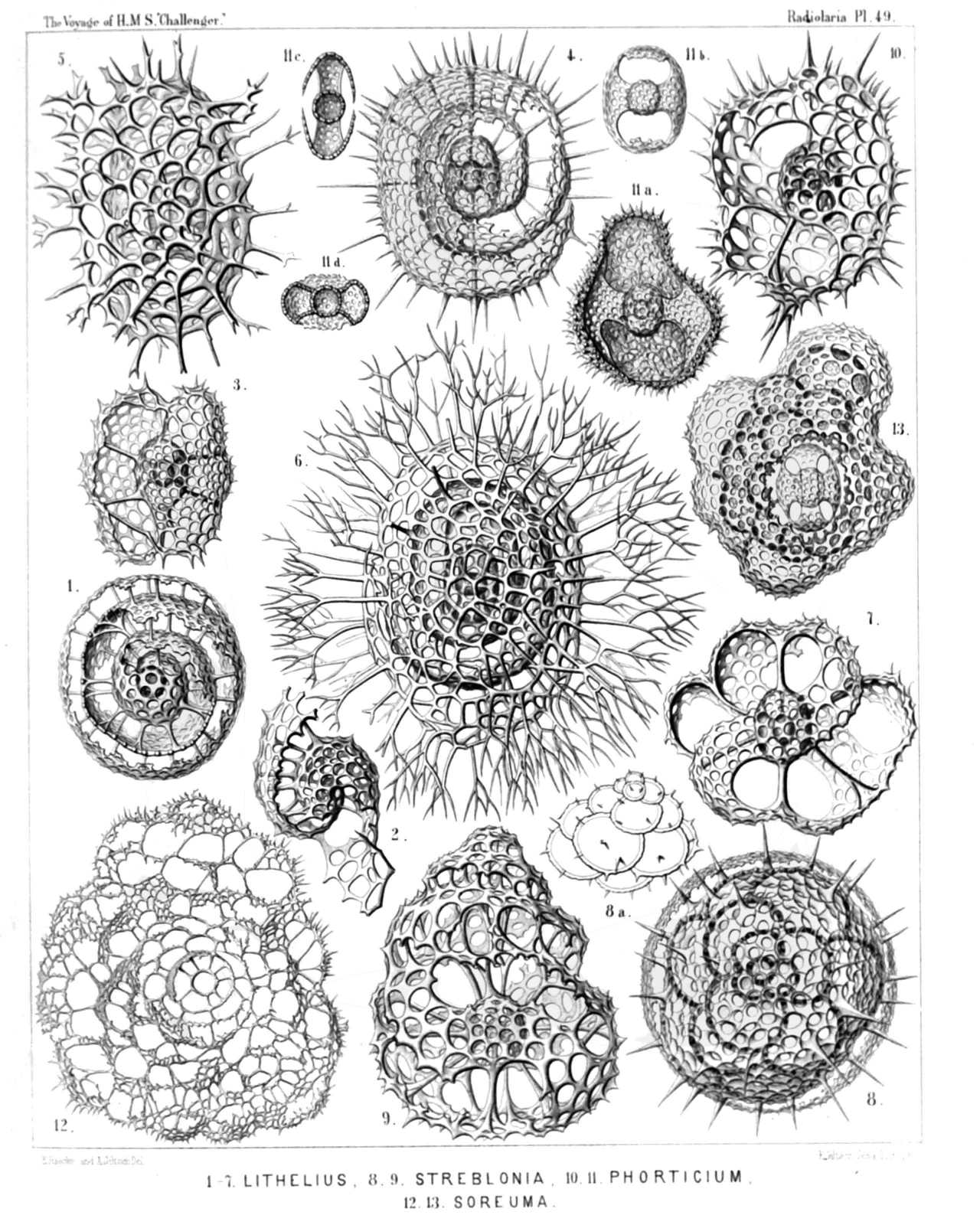 | |||||||||||||||||||||||||||||||||||||||||||||||||||||||||||||||||||||
| PLATE 49. | |||
| Lithelida, Streblonida, Phorticida et Soreumida. | |||
| Diam. | Page. | ||
Fig. 1. Spirema melonia, n. sp., | × | 300 | 692 |
| Fig. 2. Lithelius solaris, n. sp. (the first central convolutions only), | × | 300 | 695 |
| Fig. 3. Larcospira quadrangula, n. sp., | × | 300 | 696 |
| Fig. 4. Pylospira octopyle, n. sp., | × | 300 | 698 |
| Fig. 5. Tholospira cervicornis, n. sp., | × | 300 | 700 |
| Fig. 6. Tholospira dendrophora, n. sp., | × | 300 | 700 |
| Fig. 7. Spironium octonium, n. sp., | × | 300 | 701 |
| Fig. 8. Streblacantha siderolina, n. sp., | × | 300 | 706 |
| Fig. 8a. Outlines of the chambers, | × | 200 | |
| Fig. 9. Streblopyle helicina, n. sp., | × | 300 | 707 |
| Fig. 10. Phorticium pylonium, n. sp., | × | 300 | 709 |
| Fig. 11. Spongophortis larnacilla, n. sp., | × | 200 | 711 |
| Fig. 11a. The upper half of the cortical shell is removed. | |||
| Figs. 11b to 11d. The enclosed medullary Larnacilla-shell. b, Dorsal view; c, lateral view; d, basal view. | |||
| Fig. 12. Soreuma irregulare, n. sp., | × | 200 | 713 |
| Fig. 13. Sorolarcus larnacillifer, n. sp., | × | 300 | 715 |
PLATE 50.
Legion SPUMELLARIA.
Order LARCOIDEA.
Families Larcarida, Larnacida et Zonarida.
| 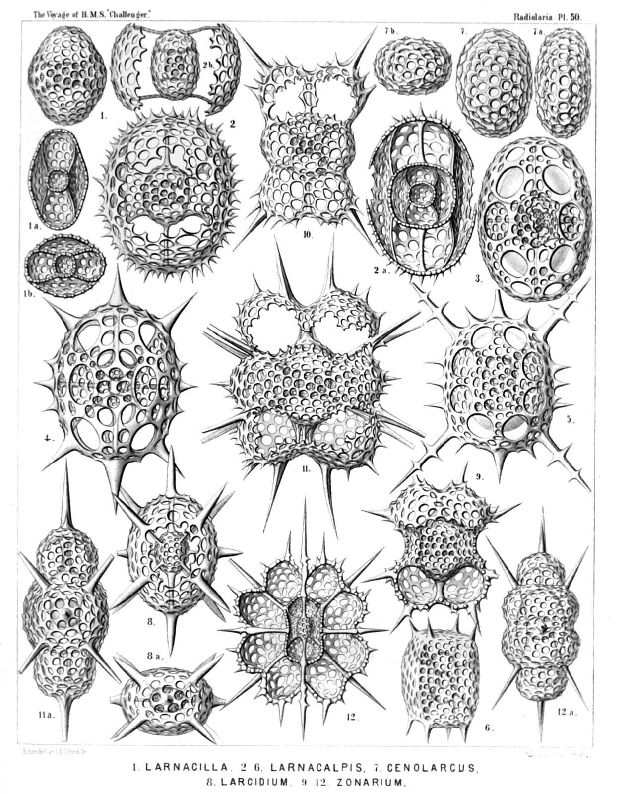 | |||||||||||||||||||||||||||||||||||||||||||||||||||||||||||||||||||||||||||||||||
| PLATE 50. | |||
| Larcarida, Larnacida et Zonarida. | |||
| Diam. | Page. | ||
Fig. 1. Larnacilla typus, n. sp., | × | 300 | 617 |
| From the sagittal pole (dorsal view). | |||
| Fig. 1a. From the lateral pole (sagittal section). | |||
| Fig. 1b. From the principal pole (equatorial section). | |||
| Fig. 2. Larnacalpis lentellipsis, n. sp., | × | 400 | 620 |
| From the sagittal pole (dorsal view). | |||
| Fig. 2a. From the lateral pole (sagittal section). | |||
| Fig. 2b. From the principal pole (equatorial section). | |||
| Fig. 3. Larnacalpis triaxonia, n. sp., | × | 400 | 621 |
| From the sagittal pole (dorsal view). | |||
| Fig. 4. Larnacantha hexacantha, n. sp., | × | 400 | 622 |
| From the sagittal pole (dorsal view). | |||
| Fig. 5. Larnacantha bicruciata, n. sp., | × | 300 | 623 |
| Frontal view. | |||
| Fig. 6. Larnacantha prismatica, n. sp., | × | 300 | 623 |
| Half frontal, half lateral view. | |||
| Fig. 7. Cenolarcus primordialis, n. sp., | × | 300 | 607 |
| From the sagittal pole. | |||
| Fig. 7a. From the lateral pole. | |||
| Fig. 7b. From the principal pole. | |||
| Fig. 8. Larcidium dodecanthum, n. sp., | × | 300 | 612 |
| From the sagittal pole. | |||
| Fig. 8a. From the principal pole. | |||
| Fig. 9. Zonarium octangulum, n. sp., | × | 300 | 685 |
| Frontal view. | |||
| Fig. 10. Zoniscus tetracanthus, n. sp., | × | 300 | 687 |
| Frontal view. | |||
| Fig. 11. Zoniscus hexatholius, n. sp., | × | 400 | 687 |
| Dorsal view (from the sagittal pole). | |||
| Fig. 11a. Lateral view (from the frontal pole). | |||
| Fig. 12. Zonidium octotholium, n. sp., | × | 300 | 688 |
| Frontal section (from the sagittal pole). | |||
| Fig. 12a. Lateral view (from the frontal pole). | |||
PLATE 51.
Legion NASSELLARIA.
Order CYRTOIDEA.
Families Tripocalpida, Phænocalpida et Cyrtocalpida.
| 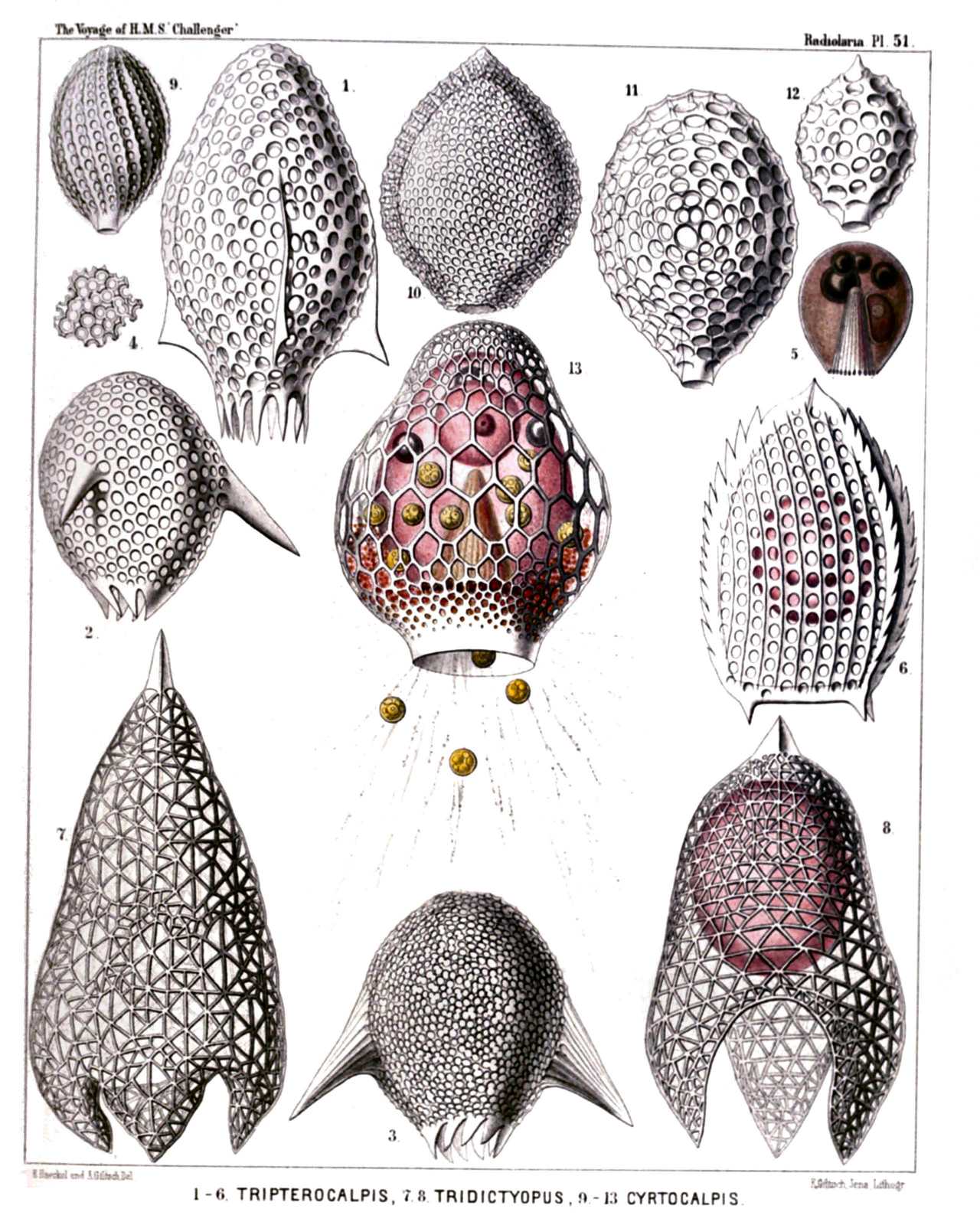 | |||||||||||||||||||||||||||||||||||||||||||||||||||||||||||||||||||
| PLATE 51. | |||
| Tripocalpida, Phænocalpida et Cyrtocalpida. | |||
| Diam. | Page. | ||
Fig. 1. Tripterocalpis phylloptera, n. sp., | × | 400 | 1138 |
| Fig. 2. Tripterocalpis conoptera, n. sp., | × | 300 | 1138 |
| Fig. 3. Tripterocalpis ogmoptera, n. sp., | × | 300 | 1138 |
| Fig. 4. Tripterocalpis ogmoptera, n. sp., | × | 500 | 1138 |
| A group of confluent pores, more enlarged. | |||
| Fig. 5. Tripterocalpis ogmoptera, n. sp., | × | 300 | 1138 |
| Central capsule. In the centre the striate podoconus, above it four oil-globules, to the right the nucleus. | |||
| Fig. 6. Tripocalpis triserrata, n. sp., | × | 600 | 1136 |
| Fig. 7. Tridictyopus conicus, n. sp., | × | 300 | 1145 |
| Fig. 8. Tridictyopus vatillum, n. sp., | × | 400 | 1145 |
| Fig. 9. Cyrtophormis spiralis, n. sp., | × | 400 | 1166 |
| Fig. 10. Archicorys ovata, n. sp., | × | 300 | 1185 |
| Fig. 11. Cyrtocalpis gromia, n. sp., | × | 400 | 1188 |
| Fig. 12. Archicorys microstoma, n. sp., | × | 400 | 1185 |
| Fig. 13. Cyrtocalpis urceolus, n. sp., | × | 500 | 1186 |
| The ovate central capsule exhibits in the lower half the podoconus, in the upper half the spherical nucleus and three oil-globules. Between the capsule and the shell numerous xanthellæ, partly protruded through the shell-mouth along the radiating pseudopodia. | |||
PLATE 52.
Legion NASSELLARIA.
Order CYRTOIDEA.
Families Tripocalpida, Phænocalpida, Cyrtocalpida et Anthocyrtida.
| 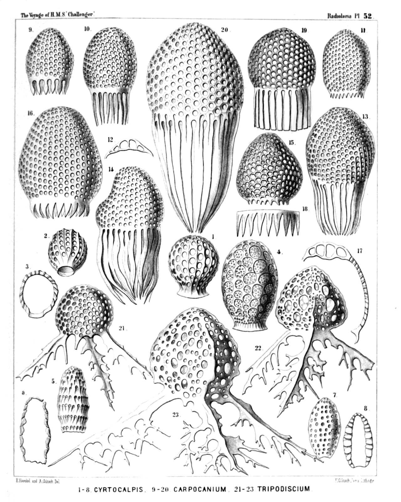 | ||||||||||||||||||||||||||||||||||||||||||||||||||||||||||||||||||||||||||||||||||||||||||||||||||||||||||||||
| PLATE 52. | |||
| Tripocalpida, Phænocalpida, Cyrtocalpida et Anthocyrtida. | |||
| Diam. | Page. | ||
Fig. 1. Cyrtophormis pila, n. sp., | × | 300 | 1165 |
| Fig. 2. Cyrtophormis ærostatica, n. sp., | × | 300 | 1166 |
| Fig. 3. Cyrtophormis ærostatica, n. sp., | × | 300 | 1166 |
| Longitudinal section. | |||
| Fig. 4. Cyrtocalpis sethopora, n. sp., | × | 600 | 1187 |
| Fig. 5. Cyrtocalpis lithomitra, n. sp., | × | 400 | 1187 |
| Fig. 6. Cyrtocalpis lithomitra, n. sp., | × | 400 | 1187 |
| Longitudinal section. | |||
| Fig. 7. Cyrtocalpis compacta, n. sp., | × | 400 | 1187 |
| Fig. 8. Cyrtocalpis compacta, n. sp., | × | 400 | 1187 |
| Longitudinal section. | |||
| Fig. 9. Carpocanistrum flosculum, n. sp., | × | 400 | 1171 |
| Fig. 10. Carpocanistrum cephalum, n. sp., | × | 300 | 1171 |
| Fig. 11. Carpocanistrum evacuatum, n. sp., | × | 400 | 1172 |
| Fig. 12. Carpocanium verecundum, n. sp., | × | 400 | 1284 |
| Vertical section through the top of the shell. | |||
| Fig. 13. Carpocanium verecundum, n. sp., | × | 400 | 1284 |
| Fig. 14. Carpocanium irregulare, n. sp., | × | 400 | 1284 |
| Fig. 15. Carpocanium hexagonale, n. sp., | × | 400 | 1282 |
| Fig. 16. Carpocanium peristomium, n. sp., | × | 500 | 1283 |
| Fig. 17. Carpocanium peristomium, n. sp., | × | 500 | 1283 |
| Vertical section. | |||
| Fig. 18. Carpocanium trepanium, n. sp., | × | 600 | 1282 |
| Peristome. | |||
| Fig. 19. Carpocanium petalospyris, n. sp., | × | 300 | 1283 |
| Fig. 20. Carpocanium virgineum, n. sp., | × | 600 | 1285 |
| Fig. 21. Tripodiscium sphærocephalum, n. sp., | × | 400 | 1144 |
| Fig. 22. Tripodiscium tristylospyris, n. sp. (vel Tristylospyris tripodiscium), | × | 600 | 1143 |
| Fig. 23. Tripodiscium ramosum, n. sp. (vel Tristylospyris ramosa), | × | 600 | 1144 |
PLATE 53.
Legion NASSELLARIA.
Orders SPYROIDEA et CYRTOIDEA.
Families Zygospyrida, Tripocalpida, Phænocalpida et Cyrtocalpida.
| 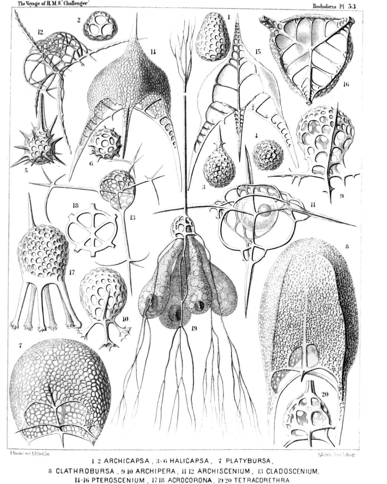 | |||||||||||||||||||||||||||||||||||||||||||||||||||||||||||||||||||||||||||||||||||||||||||||||||||||||||
| PLATE 53. | |||
| Zygospyrida, Tripocalpida, Phænocalpida et Cyrtocalpida. | |||
| Diam. | Page. | ||
Fig. 1. Archicapsa triforis, n. sp., | × | 300 | 1191 |
| Lateral view. | |||
| Fig. 2. Archicapsa triforis, n. sp., | × | 300 | 1191 |
| Basal view. | |||
| Fig. 3. Halicapsa triglochin, n. sp., | × | 200 | 1190 |
| Lateral view. | |||
| Fig. 4. Halicapsa triglochin, n. sp., | × | 200 | 1191 |
| Basal view. | |||
| Fig. 5. Halicapsa hystrix, n. sp., | × | 200 | 1191 |
| Lateral view. | |||
| Fig. 6. Halicapsa hystrix, n. sp., | × | 200 | 1191 |
| Basal view. | |||
| Fig. 7. Cantharospyris platybursa, n. sp. (vel Platybursa compressa), | × | 400 | 1051 |
| Fig. 8. Tessarospyris clathrobursa, n. sp. (vel Clathrobursa dictyopus), | × | 400 | 1045 |
| Fig. 9. Peridium spinipes, n. sp., | × | 500 | 1154 |
| Fig. 10. Peridium palmipes, n. sp., | × | 500 | 1154 |
| Fig. 11. Archiscenium quadrispinum, n. sp., | × | 500 | 1150 |
| In the spherical central capsule the dark nucleus is visible. | |||
| Fig. 12. Euscenium eucolpium, n. sp., | × | 500 | 1147 |
| Fig. 13. Cladoscenium ancoratum, n. sp., | × | 400 | 1149 |
| Fig. 14. Pteroscenium pinnatum, n. sp., | × | 400 | 1152 |
| Lateral view. | |||
| Fig. 15. Pteroscenium pinnatum, n. sp., | × | 400 | 1152 |
| Vertical section. | |||
| Fig. 16. Pteroscenium pinnatum, n. sp., | × | 400 | 1152 |
| Basal view. | |||
| Fig. 17. Calpophæna hexarrhabda, n. sp., | × | 400 | 1176 |
| Fig. 18. Calpophæna hexarrhabda, n. sp., | × | 400 | 1176 |
| Basal plate. | |||
| Fig. 19. Tetraspyris tetracorethra, n. sp., | × | 400 | 1044 |
| With the four-lobed central capsule, in each lobe an oil-globule. | |||
| Fig. 20. Tetraspyris tetracorethra, n. sp., | × | 800 | 1044 |
| Shell more enlarged. | |||
PLATE 54.
Legion NASSELLARIA.
Order CYRTOIDEA.
Families Phænocalpida, Cyrtocalpida, Anthocyrtida et Sethocyrtida.
| 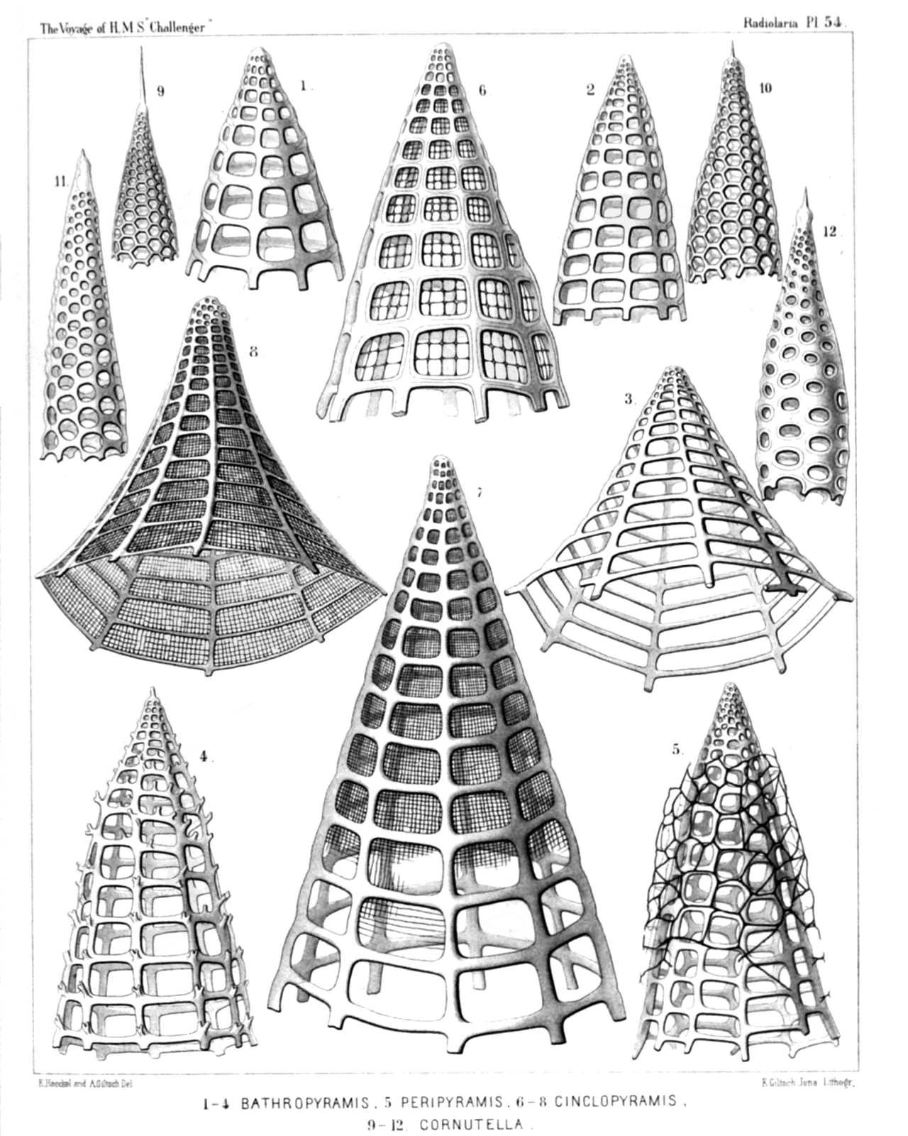 | ||||||||||||||||||||||||||||||||||||||||||||||||||||||||||||
| PLATE 54. | |||
| Phænocalpida, Cyrtocalpida, Anthocyrtida et Sethocyrtida. | |||
| Diam. | Page. | ||
Fig. 1. Bathropyramis quadrata, n. sp., | × | 300 | 1159 |
| Fig. 2. Sethopyramis quadrata, n. sp., | × | 300 | 1254 |
| Fig. 3. Bathropyramis trapezoides, n. sp., | × | 300 | 1160 |
| Fig. 4. Bathropyramis ramosa, n. sp., | × | 300 | 1161 |
| Fig. 5. Peripyramis circumtexta, n. sp., | × | 300 | 1162 |
| Fig. 6. Plectopyramis dodecomma, n. sp., | × | 400 | 1258 |
| Fig. 7. Cinclopyramis infundibulum, n. sp., | × | 300 | 1161 |
| Fig. 8. Plectopyramis trapezomma, n. sp., | × | 400 | 1258 |
| Fig. 9. Cornutella hexagona, n. sp., | × | 400 | 1180 |
| Fig. 10. Cornutella sethoconus, n. sp., | × | 400 | 1180 |
| Fig. 11. Sethoconus orthoceras, n. sp., | × | 400 | 1294 |
| Fig. 12. Sethoconus bimarginatus, n. sp., | × | 400 | 1295 |
PLATE 55.
Legion NASSELLARIA.
Order CYRTOIDEA.
Families Phænocalpida, Anthocyrtida et Sethocyrtida.
| 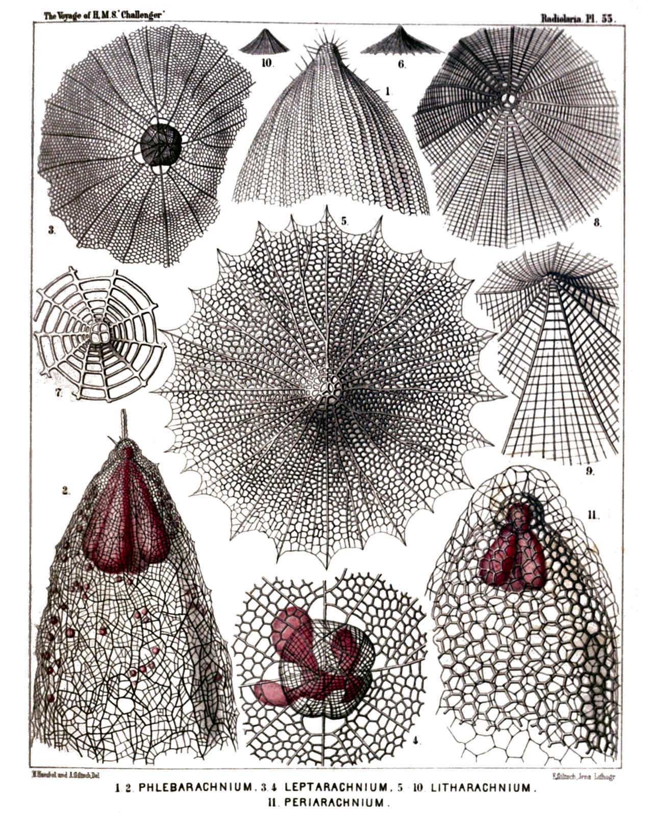 | |||||||||||||||||||||||||||||||||||||||||||||||||||||||||||||||||||
| PLATE 55. | |||
| Phænocalpida, Anthocyrtida et Sethocyrtida. | |||
| Diam. | Page. | ||
Fig. 1. Sethoconus facetus, n. sp. (vel Phlebarachnium facetum), | × | 300 | 1296 |
| Upper part of the shell. | |||
| Fig. 2. Sethoconus venosus, n. sp. (vel Phlebarachnium venosum), | × | 250 | 1297 |
| Shell including the four-lobed central capsule. | |||
| Fig. 3. Sethophormis aurelia, n. sp. (vel Leptarachnium aurelia), | × | 100 | 1248 |
| Shell seen from above. | |||
| Fig. 4. Sethophormis aurelia, n. sp., | × | 400 | 1248 |
| Cephalis more enlarged, with the enclosed four-lobed central capsule. | |||
| Fig. 5. Cladarachnium ramosum, n. sp., | × | 300 | 1165 |
| Apical view. | |||
| Fig. 6. Cladarachnium ramosum, n. sp., | × | 70 | 1165 |
| Lateral view. | |||
| Fig. 7. Bathropyramis interrupta, n. sp., | × | 300 | 1160 |
| Apical part of the shell, from above. | |||
| Fig. 8. Litharachnium araneosum, n. sp., | × | 300 | 1163 |
| Apical part of the shell, from above. | |||
| Fig. 9. Litharachnium epeira, n. sp., | × | 500 | 1164 |
| Oblique view of the shell. | |||
| Fig. 10. Litharachnium araneosum, n. sp., | × | 50 | 1163 |
| Lateral view. | |||
| Fig. 11. Periarachnium periplectum, n. sp., | × | 500 | 1297 |
| Shell enclosing the trilobed central capsule. | |||
PLATE 56.
Legion NASSELLARIA.
Order CYRTOIDEA.
Families Tripocyrtida, Anthocyrtida et Sethocyrtida.
| 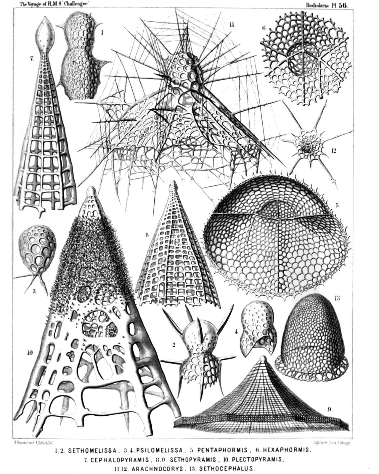 | |||||||||||||||||||||||||||||||||||||||||||||||||||||||||||||||||||
| PLATE 56. | |||
| Tripocyrtida, Anthocyrtida et Sethocyrtida. | |||
| Diam. | Page. | ||
Fig. 1. Lithomelissa bütschlii, n. sp. (vel Sethomelissa bütschlii), | × | 400 | 1207 |
| Fig. 2. Lithomelissa decacantha, n. sp. (vel Sethomelissa decacantha), | × | 400 | 1208 |
| Fig. 3. Psilomelissa calvata, n. sp., | × | 400 | 1209 |
| The cephalis alone, with the three collar beams. | |||
| Fig. 4. Lychnodictyum scaphopodium, n. sp., | × | 400 | 1231 |
| Fig. 5. Sethophormis pentalactis, n. sp. (vel Pentaphormis pentalactis), | × | 400 | 1244 |
| Oblique view of the shell, from below. | |||
| Fig. 6. Sethophormis hexalactis, n. sp. (vel Hexaphormis hexalactis), | × | 400 | 1245 |
| Central part of the shell, with the cortinar septum. | |||
| Fig. 7. Sethopyramis enneactis, n. sp. (vel Cephalopyramis enneactis), | × | 400 | 1254 |
| Fig. 8. Plectopyramis polypleura, n. sp. (vel Sethopyramis polypleura), | × | 200 | 1260 |
| Fig. 9. Sethophormis eupilium, n. sp. (vel Craspedilium eupilium), | × | 400 | 1247 |
| Fig. 10. Plectopyramis spongiosa, n. sp. (vel Spongopyramis spongiosa), | × | 400 | 1261 |
| Fig. 11. Arachnocorys araneosa, n. sp., | × | 500 | 1266 |
| Fig. 12. Sethophormis dodecaster, n. sp. (vel Astrophormis dodecaster), | × | 200 | 1248 |
| Fig. 13. Sethocephalus eucecryphalus, n. sp., | × | 400 | 1298 |
PLATE 57.
Legion NASSELLARIA.
Order CYRTOIDEA.
Families TRIPOCYRTIDA, ANTHOCYRTIDA et SETHOCYRTIDA.
| 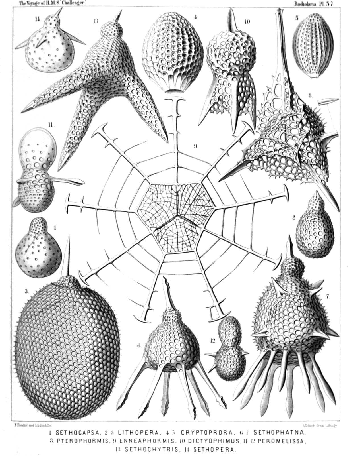 | ||||||||||||||||||||||||||||||||||||||||||||||||||||||||||||||||||||
| PLATE 57. | |||
| TRIPOCYRTIDA, ANTHOCYRTIDA et SETHOCYRTIDA. | |||
| Diam. | Page. | ||
Fig. 1. Dicolocapsa microcephala, n. sp., | × | 400 | 1312 |
| Fig. 2. Sethocapsa pyriformis, n. sp., | × | 300 | 1310 |
| Fig. 3. Lithopera ananassa, n. sp., | × | 500 | 1234 |
| Fig. 4. Sethamphora favosa, n. sp. (vel Cryptoprora favosa), | × | 400 | 1252 |
| Fig. 5. Sethamphora microstoma, n. sp. (vel Cryptoprora microstoma), | × | 300 | 1252 |
| Fig. 6. Clistophæna hexolena, n. sp., | × | 300 | 1287 |
| Fig. 7. Clistophæna armata, n. sp., | × | 300 | 1288 |
| Fig. 8. Clathromitra pterophormis, n. sp., | × | 400 | 1219 |
| Fig. 9. Sethophormis rotula, n. sp. (vel Enneaphormis rotula), | × | 400 | 1246 |
| Fig. 10. Dictyophimus sphærocephalus, n. sp., | × | 400 | 1195 |
| Fig. 11. Peromelissa phalacra, n. sp., | × | 400 | 1236 |
| Fig. 12. Peromelissa calva, n. sp., | × | 300 | 1237 |
| Fig. 13. Sethochytris triconiscus, n. sp., | × | 300 | 1239 |
| Fig. 14. Micromelissa bombus, n. sp., | × | 300 | 1235 |
PLATE 58.
Legion NASSELLARIA.
Order CYRTOIDEA.
Families Tripocyrtida, Sethocyrtida, Phormocyrtida et Theocyrtida.
| 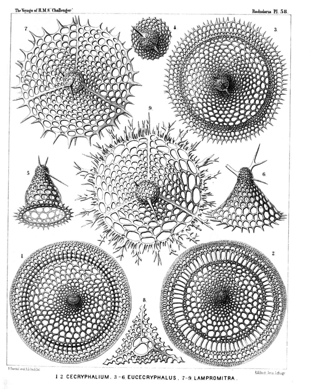 | |||||||||||||||||||||||||||||||||||||||||||||||||||||||||
| PLATE 58. | |||
| Tripocyrtida, Sethocyrtida, Phormocyrtida et Theocyrtida. | |||
| Diam. | Page. | ||
Fig. 1. Cecryphalium sestrodiscus, n. sp., | × | 400 | 1399 |
| Apical view. | |||
| Fig. 2. Cecryphalium lamprodiscus, n. sp., | × | 400 | 1398 |
| Apical view. | |||
| Fig. 3. Clathrocyclas coscinodiscus, n. sp., | × | 400 | 1389 |
| Apical view. | |||
| Fig. 4. Clathrocyclas coscinodiscus, n. sp., | × | 700 | 1389 |
| The cephalis alone, with the two horns. | |||
| Fig. 5. Clathrocyclas semeles, n. sp., | × | 400 | 1388 |
| Lateral view. | |||
| Fig. 6. Sethoconus capreolus, n. sp., | × | 400 | 1291 |
| Lateral view. | |||
| Fig. 7. Lampromitra quadricuspis, n. sp., | × | 400 | 1214 |
| Apical view. | |||
| Fig. 8. Lampromitra furcata, n. sp., | × | 400 | 1215 |
| The collar septum after removal of the cephalis. | |||
| Fig. 9. Lampromitra dendrocorona, n. sp., | × | 400 | 1216 |
| Apical view. | |||
PLATE 59.
Legion NASSELLARIA.
Order CYRTOIDEA.
Families Tripocyrtida, Podocyrtida et Phormocyrtida.
| 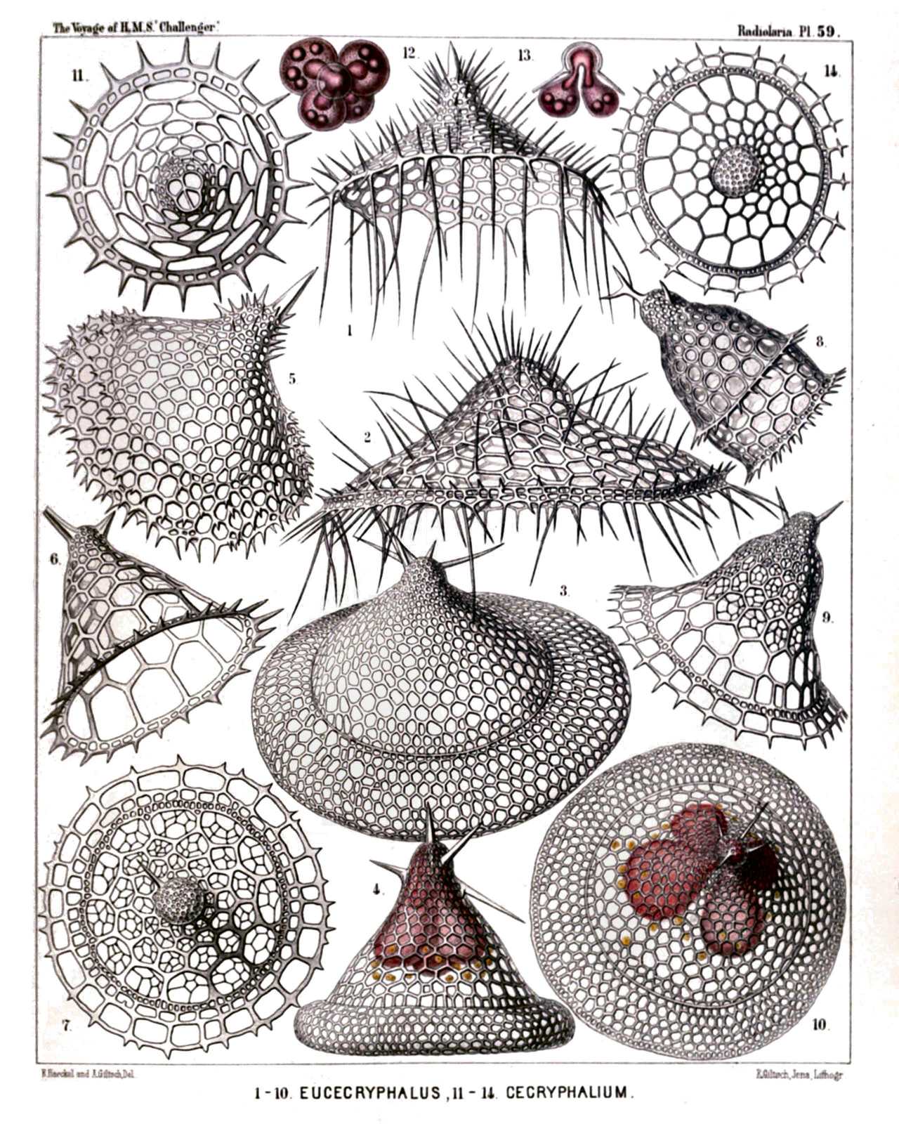 | |||||||||||||||||||||||||||||||||||||||||||||||||||||||||||||||||||||||||||
| PLATE 59. | |||
| Tripocyrtida, Podocyrtida et Phormocyrtida. | |||
| Diam. | Page. | ||
Fig. 1. Lampromitra huxleyi, n. sp., | × | 400 | 1215 |
| Fig. 2. Amphiplecta callistoma, n. sp., | × | 400 | 1224 |
| Fig. 3. Corocalyptra agnesæ, n. sp., | × | 400 | 1323 |
| Fig. 4. Corocalyptra emmæ, n. sp., | × | 400 | 1323 |
| The shell encloses the trilobate central capsule, with the trilobate nucleus. | |||
| Fig. 5. Clathrocyclas cassiopejæ, n. sp., | × | 400 | 1390 |
| Fig. 6. Clathrocyclas alcmenæ, n. sp., | × | 400 | 1388 |
| Fig. 7. Clathrocyclas latonæ, n. sp., | × | 400 | 1389 |
| Apical view. | |||
| Fig. 8. Diplocylas bicorona, n. sp., | × | 400 | 1392 |
| Fig. 9. Clathrocyclas ionis, n. sp., | × | 400 | 1389 |
| Fig. 10. Corocalyptra elisabethæ, n. sp., | × | 400 | 1323 |
| Oblique apical view of the shell, with the quadrilobate central capsule enclosed. | |||
| Fig. 11. Clathrocyclas europæ, n. sp., | × | 400 | 1388 |
| Apical view of the shell, after removal of the cephalis. | |||
| Fig. 12. Clathrocyclas europæ, n. sp., | × | 400 | 1388 |
| Central capsule, seen from above, with the quadrilobate nucleus. | |||
| Fig. 13. Clathrocyclas danaës, n. sp., | × | 300 | 1388 |
| Vertical section through the cephalis and the quadrilobate central capsule, with the quadrilobate nucleus. | |||
| Fig. 14. Clathrocyclas danaës, n. sp., | × | 300 | 1388 |
| Apical view of the shell. | |||
PLATE 60.
Legion NASSELLARIA.
Order CYRTOIDEA.
Family Tripocyrtida.
| 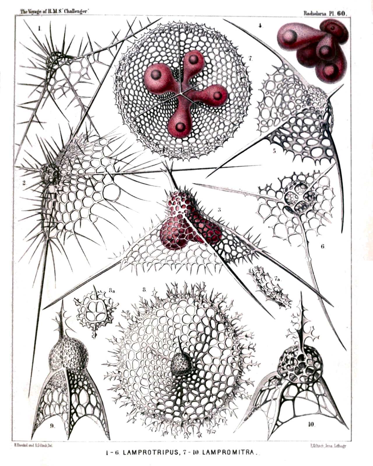 | |||||||||||||||||||||||||||||||||||||||||||||||||||||||||||||||||||
| PLATE 60. | |||
| Tripocyrtida. | |||
| Diam. | Page. | ||
Fig. 1. Dictyophimus cienkowskii, n. sp. (vel Lamprotripus squarrosus), | × | 300 | 1200 |
| Shell seen from the side. | |||
| Fig. 2. Dictyophimus bütschlii, n. sp. (vel Lamprotripus horridus), | × | 300 | 1201 |
| Fig. 3. Dictyophimus hertwigii, n. sp. (vel Lamprotripus spinosus), | × | 400 | 1201 |
| The cephalis of the shell includes the central capsule, with three lobes depending in the pyramidal thorax. | |||
| Fig. 4. Dictyophimus platycephalus, n. sp., | × | 400 | 1198 |
| Central capsule with four thoracic lobes, each of which contains an oil-globule; kidney-shaped nucleus in the cephalic lobe. | |||
| Fig. 5. Dictyophimus platycephalus, n. sp., | × | 400 | 1198 |
| Shell seen from the side. | |||
| Fig. 6. Dictyophimus brandtii, n. sp., | × | 300 | 1198 |
| Shell seen from the base, with the four large pores of the collar septum, two minor jugular and two major cardinal pores. | |||
| Fig. 7. Lampromitra coronata, n. sp., | × | 400 | 1214 |
| Shell seen from below, with the quadrilobate central capsule. | |||
| Fig. 7a. A portion of the shell-margin, | × | 800 | 1214 |
| Fig. 8. Lampromitra arborescens, n. sp., | × | 400 | 1216 |
| Shell from above. | |||
| Fig. 8a. The collar septum with the four crossed rods of the cortina, | × | 400 | 1216 |
| Fig. 9. Tripocyrtis plectaniscus, n. sp., | × | 400 | 1202 |
| Fig. 10. Tripocyrtis plagoniscus, n. sp., | × | 400 | 1201 |
PLATE 61.
Legion NASSELLARIA.
Order CYRTOIDEA.
Family Tripocyrtida.
| 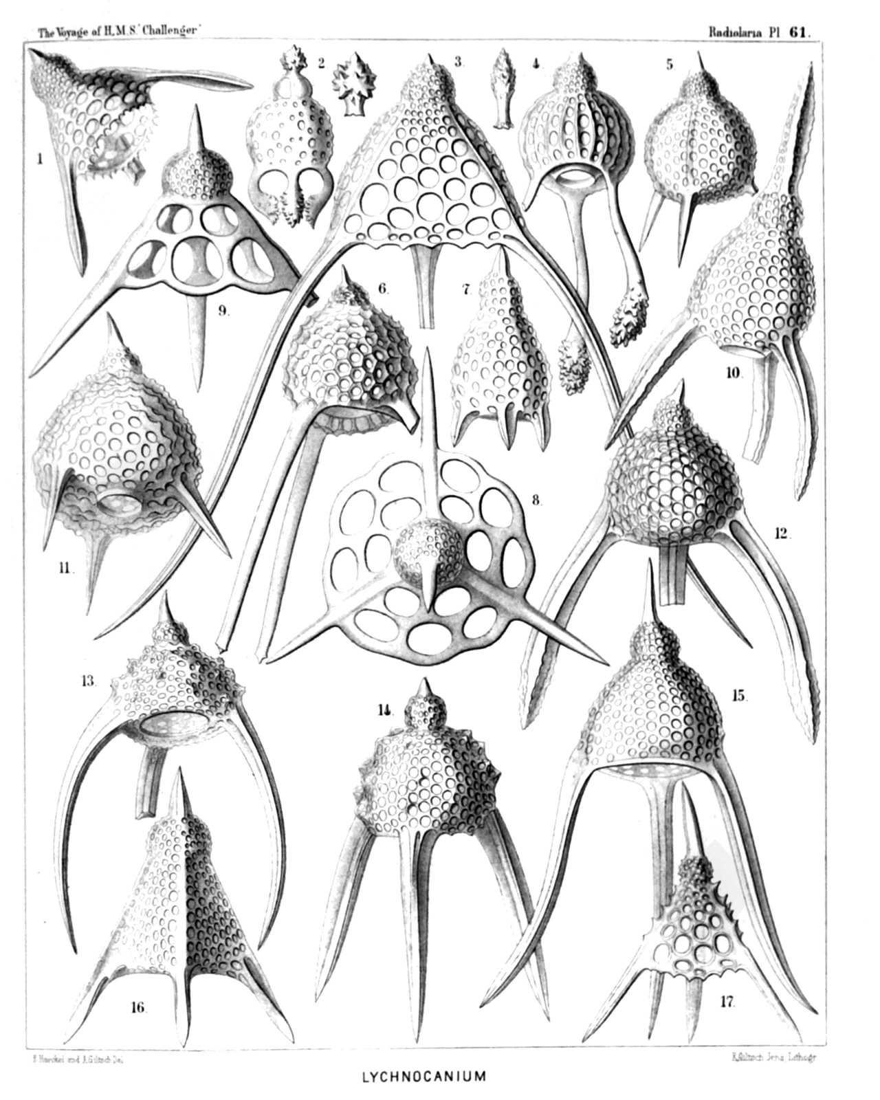 | ||||||||||||||||||||||||||||||||||||||||||||||||||||||||||||||||||||||||||||||||||
| PLATE 61. | |||
| Tripocyrtida. | |||
| Diam. | Page. | ||
Fig. 1. Dictyophimus cortina, n. sp., | × | 400 | 1197 |
| Fig. 2. Lychnocanium pudicum, n. sp., | × | 200 | 1230 |
| Fig. 3. Dictyophimus longipes, n. sp., | × | 400 | 1197 |
| Fig. 4. Lychnocanium clavigerum, n. sp., | × | 300 | 1230 |
| Fig. 5. Dictyophimus lasanum, n. sp., | × | 300 | 1197 |
| Fig. 6. Lychnocanium favosum, n. sp., | × | 300 | 1225 |
| Fig. 7. Lychnocanium lanterna, n. sp., | × | 300 | 1224 |
| Fig. 8. Dictyophimus plectaniscus, n. sp., | × | 300 | 1196 |
| Apical view. | |||
| Fig. 9. Dictyophimus plectaniscus, n. sp., | × | 300 | 1196 |
| Lateral view. | |||
| Fig. 10. Lychnocanium fenestratum, n. sp., | × | 400 | 1228 |
| Fig. 11. Lychnocanium pyriforme, n. sp., | × | 300 | 1225 |
| Fig. 12. Lychnocanium fortipes, n. sp., | × | 300 | 1227 |
| Fig. 13. Lychnocanium tuberosum, n. sp., | × | 300 | 1227 |
| Fig. 14. Lychnocanium nodosum, n. sp., | × | 300 | 1225 |
| Fig. 15. Lychnocanium sigmopodium, n. sp., | × | 400 | 1228 |
| Fig. 16. Dictyophimus pyramis, n. sp., | × | 300 | 1196 |
| Fig. 17. Dictyophimus triserratus, n. sp., | × | 300 | 1200 |
PLATE 62.
Legion NASSELLARIA.
Order CYRTOIDEA.
Families Anthocyrtida, Sethocyrtida et Phormocyrtida.
| 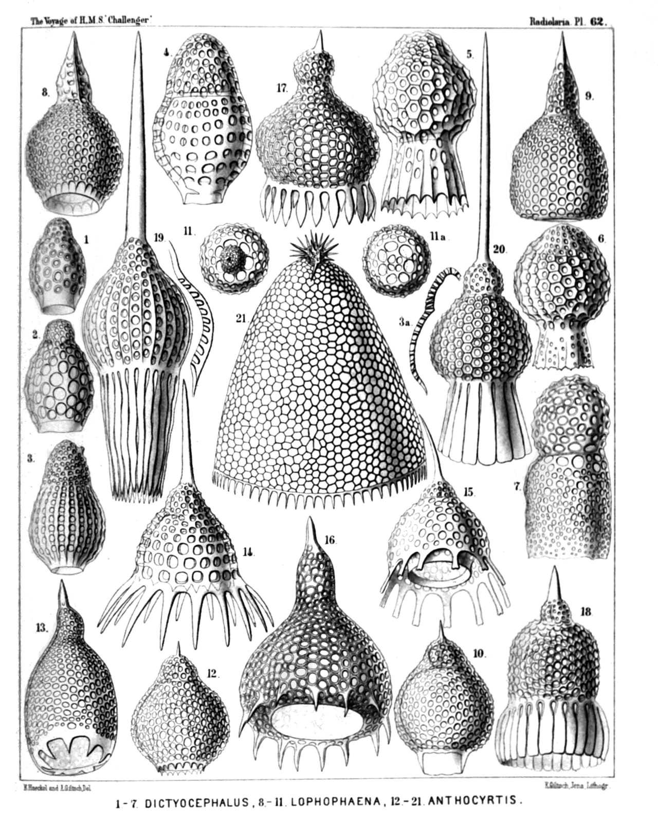 | |||||||||||||||||||||||||||||||||||||||||||||||||||||||||||||||||||||||||||||||||||||||||||||||||||||||
| PLATE 62. | |||
| Anthocyrtida, Sethocyrtida et Phormocyrtida. | |||
| Diam. | Page. | ||
Fig. 1. Dictyocephalus australis, n. sp., | × | 300 | 1306 |
| Fig. 2. Dictyocephalus mediterraneus, n. sp., | × | 300 | 1307 |
| Fig. 3. Sethamphora costata, n. sp. (vel Dictyocephalus costatus), | × | 300 | 1251 |
| Fig. 4. Dictyocephalus amphora, n. sp., | × | 400 | 1305 |
| Fig. 5. Cycladophora (?) favosa, n. sp. (an Dictyocephalus?), | × | 400 | 1380 |
| Fig. 6. Cycladophora (?) favosa, n. sp. (an Dictyocephalus?), | × | 400 | 1380 |
| A variety with obliterated ribs (?). | |||
| Fig. 7. Dictyocephalus globiceps, n. sp., | × | 400 | 1308 |
| Fig. 8. Sethocorys achillis, n. sp., | × | 400 | 1301 |
| Fig. 9. Sethocyrtis oxycephalis, n. sp., | × | 400 | 1299 |
| Fig. 10. Sethocorys odysseus, n. sp., | × | 400 | 1302 |
| Fig. 11. Sethocyrtis agamemnonis, n. sp., | × | 300 | 1300 |
| Seen from above (apical view). | |||
| Fig. 11A. Sethocyrtis agamemnonis, n. sp., | × | 300 | 1300 |
| Seen from above, after removal of the cephalis. | |||
| Fig. 12. Anthocyrtium pyrum, n. sp., | × | 400 | 1276 |
| Fig. 13. Anthocyrtis ovata, n. sp., | × | 300 | 1272 |
| Fig. 14. Anthocyrtium chrysanthemum, n. sp | × | 400 | 1272 |
| Fig. 15. Anthocyrtidium ligularia, n. sp., | × | 400 | 1278 |
| Fig. 16. Anthocyrtidium cineraria, n. sp., | × | 400 | 1278 |
| Fig. 17. Anthocyrtium campanula, n. sp., | × | 400 | 1274 |
| Fig. 18. Anthocyrtium doronicum, n. sp., | × | 300 | 1276 |
| Fig. 19. Anthocyrtium flosculus, n. sp., | × | 300 | 1277 |
| Fig. 20. Anthocyrtium adonis, n. sp., | × | 300 | 1273 |
| Fig. 21. Sethoconus anthocyrtis, n. sp. (vel Anthocyrtium sethoconium), | × | 300 | 1296 |
PLATE 63.
Legion NASSELLARIA.
Order CYRTOIDEA.
Family Tripocyrtida.
| 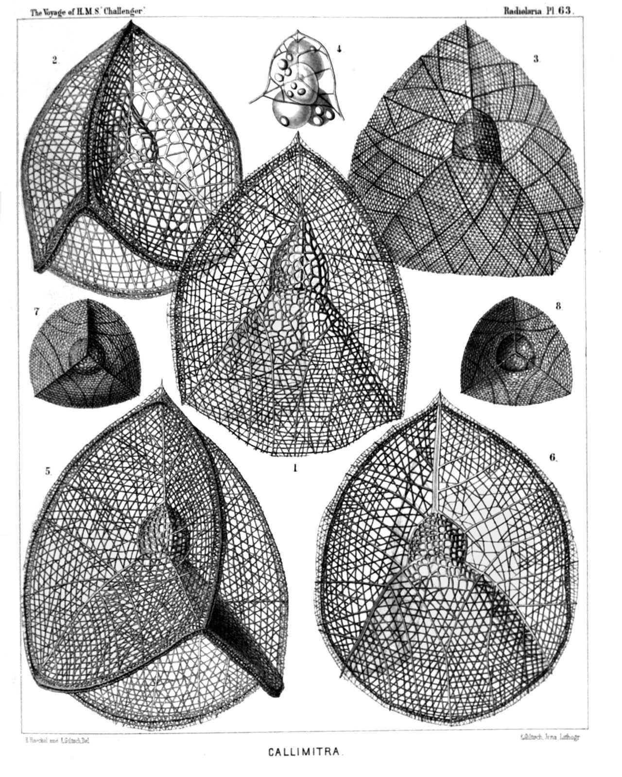 | ||||||||||||||||||||||||||||||||||||||||||||||||||||
| PLATE 63. | |||
| Tripocyrtida. | |||
| Diam. | Page. | ||
Fig. 1. Callimitra carolotæ, n. sp., | × | 400 | 1217 |
| Lateral view. | |||
| Fig. 2. Callimitra annæ, n. sp., | × | 400 | 1217 |
| Dorsal view. | |||
| Fig. 3. Callimitra emmæ, n. sp., | × | 300 | 1218 |
| Lateral view. | |||
| Fig. 4. Callimitra emmæ, n. sp., | × | 400 | 1218 |
| Cephalis alone, with the enclosed four-lobed central capsule, and the internal four divergent beams; surrounded by some scattered xanthellæ. | |||
| Fig. 5. Callimitra agnesæ, n. sp., | × | 400 | 1217 |
| Dorsal view. | |||
| Fig. 6. Callimitra elisabethæ, n. sp., | × | 400 | 1218 |
| Lateral view. | |||
| Fig. 7. Callimitra carolotæ, n. sp., | × | 200 | 1217 |
| Seen from above (from the apical pole). | |||
| Fig. 8. Callimitra carolotæ, n. sp., | × | 200 | 1217 |
| Seen from below (from the basal pole). | |||
PLATE 64.
Legion NASSELLARIA.
Order CYRTOIDEA.
Families Tripocyrtida et Podocyrtida.
| 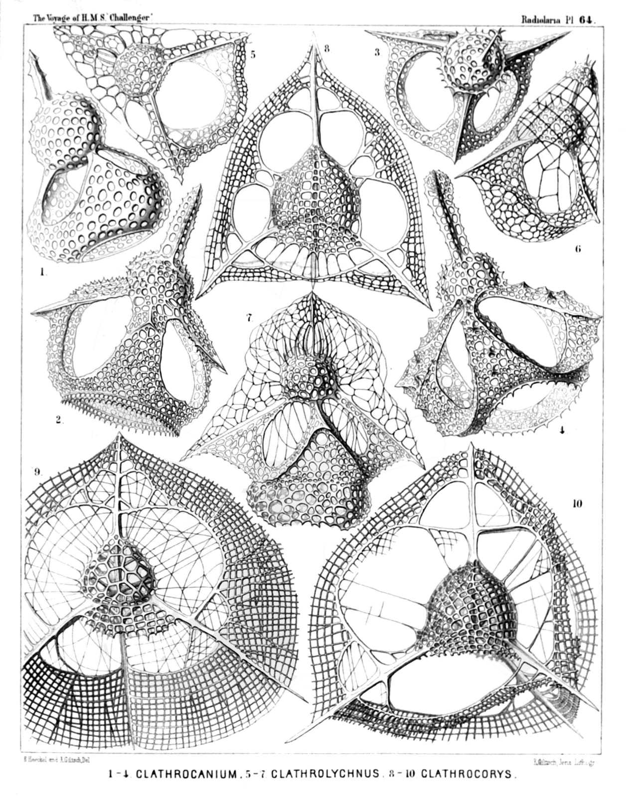 | ||||||||||||||||||||||||||||||||||||||||||||||||||||
| PLATE 64. | |||
| Tripocyrtida et Podocyrtida. | |||
| Diam. | Page. | ||
Fig. 1. Clathrocanium sphærocephalum, n. sp., | × | 600 | 1211 |
| Fig. 2. Clathrocanium diadema, n. sp., | × | 600 | 1212 |
| Fig. 3. Clathrocanium triomma, n. sp., | × | 600 | 1211 |
| Fig. 4. Clathrocanium reginæ, n. sp., | × | 600 | 1212 |
| Fig. 5. Clathrolychnus araneosus, n. sp., | × | 600 | 1240 |
| Fig. 6. Clathrolychnus periplectus, n. sp., | × | 600 | 1241 |
| Fig. 7. Pteropilium clathrocanium, n. sp., | × | 400 | 1327 |
| Fig. 8. Clathrocorys murrayi, n. sp., | × | 600 | 1219 |
| Fig. 9. Clathrocorys giltschii, n. sp., | × | 600 | 1220 |
| Fig. 10. Clathrocorys teuscheri, n. sp., | × | 600 | 1220 |
PLATE 65.
Legion NASSELLARIA.
Order CYRTOIDEA.
Family Phormocyrtida.
| 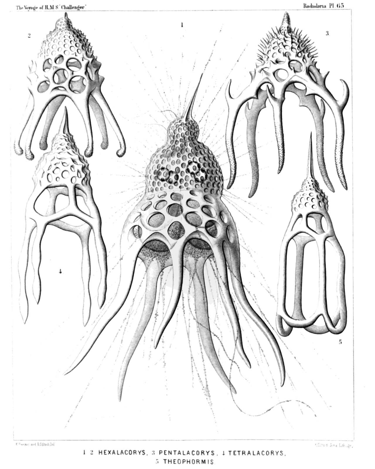 | |||||||||||||||||||||||||||||||||
| PLATE 65. | |||
| Phormocyrtida. | |||
| Diam. | Page. | ||
Fig. 1. Alacorys friderici, n. sp. (vel Hexalacorys friderici), | × | 400 | 1372 |
| The central capsule, enclosed in the fenestrated shell, exhibits in its lower half four large club-shaped lobes, each of which includes in its upper part a large oil-globule. The uppermost, undivided part of the capsule includes the nucleus, which protrudes four small nuclear lobes through the four holes of the cortinar septum into the thorax. Numerous long pseudopodia arise from the granular sarcomatrix, which the capsule surrounds, and pass through the pores of the siliceous shell. | |||
| Fig. 2. Alacorys guilelmi, n. sp. (vel Hexalacorys guilelmi), | × | 300 | 1372 |
| Fig. 3. Alacorys bismarckii, n. sp. (vel Pentalacorys bismarckii), | × | 200 | 1372 |
| Fig. 4. Alacorys lutheri, n. sp. (vel Tetralacorys lutheri), | × | 400 | 1370 |
| Fig. 5. Cycladophora goetheana, n. sp. (vel Lampterium goetheanum), | × | 300 | 1376 |
PLATE 66.
Legion NASSELLARIA.
Order CYRTOIDEA.
Family Theocyrtida.
| 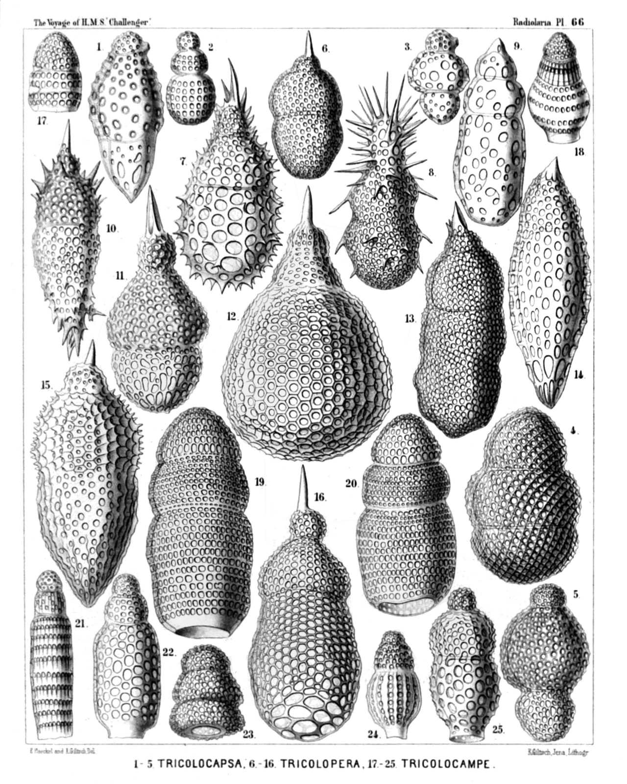 | ||||||||||||||||||||||||||||||||||||||||||||||||||||||||||||||||||||||||||||||||||||||||||||||||||||||||||||||||
| PLATE 66. | |||
| Theocyrtida. | |||
| Diam. | Page. | ||
Fig. 1. Tricolocapsa theophrasti, n. sp., | × | 400 | 1432 |
| Fig. 2. Tricolocapsa schleidenii, n. sp., | × | 300 | 1433 |
| Fig. 3. Tricolocapsa dioscoridis, n. sp., | × | 300 | 1432 |
| Fig. 4. Tricolocapsa decandollei, n. sp., | × | 300 | 1433 |
| Fig. 5. Tricolocapsa linnæi, n. sp., | × | 400 | 1432 |
| Fig. 6. Theocapsa aristotelis, n. sp., | × | 300 | 1427 |
| Fig. 7. Theocapsa mülleri, n. sp., | × | 400 | 1431 |
| Fig. 8. Theocapsa democriti, n. sp., | × | 400 | 1427 |
| Fig. 9. Theocapsa forskalii, n. sp., | × | 400 | 1429 |
| Fig. 10. Theocapsa cuvieri, n. sp., | × | 400 | 1430 |
| Fig. 11. Theocapsa wottonis, n. sp., | × | 400 | 1428 |
| Fig. 12. Theocapsa darwinii, n. sp., | × | 300 | 1431 |
| Fig. 13. Theocapsa linnæi, n. sp., | × | 400 | 1429 |
| Fig. 14. Theocapsa wolffii, n. sp., | × | 400 | 1429 |
| Fig. 15. Theocapsa malpighii, n. sp., | × | 400 | 1428 |
| Fig. 16. Theocapsa lamarckii, n. sp., | × | 400 | 1430 |
| Fig. 17. Tricolocampe amphizona, n. sp., | × | 400 | 1413 |
| Fig. 18. Theocampe collaris, n. sp., | × | 300 | 1425 |
| Fig. 19. Tricolocampe polyzona, n. sp., | × | 400 | 1412 |
| Fig. 20. Tricolocampe stenozona, n. sp., | × | 400 | 1413 |
| Fig. 21. Tricolocampe cylindrica, n. sp., | × | 300 | 1412 |
| Fig. 22. Tricolocampe urnula, n. sp., | × | 400 | 1422 |
| Fig. 23. Theocampe stenostoma, n. sp., | × | 300 | 1423 |
| Fig. 24. Theocampe costata, n. sp., | × | 300 | 1424 |
| Fig. 25. Theocampe sphærothorax, n. sp., | × | 300 | 1424 |
PLATE 67.
Legion NASSELLARIA.
Order CYRTOIDEA.
Family Podocyrtida.
| 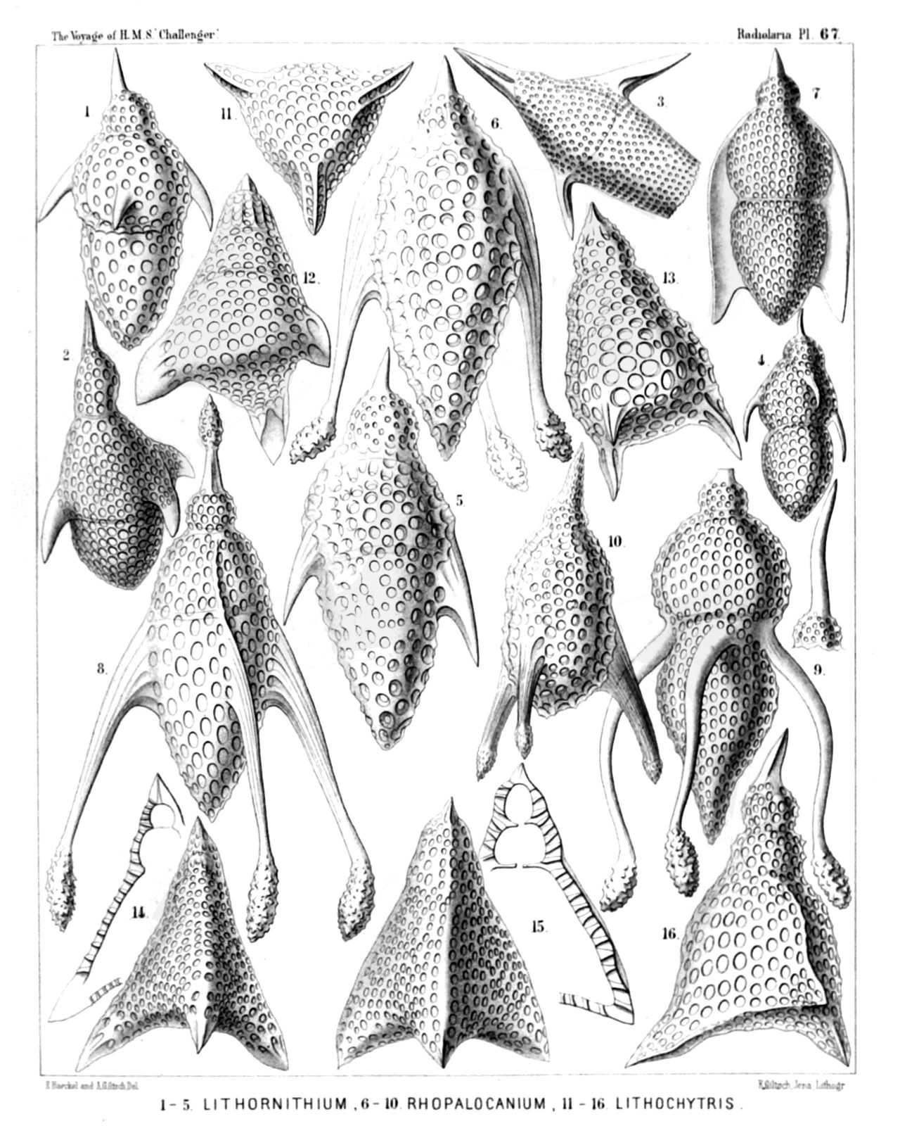 | ||||||||||||||||||||||||||||||||||||||||||||||||||||||||||||||||||||||||||||
| PLATE 67. | |||
| Podocyrtida. | |||
| Diam. | Page. | ||
Fig. 1. Lithornithium falco, n. sp., | × | 400 | 1355 |
| Fig. 2. Lithornithium fringilla, n. sp., | × | 400 | 1355 |
| Fig. 3. Lithornithium ciconia, n. sp., | × | 400 | 1354 |
| Fig. 4. Lithornithium trochilus, n. sp., | × | 400 | 1355 |
| Fig. 5. Theopera fusiformis, n. sp., | × | 400 | 1357 |
| Fig. 6. Theopera chytropus, n. sp., | × | 400 | 1358 |
| Fig. 7. Theopera prismatica, n. sp., | × | 300 | 1357 |
| Fig. 8. Theopera cortina, n. sp., | × | 400 | 1358 |
| Fig. 9. Rhopalocanium delphicum, n. sp., | × | 400 | 1360 |
| Fig. 10. Rhopalocanium lasanum, n. sp., | × | 300 | 1359 |
| Fig. 11. Lithochytris lanterna, n. sp., | × | 300 | 1364 |
| Fig. 12. Lithochytris cortina, n. sp., | × | 300 | 1362 |
| Fig. 13. Lithochytris pyriformis, n. sp., | × | 400 | 1362 |
| Fig. 14. Lithochytris lucerna, n. sp., | × | 300 | 1364 |
| Fig. 15. Lithochytris pteropus, n. sp., | × | 300 | 1364 |
| Fig. 16. Lithochytris galeata, n. sp., | × | 400 | 1363 |
PLATE 68.
Legion NASSELLARIA.
Order CYRTOIDEA.
Families Podocyrtida, Phormocyrtida et Theocyrtida.
| 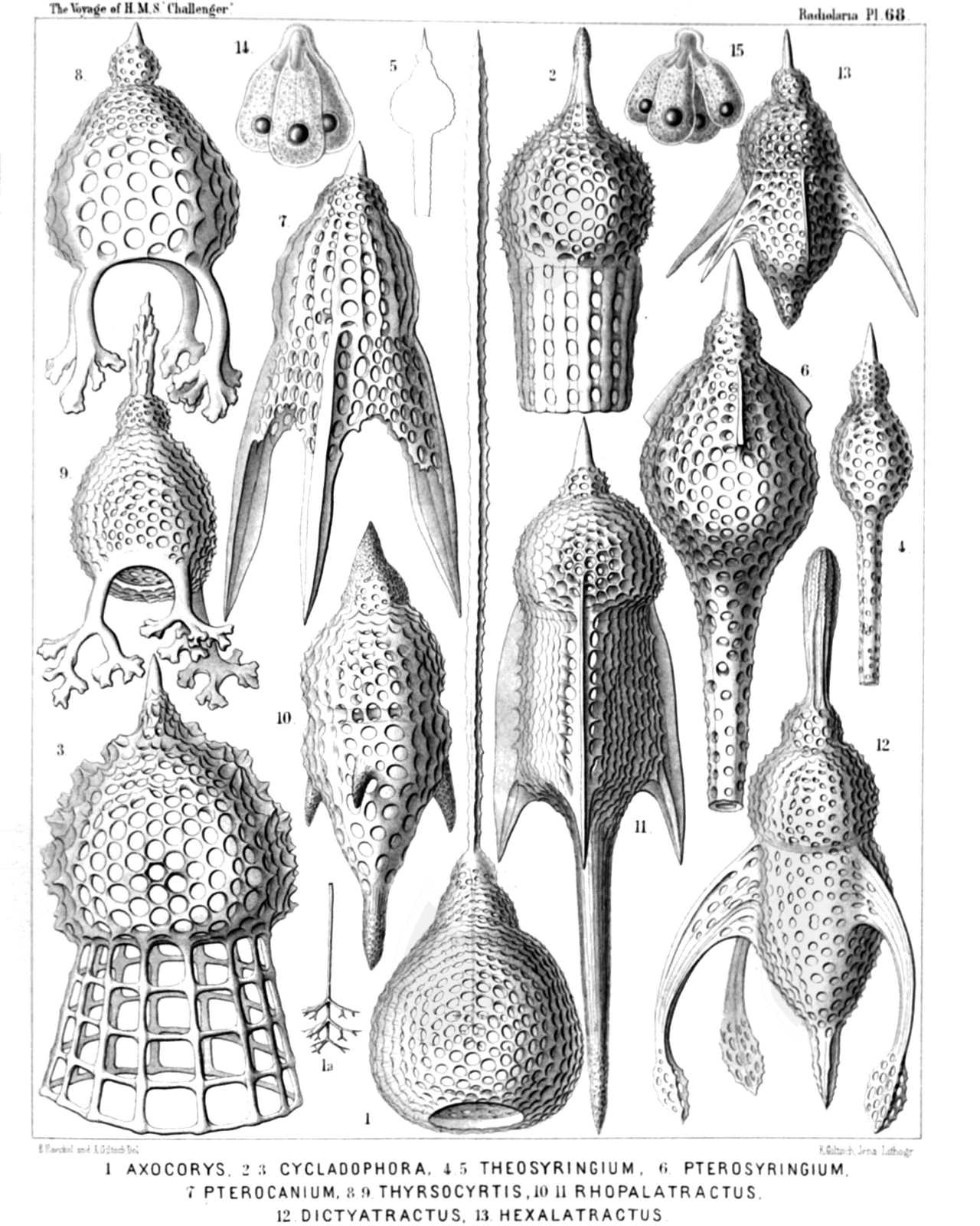 | |||||||||||||||||||||||||||||||||||||||||||||||||||||||||||||||||||||||||||||
| PLATE 68. | |||
| Podocyrtida, Phormocyrtida et Theocyrtida. | |||
| Diam. | Page. | ||
Fig. 1. Axocorys macroceros, n. sp., | × | 300 | 1420 |
| Fig. 1a. The internal axial rod of the shell, which bears on its basal part three verticils of three diverging forked spines, | × | 300 | |
| Fig. 2. Cycladophora fenestrata, n. sp., | × | 300 | 1380 |
| Fig. 3. Cycladophora pantheon, n. sp., | × | 400 | 1379 |
| Fig. 4. Theosyringium tibia, n. sp., | × | 300 | 1409 |
| Fig. 5. Theosyringium pipetta, n. sp., | × | 200 | 1409 |
| Fig. 6. Pterocorys tubulosa, n. sp., | × | 400 | 1319 |
| Fig. 7. Pterocanium pyramis, n. sp., | × | 400 | 1330 |
| Fig. 8. Thyrsocyrtis rhizopodium, n. sp., | × | 300 | 1351 |
| Fig. 9. Thyrsocyrtis arborescens, n. sp., | × | 400 | 1350 |
| Fig. 10. Rhopalatractus foveolatus, n. sp., | × | 400 | 1361 |
| Fig. 11. Rhopalatractus pentacanthus, n. sp., | × | 300 | 1361 |
| Fig. 12. Rhopalatractus fenestratus, n. sp. (vel Dictyatractus fenestratus), | × | 300 | 1361 |
| Fig. 13. Hexalatractus fusiformis, n. sp., | × | 300 | 1391 |
| Fig. 14. Sethornithium dictyopterum, n. sp., | × | 300 | 1356 |
| The trilobate central capsule, which contains in its uppermost part the trilobate nucleus, and in the basal part of each lobe an oil-globule. | |||
| Fig. 15. Lophocyrtis synapta, n. sp., | × | 300 | 1411 |
| The quadrilobate central capsule, which contains in its uppermost part the quadrilobate nucleus, and in the basal part of each lobe an oil-globule. | |||
PLATE 69.
Legion NASSELLARIA.
Order CYRTOIDEA.
Families Phormocyrtida et Theocyrtida.
| 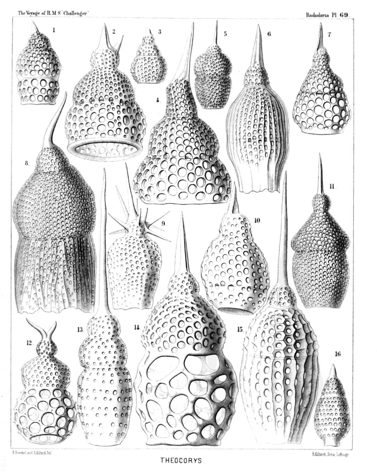 | ||||||||||||||||||||||||||||||||||||||||||||||||||||||||||||||||||||||||||||
| PLATE 69. | |||
| Phormocyrtida et Theocyrtida. | |||
| Diam. | Page. | ||
Fig. 1. Theocorys plutonis, n. sp., | × | 400 | 1416 |
| Fig. 2. Lophoconus rhinoceros, n. sp., | × | 400 | 1405 |
| Fig. 3. Theocorys apollinis, n. sp., | × | 300 | 1418 |
| Fig. 4. Theoconus jovis, n. sp., | × | 400 | 1401 |
| Fig. 5. Theocorys veneris, n. sp., | × | 300 | 1415 |
| Fig. 6. Phormocyrtis costata, n. sp., | × | 300 | 1369 |
| Fig. 7. Theoconus junonis, n. sp., | × | 300 | 1401 |
| Fig. 8. Theocyrtis ptychodes, n. sp., | × | 400 | 1408 |
| Fig. 9. Lophocorys astrocephala, n. sp., | × | 300 | 1421 |
| Fig. 10. Theocorys obliqua, n. sp., | × | 400 | 1417 |
| Fig. 11. Theocorys dianæ, n. sp., | × | 400 | 1416 |
| Fig. 12. Lophocorys bovicornis, n. sp., | × | 300 | 1422 |
| Fig. 13. Theocyrtis macroceros, n. sp., | × | 400 | 1407 |
| Fig. 14. Theocorys minervæ, n. sp., | × | 300 | 1419 |
| Fig. 15. Phormocyrtis longicornis, n. sp., | × | 400 | 1370 |
| Fig. 16. Theocorys ovata, n. sp., | × | 300 | 1416 |
PLATE 70.
Legion NASSELLARIA.
Order CYRTOIDEA.
Families Anthocyrtida, Podocyrtida, Phormocyrtida et Theocyrtida.
| 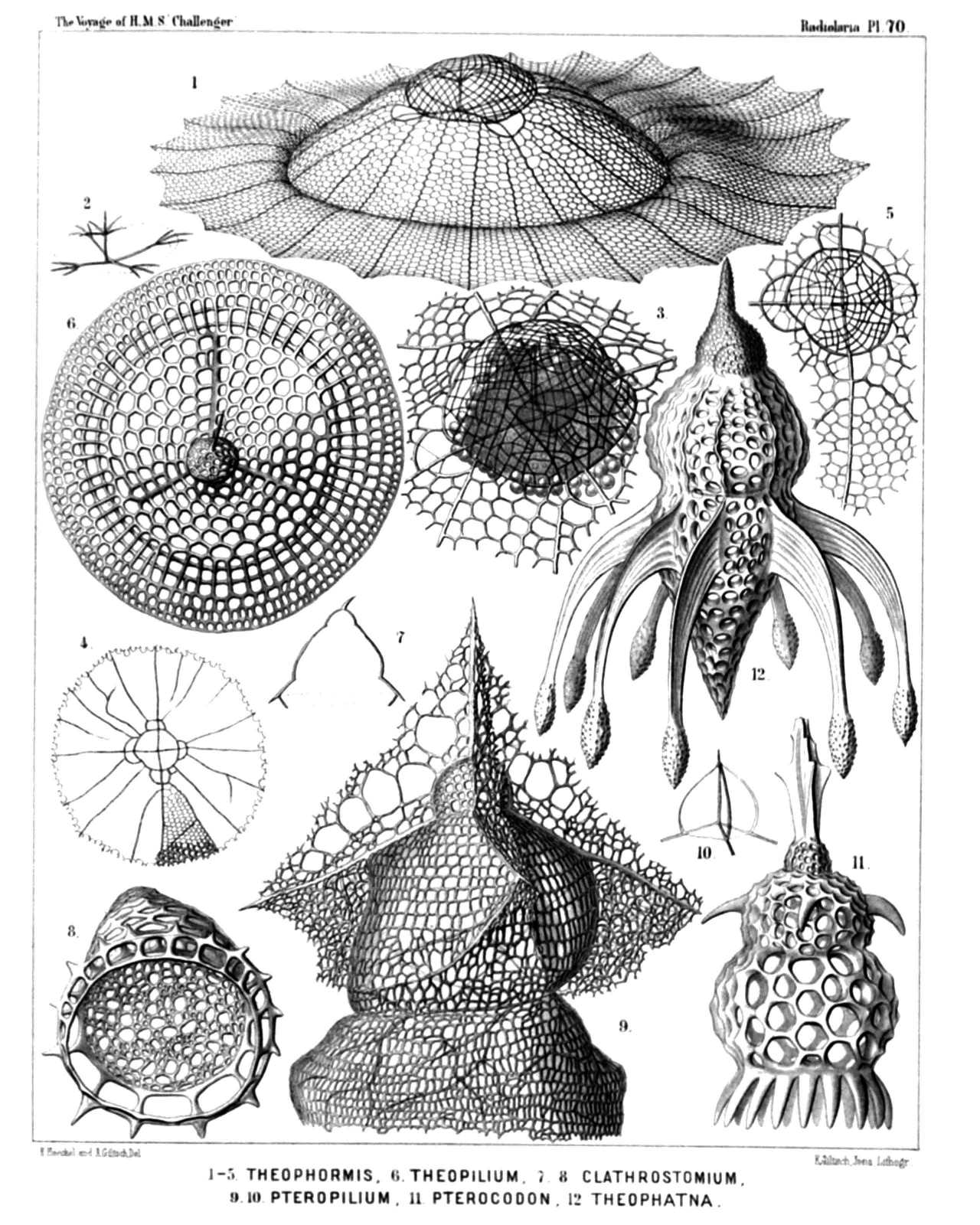 | |||||||||||||||||||||||||||||||||||||||||||||||||||||||||||||||||||
| PLATE 70. | |||
| Anthocyrtida, Podocyrtida, Phormocyrtida et Theocyrtida. | |||
| Diam. | Page. | ||
Fig. 1. Theophormis callipilium, n. sp., | × | 300 | 1367 |
| Fig. 2. Theophormis callipilium, n. sp., | × | 300 | 1367 |
| The four cruciate rods of the cortinar septum and the vertical columella in its centre. | |||
| Fig. 3. Theophormis callipilium, n. sp., | × | 400 | 1367 |
| The cephalis alone with the enclosed quadrilobate central capsule, which is surrounded by numerous xanthellæ. | |||
| Fig. 4. Sethophormis umbrella, n. sp., | × | 150 | 1248 |
| Fig. 5. Sethophormis umbrella, n. sp., | × | 400 | 1248 |
| Cephalis with the cruciform cortinar septum. | |||
| Fig. 6. Theopilium tricostatum, n. sp., | × | 400 | 1322 |
| Seen from above. | |||
| Fig. 7. Phrenocodon clathrostomium, n. sp., | × | 250 | 1434 |
| Vertical section through the shell. | |||
| Fig. 8. Phrenocodon clathrostomium, n. sp., | × | 500 | 1434 |
| Shell seen half from below, and exhibiting the fenestrated septum between thorax and abdomen. | |||
| Fig. 9. Pteropilium stratiotes, n. sp., | × | 400 | 1326 |
| Fig. 10. Pteropilium stratiotes, n. sp., | × | 400 | 1326 |
| The three rods of the cortinar septum and the three arches connecting them with the central axial columella. | |||
| Fig. 11. Pterocodon ornatus, n. sp., | × | 300 | 1333 |
| Fig. 12. Theophæna corona, n. sp., | × | 300 | 1394 |
PLATE 71.
Legion NASSELLARIA.
Order CYRTOIDEA.
Family Podocyrtida.
| 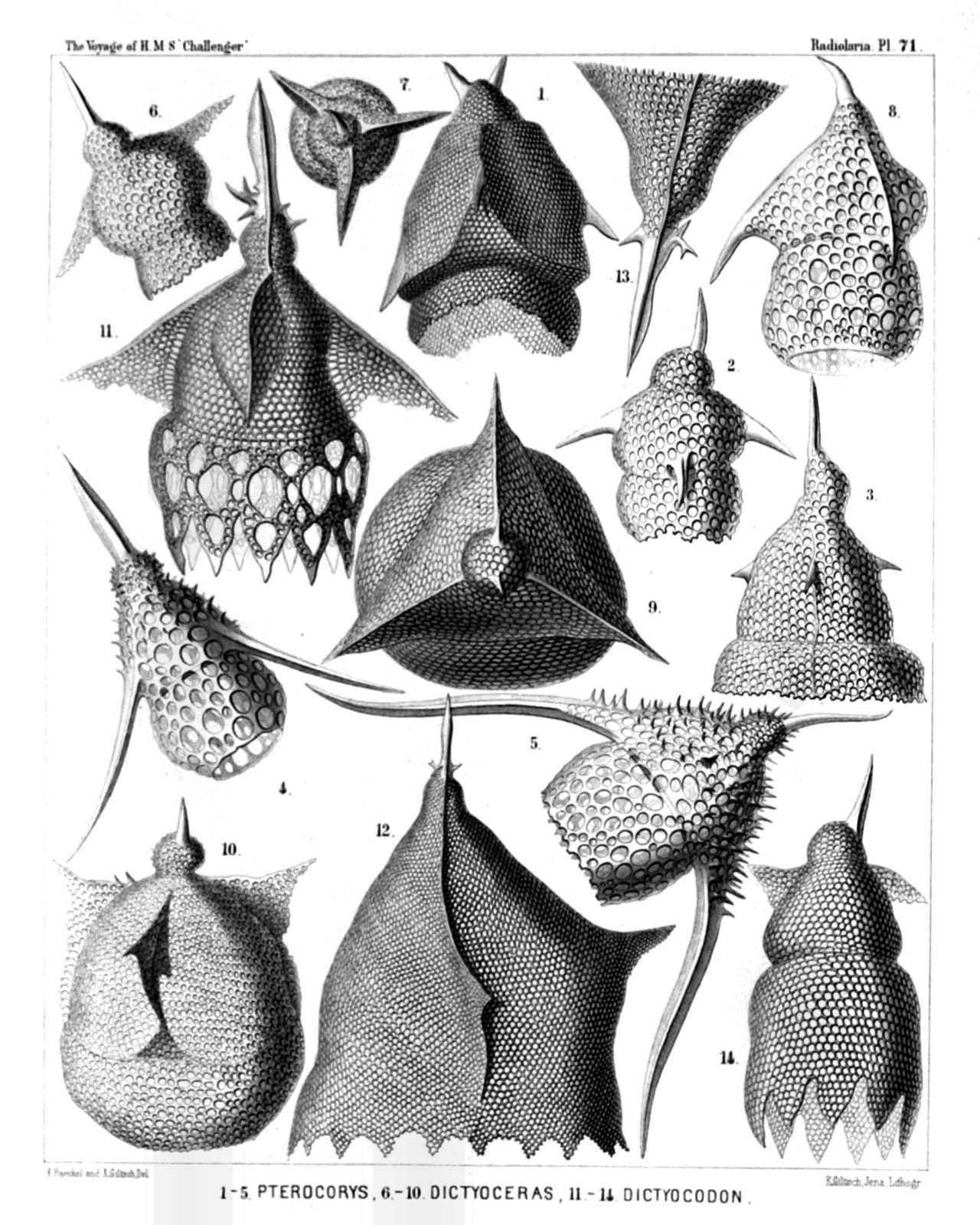 | |||||||||||||||||||||||||||||||||||||||||||||||||||||||||||||||||||||||
| PLATE 71. | |||
| Podocyrtida. | |||
| Diam. | Page. | ||
Fig. 1. Pterocorys rhinoceros, n. sp., | × | 400 | 1320 |
| Fig. 2. Pterocorys columba, n. sp., | × | 400 | 1317 |
| Fig. 3. Pterocorys campanula, n. sp., | × | 400 | 1316 |
| Fig. 4. Pterocorys hirundo, n. sp., | × | 300 | 1318 |
| Fig. 5. Pterocorys aquila, n. sp., | × | 300 | 1317 |
| Fig. 6. Dictyoceras insectum, n. sp., | × | 400 | 1324 |
| Fig. 7. Dictyoceras insectum, n. sp., | × | 400 | 1324 |
| Seen from the apex. | |||
| Fig. 8. Dictyoceras formica, n. sp., | × | 400 | 1325 |
| Fig. 9. Dictyoceras melitta, n. sp., | × | 400 | 1325 |
| Seen from the apex. | |||
| Fig. 10. Dictyoceras bombus, n. sp., | × | 400 | 1325 |
| Fig. 11. Dictyocodon annasethe, n. sp., | × | 400 | 1334 |
| Fig. 12. Dictyocodon palladius, n. sp., | × | 300 | 1335 |
| Fig. 13. Dictyocodon palladius, n. sp., | × | 600 | 1335 |
| Apical part of the shell alone. | |||
| Fig. 14. Dictyocodon carolotæ, n. sp., | × | 300 | 1335 |
PLATE 72.
Legion NASSELLARIA.
Order CYRTOIDEA.
Family Podocyrtida.
| 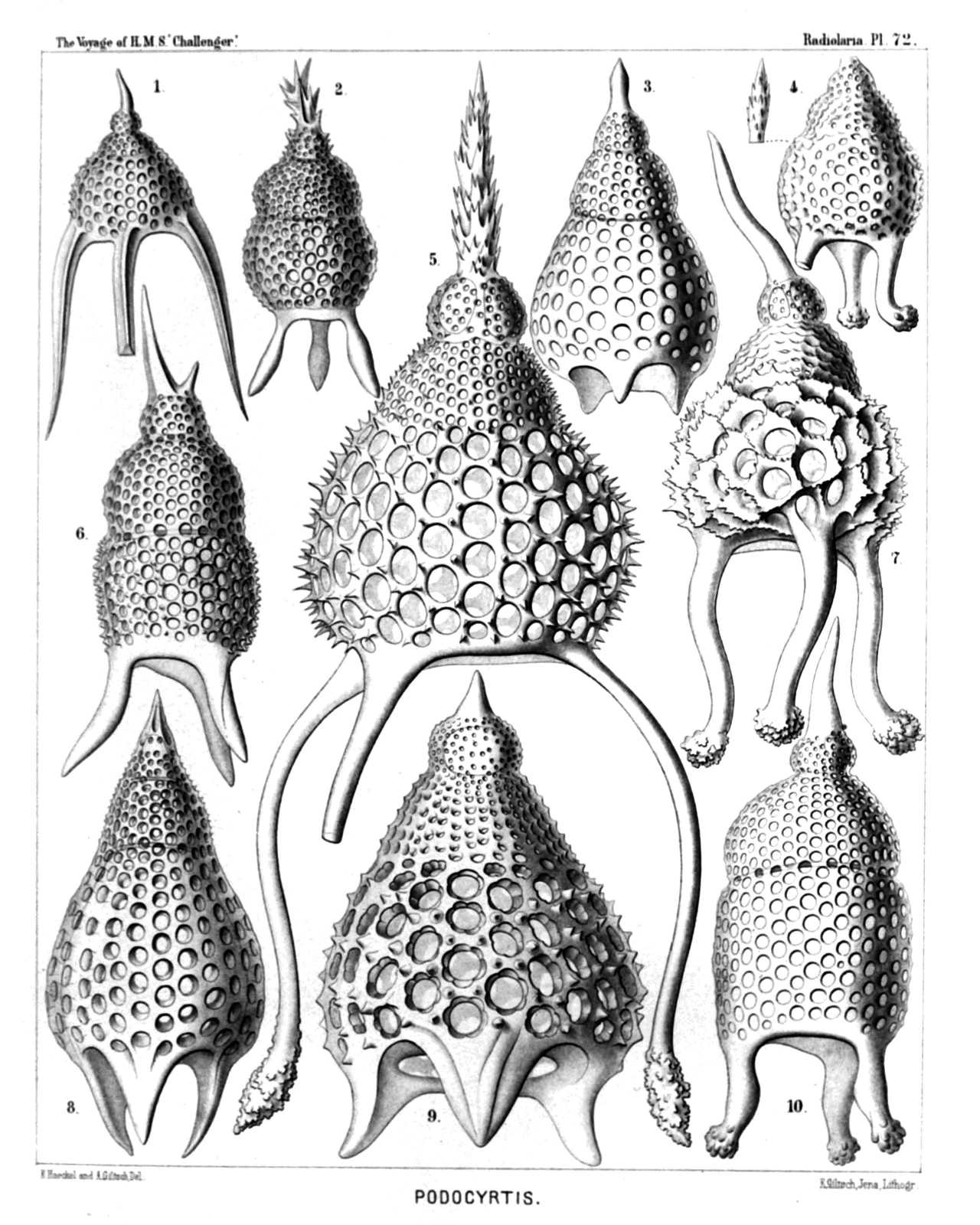 | ||||||||||||||||||||||||||||||||||||||||||||||||||||
| PLATE 72. | |||
| Podocyrtida. | |||
| Diam. | Page. | ||
Fig. 1. Podocyrtis prismatica, n. sp., | × | 300 | 1340 |
| Fig. 2. Podocyrtis corythæola, n. sp., | × | 300 | 1339 |
| Fig. 3. Podocyrtis lithoconus, n. sp., | × | 300 | 1348 |
| Fig. 4. Podocyrtis tripodiscus, n. sp., | × | 300 | 1338 |
| Fig. 5. Podocyrtis magnifica, n. sp., | × | 500 | 1341 |
| Fig. 6. Podocyrtis divergens, n. sp., | × | 400 | 1340 |
| Fig. 7. Podocyrtis cristata, n. sp., | × | 400 | 1342 |
| Fig. 8. Podocyrtis pedicellaria, n. sp., | × | 300 | 1347 |
| Fig. 9. Podocyrtis flosculata, n. sp., | × | 500 | 1341 |
| Fig. 10. Podocyrtis surena, n. sp., | × | 400 | 1339 |
PLATE 73.
Legion NASSELLARIA.
Order CYRTOIDEA.
Families Podocyrtida et Phormocyrtida.
| 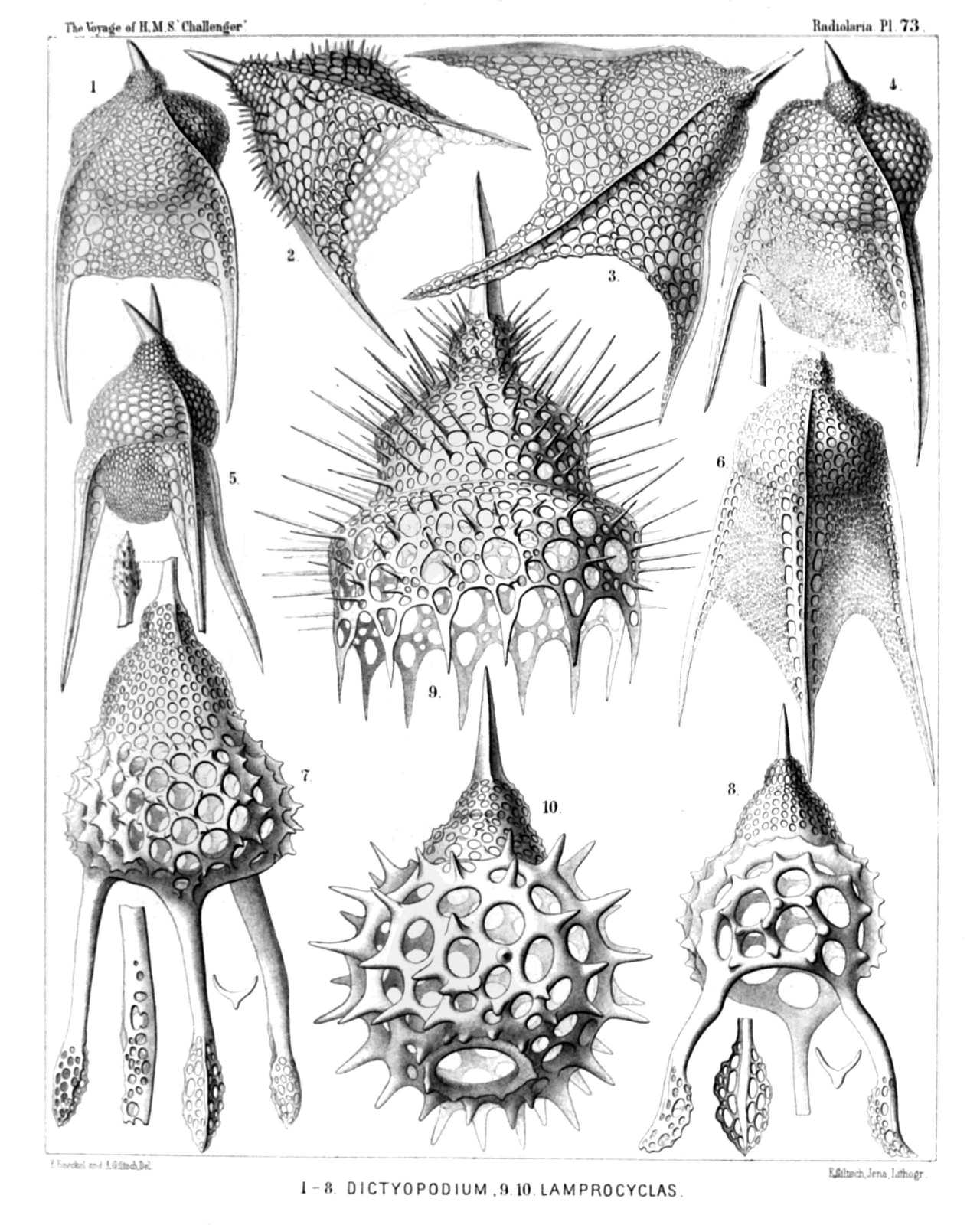 | ||||||||||||||||||||||||||||||||||||||||||||||||||||
| PLATE 73. | |||
| Podocyrtida et Phormocyrtida. | |||
| Diam. | Page. | ||
Fig. 1. Pterocanium tricolpum, n. sp., | × | 400 | 1331 |
| Fig. 2. Pterocanium orcinum, n. sp., | × | 400 | 1329 |
| Fig. 3. Pterocanium gravidum, n. sp., | × | 400 | 1329 |
| Fig. 4. Pterocanium eucolpum, n. sp., | × | 400 | 1332 |
| Fig. 5. Pterocanium bicorne, n. sp., | × | 400 | 1332 |
| Fig. 6. Pterocanium virgineum, n. sp., | × | 400 | 1330 |
| Fig. 7. Dictyopodium thyrsolophus, n. sp., | × | 300 | 1354 |
| Fig. 8. Dictyopodium scaphopodium, n. sp., | × | 300 | 1353 |
| Fig. 9. Calocyclas monumentum, n. sp., | × | 400 | 1385 |
| Fig. 10. Calocyclas casta, n. sp., | × | 400 | 1384 |
PLATE 74.
Legion NASSELLARIA.
Order CYRTOIDEA.
Family Phormocyrtida.
| 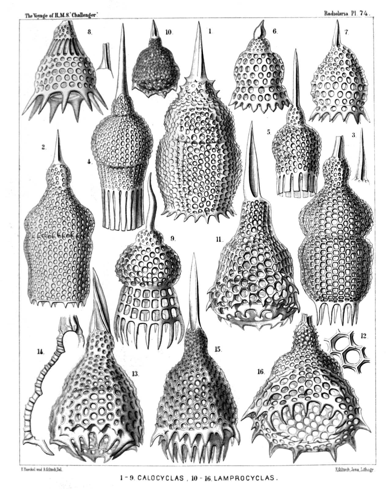 | ||||||||||||||||||||||||||||||||||||||||||||||||||||||||||||||||||||||||||||||
| PLATE 74. | |||
| Phormocyrtida. | |||
| Diam. | Page. | ||
Fig. 1. Calocyclas parthenia, n. sp., | × | 400 | 1385 |
| Fig. 2. Calocyclas amicæ, n. sp., | × | 400 | 1382 |
| Fig. 3. Calocyclas vestalis, n. sp., | × | 400 | 1382 |
| Fig. 4. Calocyclas virginis, n. sp., | × | 300 | 1381 |
| Fig. 5. Calocyclas veneris, n. sp., | × | 300 | 1381 |
| Fig. 6. Clathrocyclas basilea, n. sp. (vel Calocyclas basilea), | × | 400 | 1386 |
| Fig. 7. Clathrocyclas principessa, n. sp. (vel Calocyclas principessa), | × | 400 | 1386 |
| Fig. 8. Clathrocyclas collaris, n. sp. (vel Calocyclas collaris), | × | 400 | 1387 |
| Fig. 9. Alacorys carcinus, n. sp. (vel Calocyclas carcinus), | × | 300 | 1375 |
| Fig. 10. Lamprocyclas deflorata, n. sp., | × | 200 | 1391 |
| Fig. 11. Lamprocyclas reginæ, n. sp., | × | 400 | 1391 |
| Fig. 12. Lamprocyclas reginæ, n. sp., | × | 800 | 1391 |
| Two meshes of the network. | |||
| Fig. 13. Lamprocyclas maritalis, n. sp., | × | 400 | 1390 |
| Fig. 14. Lamprocyclas maritalis, n. sp., | × | 400 | 1390 |
| Vertical section. | |||
| Fig. 15. Lamprocyclas nuptialis, n. sp., | × | 400 | 1390 |
| Fig. 16. Lamprocyclas saltatricis, n. sp., | × | 400 | 1391 |
PLATE 75.
Legion NASSELLARIA.
Order CYRTOIDEA.
Families Podocampida et Phormocampida.
| 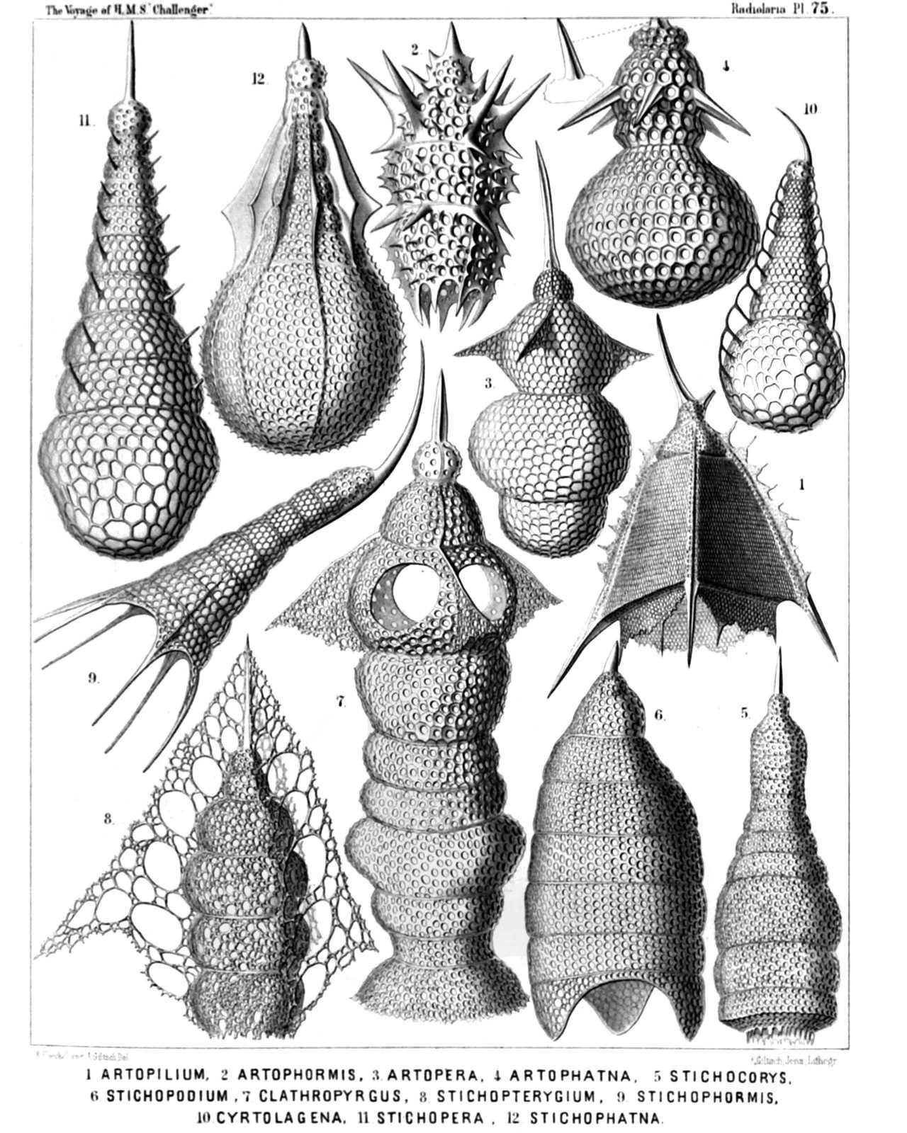 | ||||||||||||||||||||||||||||||||||||||||||||||||||||||||||||
| PLATE 75. | |||
| Podocampida et Phormocampida. | |||
| Diam. | Page. | ||
Fig. 1. Artopilium elegans, n. sp. (vel Trictenartus elegans), | × | 200 | 1440 |
| Fig. 2. Artophormis horrida, n. sp., | × | 300 | 1458 |
| Fig. 3. Cyrtopera thoracoptera, n. sp. (vel Artopera thoracoptera), | × | 300 | 1450 |
| Fig. 4. Stichophæna ærostatica, n. sp. (vel Artophæna ærostatica), | × | 400 | 1463 |
| Fig. 5. Cyrtophormis turricula, n. sp., | × | 300 | 1463 |
| Fig. 6. Stichopodium dictyopodium, n. sp., | × | 400 | 1447 |
| Fig. 7. Artopilium trifenestra, n. sp. (vel Clathropyrgus trifenestra), | × | 500 | 1441 |
| Fig. 8. Artopilium stichopterygium, n. sp., | × | 400 | 1442 |
| Fig. 9. Stichophormis cornutella, n. sp., | × | 400 | 1455 |
| Fig. 10. Cyrtopera laguncula, n. sp. (vel Cyrtolagena laguncula), | × | 400 | 1451 |
| Fig. 11. Stichopera pectinata, n. sp., | × | 500 | 1449 |
| Fig. 12. Stichophæna ritteriana, n. sp., | × | 400 | 1465 |
PLATE 76.
Legion NASSELLARIA.
Order CYRTOIDEA.
Families Phormocampida et Lithocampida.
| 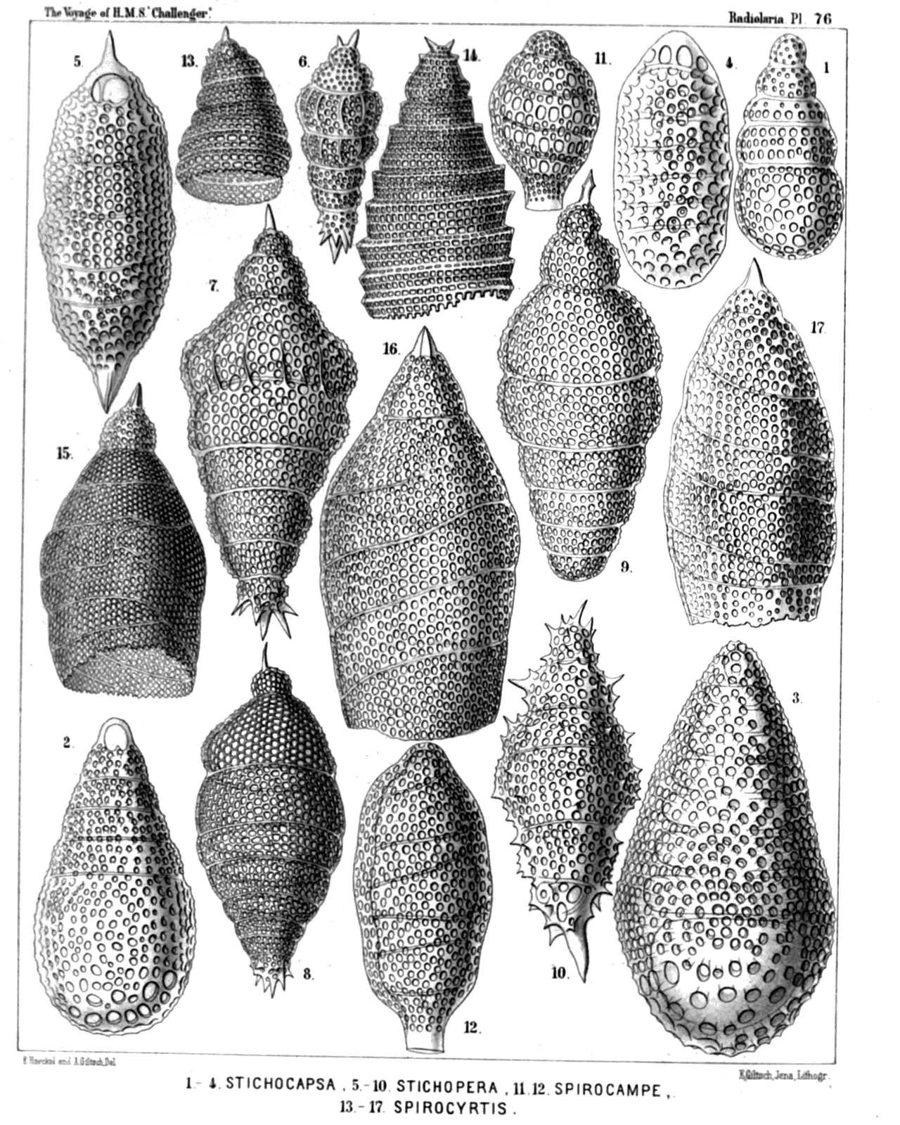 | ||||||||||||||||||||||||||||||||||||||||||||||||||||||||||||||||||||||||||||||||
| PLATE 76. | |||
| Phormocampida et Lithocampida. | |||
| Diam. | Page. | ||
Fig. 1. Stichocapsa pentacola, n. sp., | × | 400 | 1517 |
| Fig. 2. Stichocapsa hexacola, n. sp., | × | 400 | 1517 |
| Fig. 3. Stichocapsa compacta, n. sp., | × | 400 | 1517 |
| Fig. 4. Stichocapsa paniscus, n. sp., | × | 400 | 1518 |
| Fig. 5. Artocapsa fusiformis, n. sp., | × | 400 | 1519 |
| Fig. 6. Stichophæna nonaria, n. sp., | × | 200 | 1466 |
| Fig. 7. Stichophæna novena, n. sp., | × | 400 | 1466 |
| Fig. 8. Artocapsa elegans, n. sp., | × | 400 | 1520 |
| Fig. 9. Cyrtocapsa chrysalidium, n. sp., | × | 400 | 1515 |
| Fig. 10. Artocapsa spinosa, n. sp., | × | 400 | 1519 |
| Fig. 11. Spirocampe callispira, n. sp., | × | 300 | 1511 |
| Fig. 12. Spirocampe allospira, n. sp., | × | 400 | 1511 |
| Fig. 13. Spirocyrtis cornutella, n. sp., | × | 400 | 1509 |
| Fig. 14. Spirocyrtis scalaris, n. sp., | × | 400 | 1509 |
| Fig. 15. Spirocyrtis merospira, n. sp., | × | 500 | 1510 |
| Fig. 16. Spirocyrtis holospira, n. sp., | × | 400 | 1509 |
| Fig. 17. Spirocyrtis diplospira, n. sp., | × | 400 | 1510 |
PLATE 77.
Legion NASSELLARIA.
Order CYRTOIDEA.
Families Podocampida, Phormocampida et Lithocampida.
| 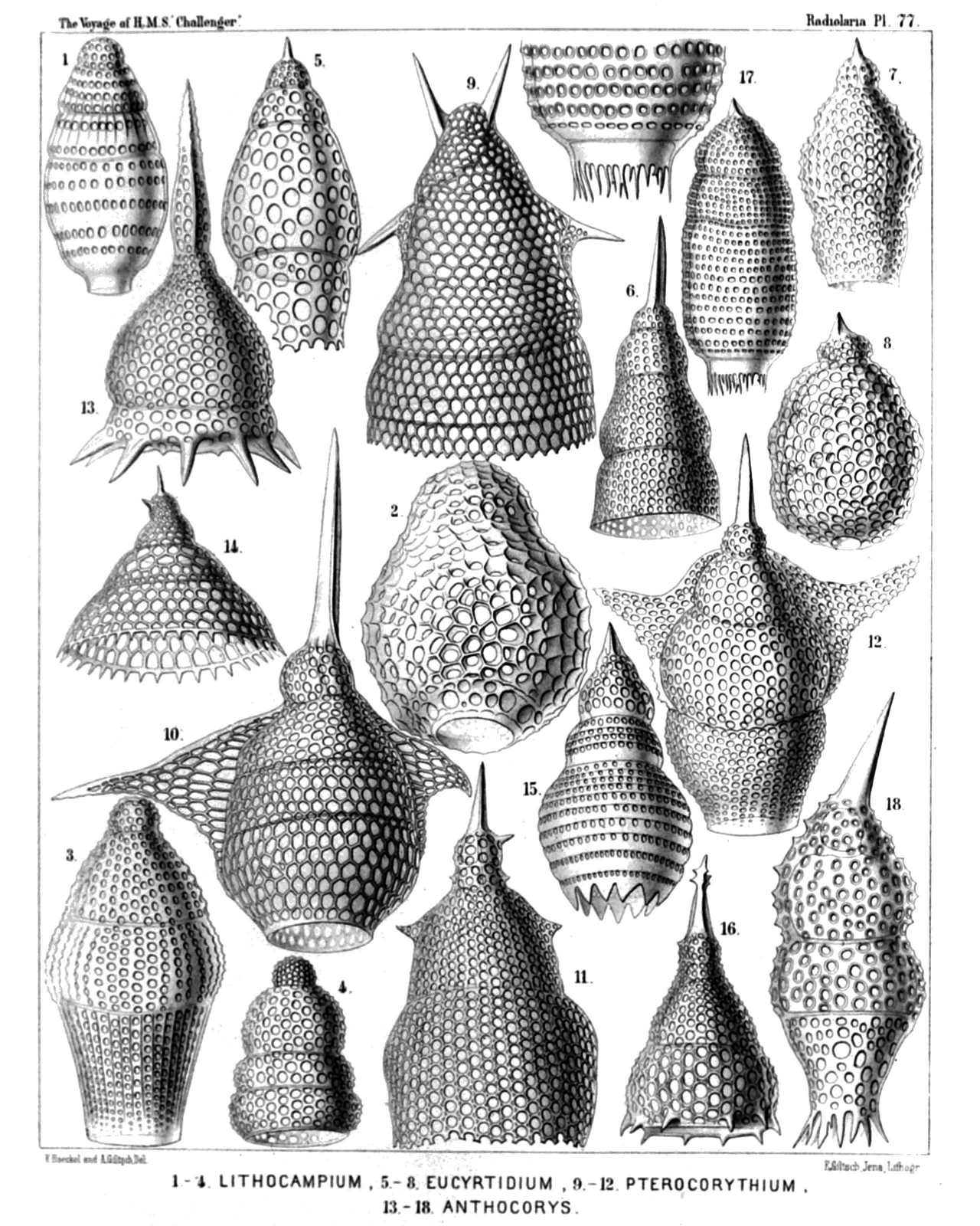 | ||||||||||||||||||||||||||||||||||||||||||||||||||||||||||||||||||||||||||||||||||||
| PLATE 77. | |||
| Podocampida, Phormocampida et Lithocampida. | |||
| Diam. | Page. | ||
Fig. 1. Lithocampe ovata, n. sp., | × | 500 | 1504 |
| Fig. 2. Lithocampe urceolata, n. sp., | × | 400 | 1507 |
| Fig. 3. Lithocampe diploconus, n. sp., | × | 400 | 1505 |
| Fig. 4. Dictyomitra eurythorax, n. sp., | × | 300 | 1477 |
| Fig. 5. Eucyrtidium teuscheri, n. sp., | × | 400 | 1491 |
| Fig. 6. Lithostrobus cornutus, n. sp., | × | 400 | 1474 |
| Fig. 7. Eucyrtidium bütschlii, n. sp., | × | 400 | 1492 |
| Fig. 8. Cyrtocapsa compacta, n. sp., | × | 300 | 1512 |
| Fig. 9. Stichopilium bicorne, n. sp., | × | 600 | 1437 |
| Fig. 10. Artopilium longicorne, n. sp., | × | 500 | 1440 |
| Fig. 11. Stichopilium campanulatum, n. sp., | × | 400 | 1438 |
| Fig. 12. Artopilium cyrtopterum, n. sp., | × | 400 | 1440 |
| Fig. 13. Phormocampe campanula, n. sp., | × | 400 | 1456 |
| Fig. 14. Phormocampe eucalyptra, n. sp., | × | 300 | 1457 |
| Fig. 15. Cyrtophormis corona, n. sp., | × | 300 | 1462 |
| Fig. 16. Phormocampe lamprocyclas, n. sp., | × | 300 | 1457 |
| Fig. 17. Cyrtophormis cylindrica, n. sp., | × | 300 | 1461 |
| Fig. 18. Cyrtophormis cornuta, n. sp., | × | 500 | 1462 |
PLATE 78.
Legion NASSELLARIA.
Order CYRTOIDEA.
Families Phormocampida et Lithocampida.
| 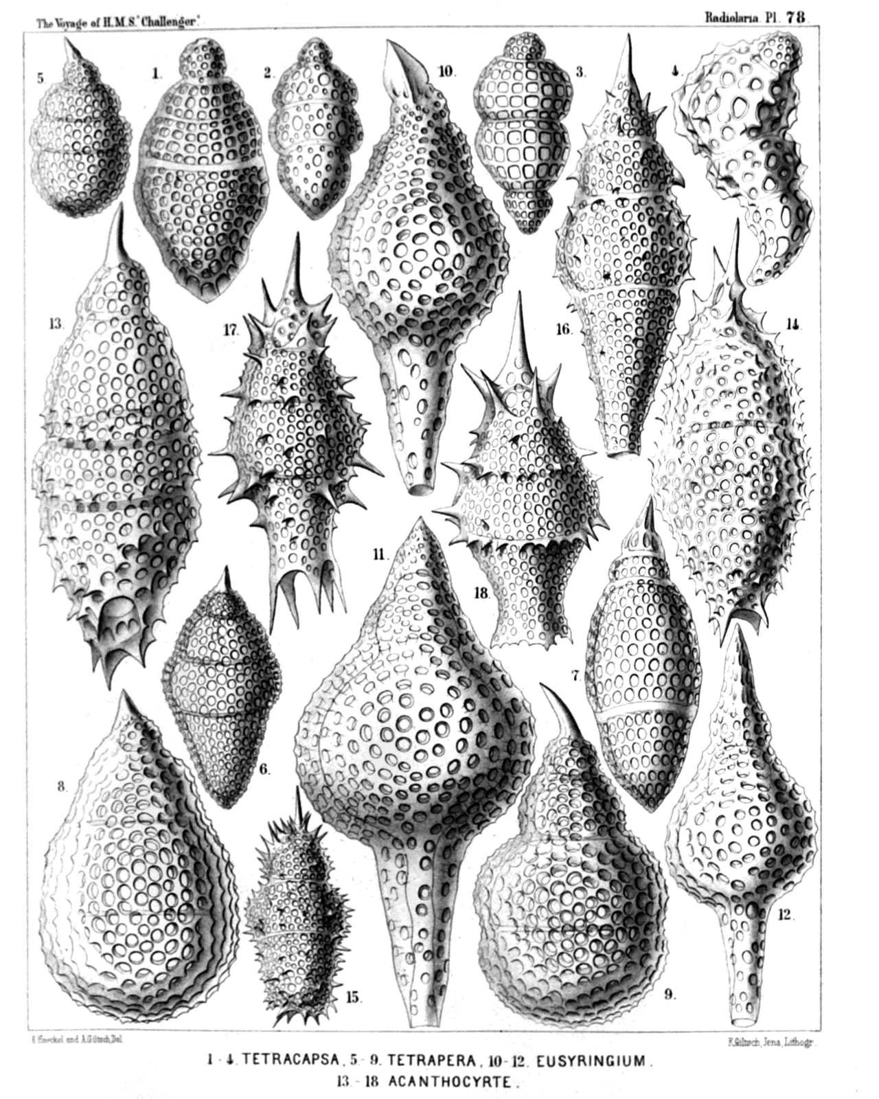 | ||||||||||||||||||||||||||||||||||||||||||||||||||||||||||||||||||||||||||||||||||||
| PLATE 78. | |||
| Phormocampida et Lithocampida. | |||
| Diam. | Page. | ||
Fig. 1. Stichocapsa tetracola, n. sp., | × | 600 | 1515 |
| Fig. 2. Stichocapsa tricincta, n. sp., | × | 400 | 1516 |
| Fig. 3. Stichocapsa quadrigata, n. sp., | × | 400 | 1515 |
| Fig. 4. Stichocapsa monstrosa, n. sp., | × | 400 | 1517 |
| Fig. 5. Cyrtocapsa tetrapera, n. sp., | × | 300 | 1512 |
| Fig. 6. Cyrtocapsa diploconus, n. sp., | × | 300 | 1513 |
| Fig. 7. Cyrtocapsa fusulus, n. sp., | × | 400 | 1514 |
| Fig. 8. Cyrtocapsa pyrum, n. sp., | × | 400 | 1513 |
| Fig. 9. Cyrtocapsa cornuta, n. sp., | × | 400 | 1513 |
| Fig. 10. Eusyringium conosiphon, n. sp., | × | 400 | 1496 |
| Fig. 11. Eusyringium pachysiphon, n. sp., | × | 400 | 1496 |
| Fig. 12. Eusyringium macrosiphon, n. sp., | × | 400 | 1497 |
| Fig. 13. Eucyrtidium tricinctum, n. sp., | × | 400 | 1494 |
| Fig. 14. Eucyrtidium armatum, n. sp., | × | 400 | 1495 |
| Fig. 15. Eucyrtidium ehrenbergii, n. sp., | × | 300 | 1495 |
| Fig. 16. Eucyrtidium conostoma, n. sp., | × | 400 | 1495 |
| Fig. 17. Cyrtophormis armata, n. sp., | × | 400 | 1460 |
| Fig. 18. Cyrtophormis cingulata, n. sp., | × | 400 | 1460 |
PLATE 79.
Legion NASSELLARIA.
Order CYRTOIDEA.
Families Phormocampida et Lithocampida.
| 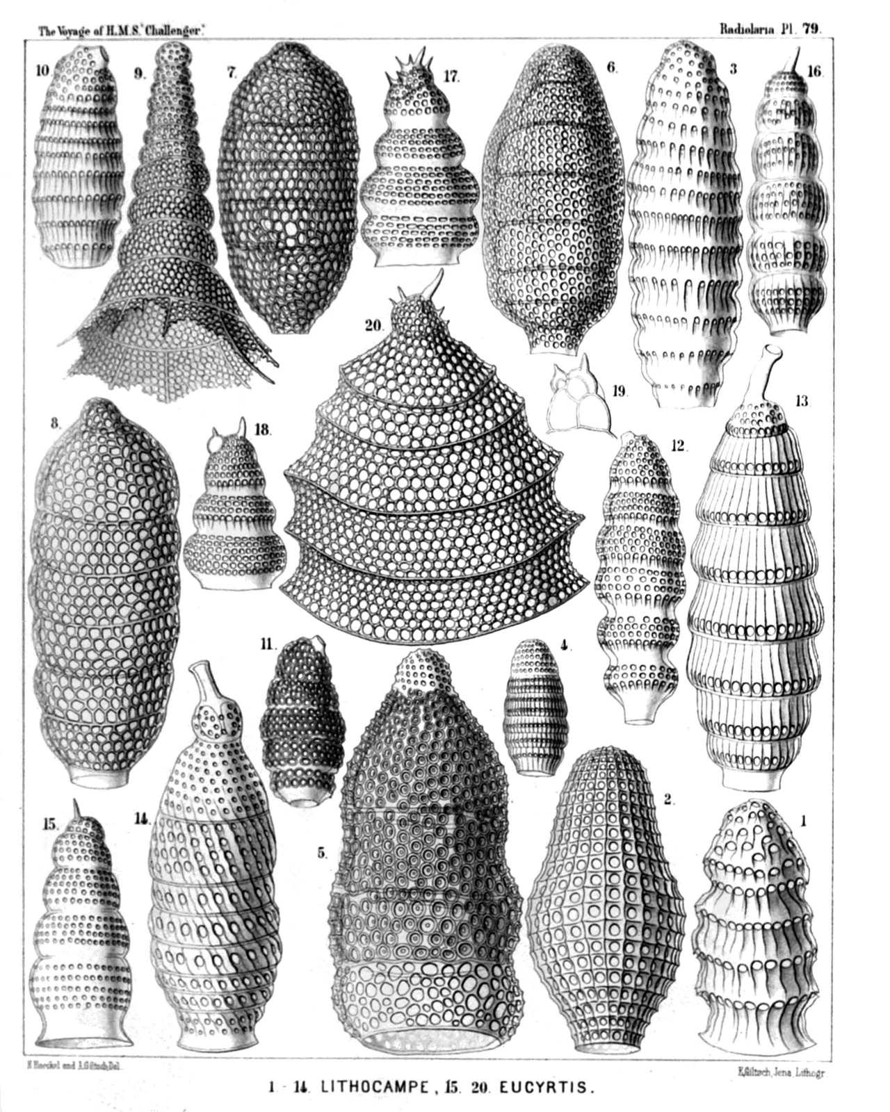 | |||||||||||||||||||||||||||||||||||||||||||||||||||||||||||||||||||||||||||||||||||||||||||||
| PLATE 79. | |||
| Phormocampida et Lithocampida. | |||
| Diam. | Page. | ||
Fig. 1. Lithomitra nodosaria, n. sp., | × | 600 | 1484 |
| Fig. 2. Cyrtophormis tabulata, n. sp., | × | 400 | 1166 |
| Fig. 3. Lithomitra eruca, n. sp., | × | 500 | 1485 |
| Fig. 4. Lithomitra chrysalis, n. sp., | × | 300 | 1485 |
| Fig. 5. Lithomitra infundibulum, n. sp., | × | 500 | 1487 |
| Fig. 6. Lithocampe octocola, n. sp., | × | 400 | 1508 |
| Fig. 7. Lithocampe hexacola, n. sp., | × | 400 | 1507 |
| Fig. 8. Lithocampe heptacola, n. sp., | × | 400 | 1508 |
| Fig. 9. Stichophormis novena, n. sp., | × | 400 | 1455 |
| Fig. 10. Siphocampe annulosa, n. sp., | × | 300 | 1500 |
| Fig. 11. Siphocampe erucosa, n. sp., | × | 300 | 1500 |
| Fig. 12. Siphocampe caminosa, n. sp., | × | 400 | 1500 |
| Fig. 13. Siphocampe tubulosa, n. sp., | × | 400 | 1500 |
| Fig. 14. Siphocampe spiralis, n. sp., | × | 500 | 1501 |
| Fig. 15. Lithostrobus seriatus, n. sp., | × | 400 | 1474 |
| Fig. 16. Artostrobus articulatus, n. sp., | × | 400 | 1483 |
| Fig. 17. Lithostrobus lithobotrys, n. sp., | × | 400 | 1475 |
| Fig. 18. Lithostrobus botryocyrtis, n. sp., | × | 400 | 1475 |
| Fig. 19. Lithostrobus botryocyrtis, n. sp., | × | 400 | 1475 |
| Vertical section through the cephalis. | |||
| Fig. 20. Lithostrobus hexagonalis, n. sp., | × | 400 | 1475 |
PLATE 80.
Legion NASSELLARIA.
Order CYRTOIDEA.
Family Lithocampida.
| 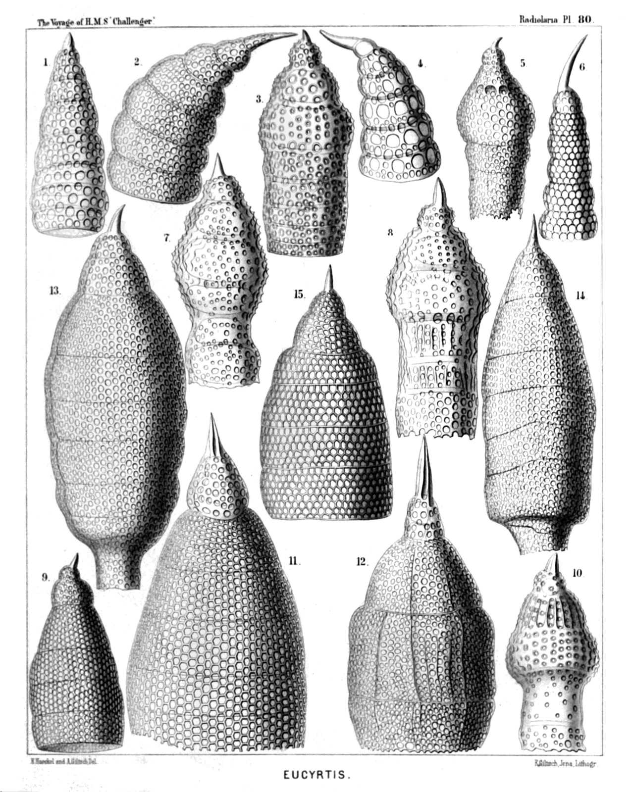 | ||||||||||||||||||||||||||||||||||||||||||||||||||||||||||||||||||||||||
| PLATE 80. | |||
| Lithocampida. | |||
| Diam. | Page. | ||
Fig. 1. Lithostrobus conulus, n. sp. (vel Cyrtostrobus conulus), | × | 400 | 1472 |
| Fig. 2. Lithostrobus cyrtoceras, n. sp. (vel Cornustrobus cyrtoceras), | × | 400 | 1470 |
| Fig. 3. Stichocorys huschkei, n. sp., | × | 400 | 1480 |
| Fig. 4. Lithostrobus caloceras, n. sp. (vel Cornustrobus caloceras), | × | 400 | 1471 |
| Fig. 5. Stichocorys okenii, n. sp., | × | 300 | 1480 |
| Fig. 6. Lithostrobus tetrastichus, n. sp. (vel Conostrobus tetrastichus), | × | 500 | 1470 |
| Fig. 7. Stichocorys panderi, n. sp., | × | 400 | 1479 |
| Fig. 8. Stichocorys baerii, n. sp., | × | 400 | 1479 |
| Fig. 9. Eucyrtidium cienkowskii, n. sp., | × | 400 | 1493 |
| Fig. 10. Stichocorys wolffii, n. sp., | × | 400 | 1479 |
| Fig. 11. Eucyrtidium hexagonatum, n. sp., | × | 600 | 1489 |
| Fig. 12. Eucyrtidium hertwigii, n. sp., | × | 400 | 1491 |
| Fig. 13. Eusyringium cannostoma, n. sp., | × | 600 | 1499 |
| Fig. 14. Eusyringium siphonostoma, n. sp., | × | 500 | 1499 |
| Fig. 15. Lithostrobus hexastichus, n. sp. (vel Artostrobus hexastichus), | × | 500 | 1470 |
PLATE 81.
Legion NASSELLARIA.
Order STEPHOIDEA.
Family Stephanida.
| 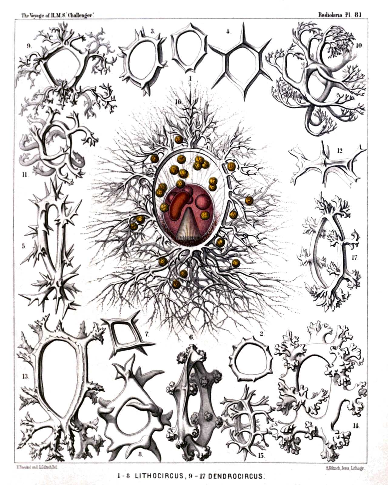 | |||||||||||||||||||||||||||||||||||||||||||||||||||||||||||||||||||||||||||||||||
| PLATE 81. | |||
| Stephanida. | |||
| Diam. | Page. | ||
Fig. 1. Archicircus primordialis, n. sp., | × | 200 | 942 |
| Fig. 2. Zygocircus polygonus, n. sp., | × | 200 | 947 |
| Fig. 3. Zygocircus triquetrus, n. sp., | × | 300 | 947 |
| Fig. 4. Archicircus hexacanthus, n. sp., | × | 300 | 942 |
| Fig. 5. Zygocircus acacia, n. sp., | × | 300 | 947 |
| Fig. 6. Lithocircus crambessa, n. sp., | × | 400 | 944 |
| Fig. 7. Archicircus rhombus, n. sp., | × | 300 | 942 |
| Fig. 8. Zygocircus pentagonus, n. sp., | × | 300 | 946 |
| Fig. 9. Lithocircus quadricornis, n. sp., | × | 300 | 944 |
| Fig. 10. Dendrocircus arborescens, n. sp., | × | 300 | 949 |
| Fig. 11. Dendrocircus dodecancistra, n. sp., | × | 300 | 949 |
| Fig. 12. Archicircus sexangularis, n. sp., | × | 300 | 943 |
| Fig. 13. Dendrocircus elegans, n. sp., | × | 400 | 949 |
| Fig. 14. Dendrocircus stalactites, n. sp., | × | 400 | 950 |
| Fig. 15. Lithocircus decimalis, n. sp., | × | 300 | 944 |
| Fig. 16. Lithocircus magnificus, n. sp., | × | 400 | 945 |
| The ovate, red-coloured central capsule exhibits in the lower half the striate podoconus, in the upper half four oil-globules, and at the left the kidney-shaped nucleus. Numerous "yellow cells" or xanthellæ are scattered in the calymma, which contains brown pigment around the porochora. Numerous pseudopodia radiate from the supporting spines of the sagittal ring. | |||
| Fig. 17. Lithocircus hexablastus, n. sp., | × | 400 | 944 |
PLATE 82.
Legion NASSELLARIA.
Order STEPHOIDEA.
Families Coronida et Tympanida.
| 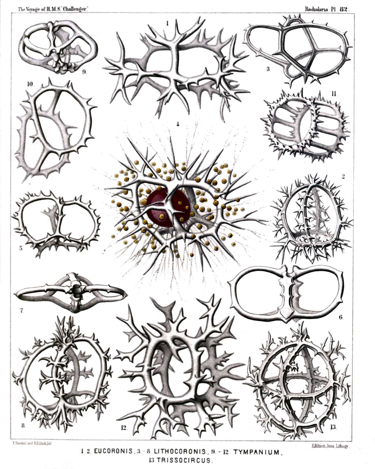 | |||||||||||||||||||||||||||||||||||||||||||||||||||||||||||||||||||||
| PLATE 82. | |||
| Coronida et Tympanida. | |||
| Diam. | Page. | ||
Fig. 1. Coronidium cervicorne, n. sp., | × | 400 | 974 |
| Seen from the apical pole. | |||
| Fig. 2. Coronidium acacia, n. sp., | × | 300 | 975 |
| Fig. 3. Eucoronis angulata, n. sp., | × | 400 | 978 |
| Half from the apical, half from the dorsal side. | |||
| Fig. 4. Eucoronis challengeri, n. sp., | × | 400 | 978 |
| The red central capsule encloses a large ovate nucleus and is surrounded by numerous xanthellæ. | |||
| Fig. 5. Eucoronis nephrospyris, n. sp., | × | 300 | 977 |
| Fig. 6. Eucoronis perspicillum, n. sp., | × | 300 | 977 |
| Fig. 7. Coronidium dyostephanus, n. sp., | × | 400 | 974 |
| Seen from the apical pole. | |||
| Fig. 8. Coronidium diadema, n. sp., | × | 300 | 974 |
| Fig. 9. Acrocubus octopylus, n. sp., | × | 300 | 993 |
| Fig. 10. Parastephanus asymmetricus, n. sp., | × | 400 | 1008 |
| Fig. 11. Eutympanium militare, n. sp., | × | 400 | 1014 |
| Oblique view. | |||
| Fig. 12. Lithocubus astragalus, n. sp., | × | 400 | 1012 |
| Fig. 13. Trissocircus globus, n. sp., | × | 400 | 986 |
PLATE 83.
Legion NASSELLARIA.
Orders STEPHOIDEA et SPYROIDEA.
Families Stephanida, Semantida, Coronida, Tympanida, Zygospyrida, Phormospyrida et Androspyrida.
| 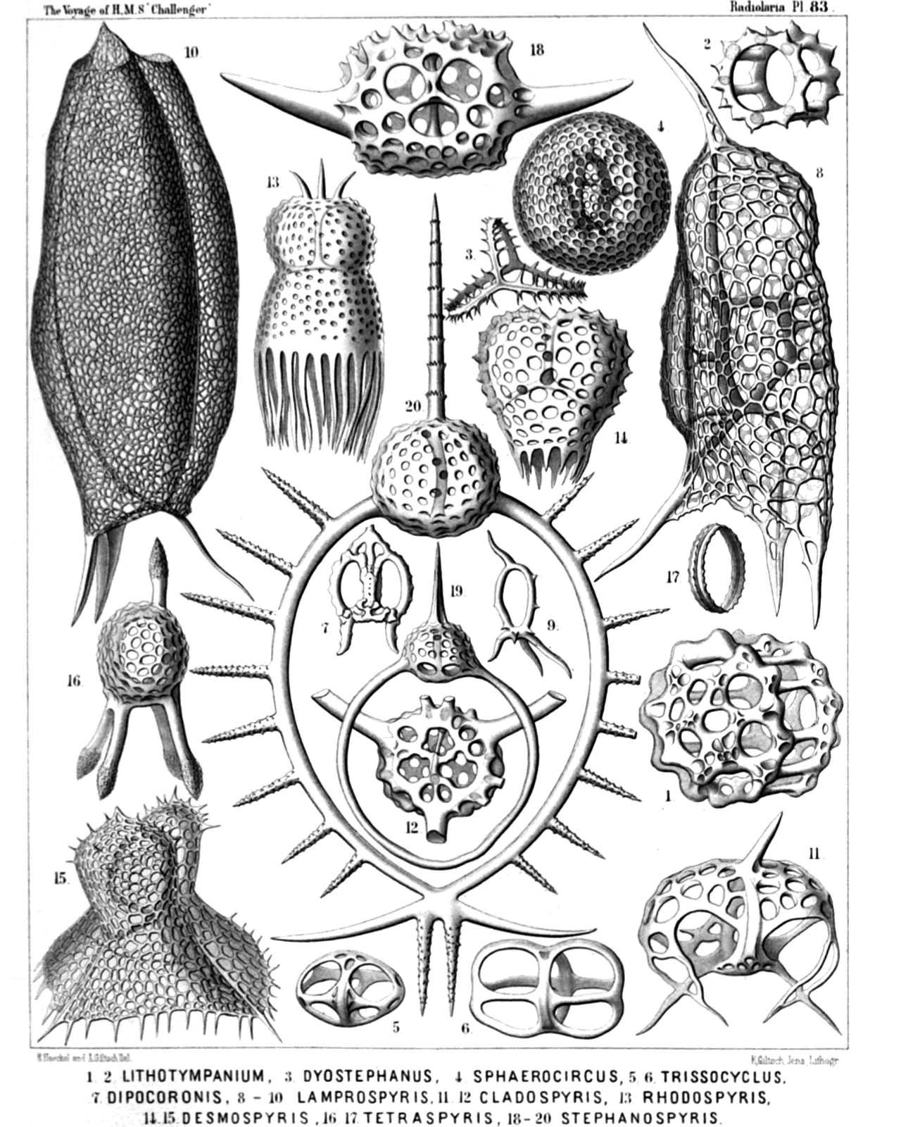 | ||||||||||||||||||||||||||||||||||||||||||||||||||||||||||||||||||||||||||||||||||||||||||||||
| PLATE 83. | |||
| Stephanida, Semantida, Coronida, Tympanida, Zygospyrida, Phormospyrida et Androspyrida. | |||
| Diam. | Page. | ||
Fig. 1. Lithotympanum tuberosum, n. sp., | × | 400 | 1006 |
| Fig. 2. Eutympanium musicantum, n. sp., | × | 300 | 1013 |
| Fig. 3. Semantis distephanus, n. sp., | × | 300 | 957 |
| Fig. 4. Sphærospyris globosa, n. sp., | × | 300 | 1100 |
| Fig. 5. Trissocyclus stauroporus, n. sp., | × | 200 | 987 |
| Fig. 6. Trissocircus binellipsis, n. sp., | × | 300 | 985 |
| Fig. 7. Podocoronis toxarium, n. sp., | × | 200 | 980 |
| Fig. 8. Androspyris anthropiscus, n. sp., | × | 400 | 1093 |
| Fig. 9. Cortina tripus, n. sp., | × | 200 | 950 |
| Fig. 10. Cephalospyris cancellata, n. sp., | × | 400 | 1035 |
| Fig. 11. Tripospyris furcata, n. sp., | × | 400 | 1029 |
| Fig. 12. Petalospyris novena, n. sp., | × | 400 | 1062 |
| Basal view of the shell, with the cortinar septum. | |||
| Fig. 13. Rhodospyris tricornis, n. sp., | × | 400 | 1089 |
| Fig. 14. Desmospyris mammillata, n. sp., | × | 400 | 1089 |
| Fig. 15. Phormospyris tricostata, n. sp., | × | 400 | 1087 |
| Fig. 16. Zygospyris equus, n. sp., | × | 300 | 1056 |
| Fig. 17. Archicircus monostephus, n. sp., | × | 300 | 941 |
| Fig. 18. Dipospyris cubus, n. sp., | × | 400 | 1036 |
| Basal view of the shell, with the cortinar septum. | |||
| Fig. 19. Gamospyris circulus, n. sp., | × | 200 | 1042 |
| Fig. 20. Stephanospyris excellens, n. sp., | × | 300 | 1043 |
PLATE 84.
Legion NASSELLARIA.
Order SPYROIDEA.
Family Zygospyrida.
| 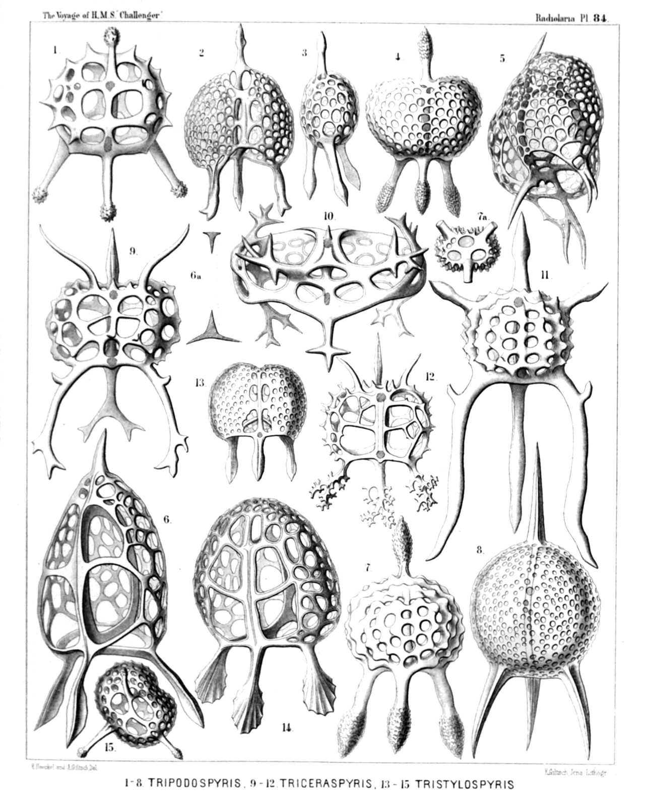 | |||||||||||||||||||||||||||||||||||||||||||||||||||||||||||||||||||||||||||||||||||||||||||||
| PLATE 84. | |||
| Zygospyrida. | |||
| Diam. | Page. | ||
Fig. 1. Tripospyris capitata, n. sp., | × | 400 | 1028 |
| Seen from the dorsal side. | |||
| Fig. 2. Tripospyris semantis, n. sp., | × | 300 | 1026 |
| Seen from the ventral side. | |||
| Fig. 3. Tripospyris semantis, n. sp., | × | 300 | 1026 |
| Seen from the lateral side. | |||
| Fig. 4. Tripospyris eucolpos, n. sp., | × | 300 | 1029 |
| Seen from the dorsal side. | |||
| Fig. 5. Tripospyris diomma, n. sp., | × | 400 | 1026 |
| Half from the right side, half from the basal side. | |||
| Fig. 6. Tripospyris cortiniscus, n. sp., | × | 500 | 1026 |
| Half from the dorsal, half from the right side. | |||
| Fig. 6a. Frontal section through the ring, | × | 500 | |
| Fig. 7. Tripospyris conifera, n. sp., | × | 400 | 1027 |
| Seen from the dorsal side. | |||
| Fig. 7a. From the basal side, | × | 200 | |
| Fig. 8. Tripospyris euscenium, n. sp. (vel Euscenium tripospyris), | × | 400 | 1147 |
| Seen from the frontal or ventral side. | |||
| Fig. 9. Triceraspyris gazella, n. sp., | × | 500 | 1031 |
| Seen from the ventral side. | |||
| Fig. 10. Triceraspyris damæcornis, n. sp., (vel Elaphospyris damæcornis?); compare p. 1032, | × | 400 | 1057 |
| Seen from the apical (or basal?) side. | |||
| Fig. 11. Triceraspyris giraffa, n. sp., | × | 400 | 1031 |
| Seen from the frontal side. | |||
| Fig. 12. Triceraspyris corallorrhiza, n. sp., | × | 400 | 1031 |
| Seen from the frontal side. | |||
| Fig. 13. Tristylospyris scaphipes, n. sp., | × | 400 | 1033 |
| Seen from the dorsal side. | |||
| Fig. 14. Tristylospyris palmipes, n. sp., | × | 400 | 1033 |
| Seen from the dorsal side. | |||
| Fig. 15. Tristylospyris clavipes, n. sp., | × | 400 | 1033 |
| Seen from the basal side. | |||
PLATE 85.
Legion NASSELLARIA.
Order SPYROIDEA.
Family Zygospyrida.
| 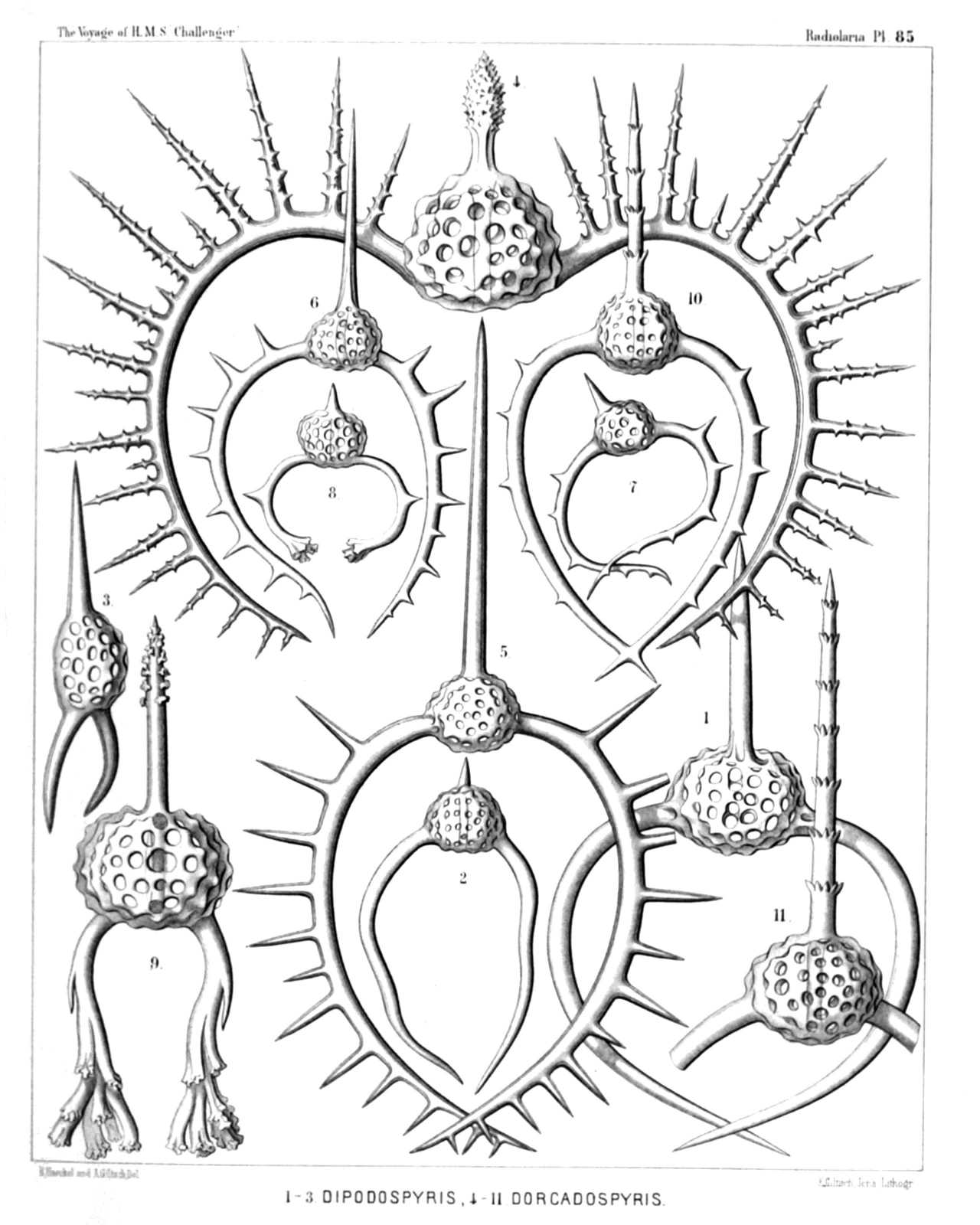 | ||||||||||||||||||||||||||||||||||||||||||||||||||||||||
| PLATE 85. | |||
| Zygospyrida. | |||
| Diam. | Page. | ||
Fig. 1. Dipospyris forcipata, n. sp., | × | 300 | 1037 |
| Fig. 2. Dipospyris irregularis, n. sp., | × | 200 | 1037 |
| Fig. 3. Dipospyris chelifer, n. sp., | × | 300 | 1037 |
| Fig. 4. Dorcadospyris dinoceras, n. sp., | × | 400 | 1041 |
| Fig. 5. Dorcadospyris antilope, n. sp., | × | 200 | 1041 |
| Fig. 6. Dorcadospyris dentata, n. sp., | × | 200 | 1040 |
| Fig. 7. Dorcadospyris decussata, n. sp., | × | 200 | 1041 |
| Fig. 8. Dendrospyris polyrrhiza, n. sp., | × | 200 | 1039 |
| Fig. 9. Dendrospyris arborescens, n. sp., | × | 400 | 1040 |
| Fig. 10. Stephanospyris cordata, n. sp., | × | 200 | 1042 |
| Fig. 11. Stephanospyris verticillata, n. sp., | × | 300 | 1043 |
PLATE 86.
Legion NASSELLARIA.
Order SPYROIDEA.
Family Zygospyrida.
| 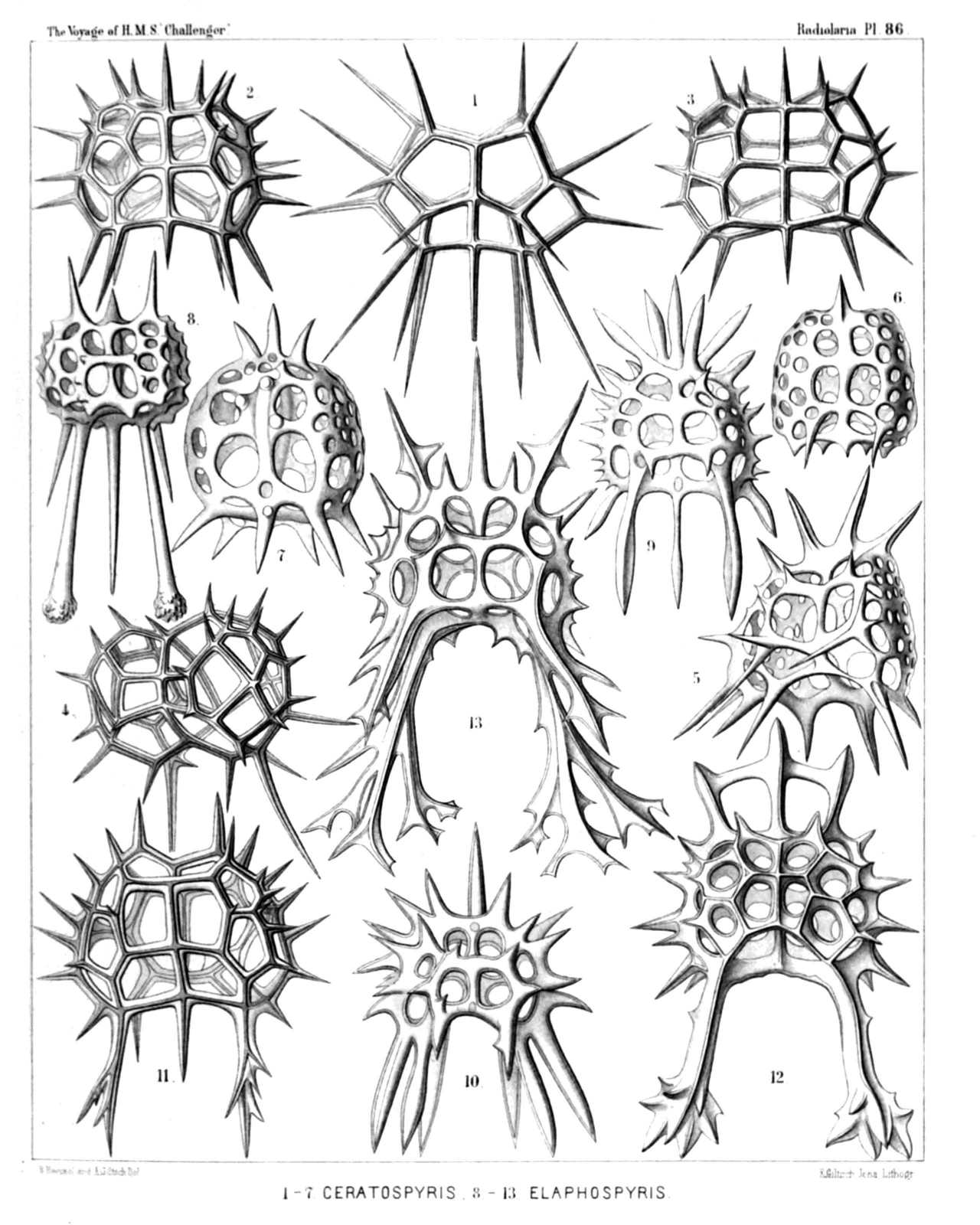 | ||||||||||||||||||||||||||||||||||||||||||||||||||||||||||||||||
| PLATE 86. | |||
| Zygospyrida. | |||
| Diam. | Page. | ||
Fig. 1. Ceratospyris polygona, n. sp., | × | 400 | 1066 |
| Fig. 2. Ceratospyris strasburgeri, n. sp., | × | 400 | 1067 |
| Fig. 3. Ceratospyris allmersii, n. sp., | × | 400 | 1067 |
| Fig. 4. Ceratospyris mulderi, n. sp., | × | 400 | 1067 |
| Fig. 5. Anthospyris aculeata, n. sp., | × | 400 | 1065 |
| Fig. 6. Petalospyris dictyocubus, n. sp., | × | 400 | 1063 |
| Fig. 7. Liriospyris hexapoda, n. sp., | × | 400 | 1049 |
| Fig. 8. Aegospyris caprina, n. sp., | × | 400 | 1054 |
| Fig. 9. Ceratospyris preyeri, n. sp., | × | 400 | 1068 |
| Fig. 10. Ceratospyris krausei, n. sp., | × | 400 | 1068 |
| Fig. 11. Ceratospyris carnerii, n. sp., | × | 400 | 1069 |
| Fig. 12. Elaphospyris alcicornis, n. sp., | × | 400 | 1057 |
| Fig. 13. Elaphospyris cervicornis, n. sp., | × | 400 | 1057 |
PLATE 87.
Legion NASSELLARIA.
Order SPYROIDEA.
Families Zygospyrida et Tholospyrida.
| 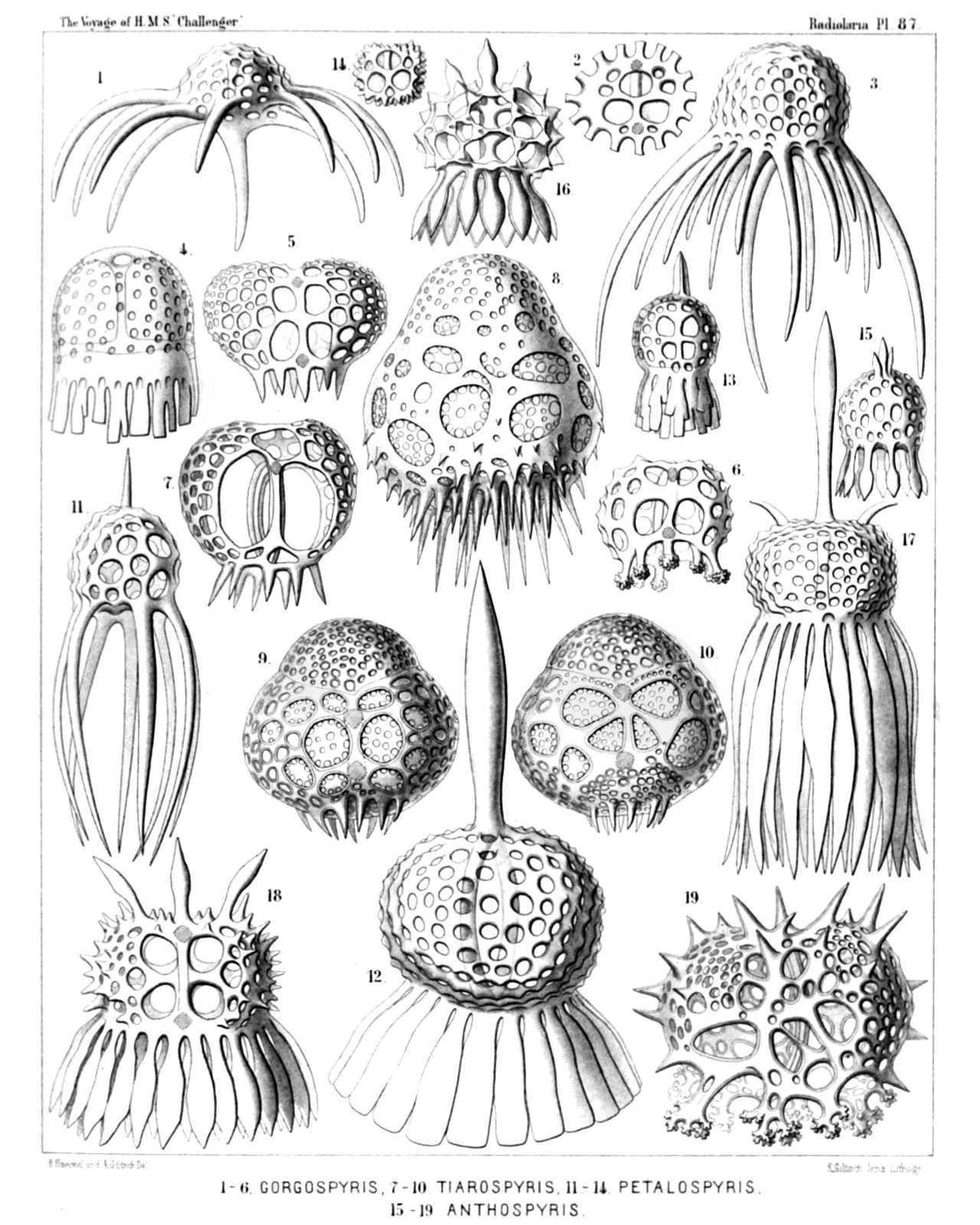 | ||||||||||||||||||||||||||||||||||||||||||||||||||||||||||||||||||||||||||||||||||||||||||||
| PLATE 87. | |||
| Zygospyrida et Tholospyrida. | |||
| Diam. | Page. | ||
Fig. 1. Gorgospyris medusa, n. sp., | × | 300 | 1070 |
| Fig. 2. Gorgospyris medusetta, n. sp., | × | 300 | 1070 |
| From the basal side, with the nine cortinar pores. | |||
| Fig. 3. Gorgospyris polypus, n. sp., | × | 300 | 1070 |
| Fig. 4. Gorgospyris schizopodia, n. sp., | × | 400 | 1071 |
| Fig. 5. Gorgospyris eurycolpos, n. sp., | × | 300 | 1071 |
| Fig. 6. Gorgospyris liriope, n. sp., | × | 300 | 1071 |
| Fig. 7. Tiarospyris pervia, n. sp., | × | 400 | 1082 |
| Fig. 8. Tiarospyris amphora, n. sp., | × | 400 | 1083 |
| Fig. 9. Tiarospyris mitra, n. sp., | × | 400 | 1082 |
| From the ventral side. | |||
| Fig. 10. Tiarospyris mitra, n. sp., | × | 400 | 1082 |
| From the dorsal side. | |||
| Fig. 11. Petalospyris octopus, n. sp., | × | 400 | 1061 |
| Fig. 12. Petalospyris dinoceras, n. sp., | × | 400 | 1063 |
| Fig. 13. Petalospyris lobata, n. sp., | × | 300 | 1064 |
| Fig. 14. Petalospyris triomma, n. sp., | × | 200 | 1060 |
| From the basal side, with the six cortinar pores. | |||
| Fig. 15. Anthospyris spathulata, n. sp., | × | 400 | 1065 |
| Fig. 16. Anthospyris mammillata, n. sp., | × | 400 | 1064 |
| Fig. 17. Anthospyris tragopogon, n. sp., | × | 300 | 1066 |
| Fig. 18. Anthospyris doronicum, n. sp., | × | 300 | 1065 |
| Fig. 19. Ceratospyris calorrhiza, n. sp., | × | 400 | 1069 |
PLATE 88.
Legion NASSELLARIA.
Orders STEPHOIDEA et SPYROIDEA.
Families Tympanida et Androspyrida.
| 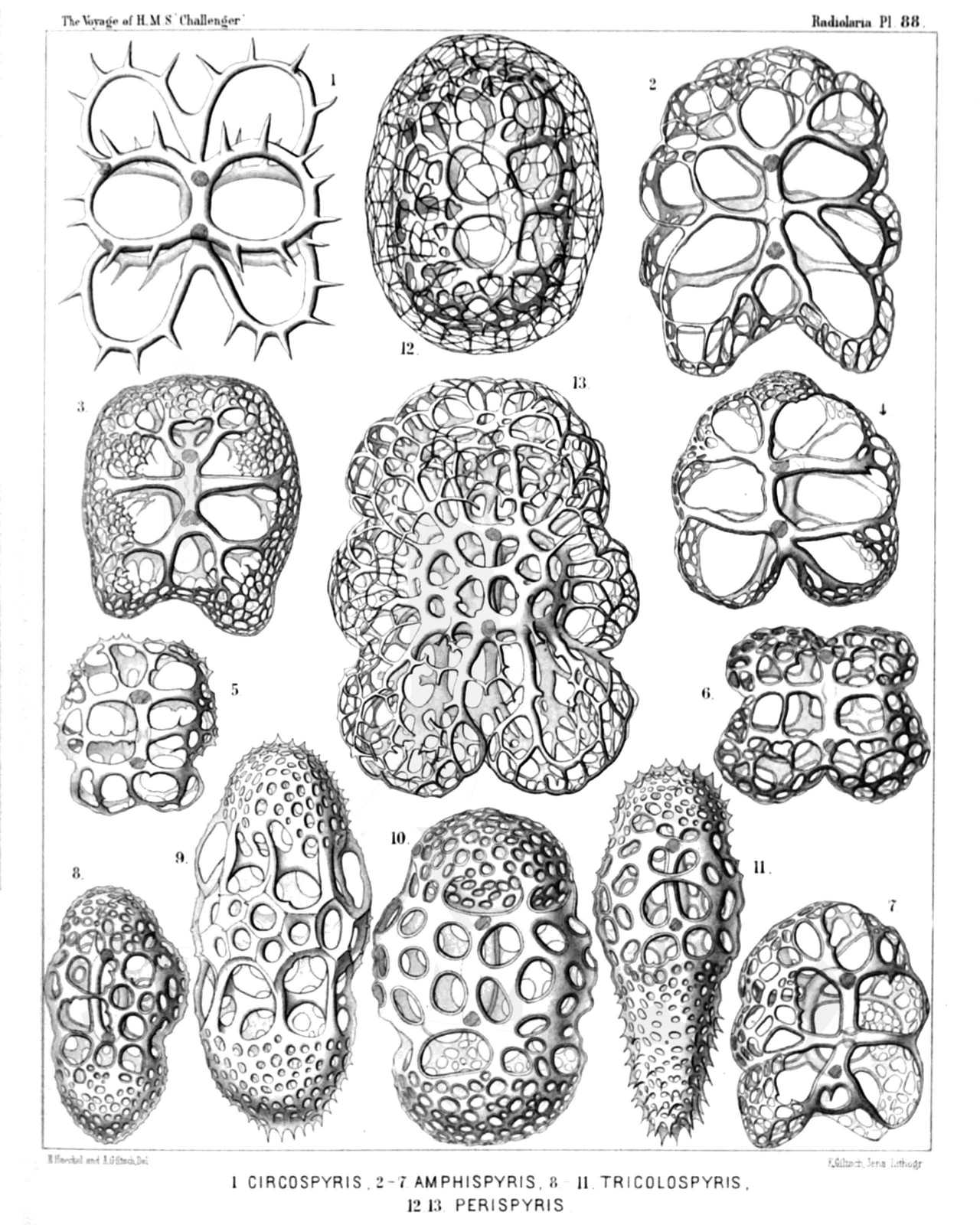 | ||||||||||||||||||||||||||||||||||||||||||||||||||||||||||||||||
| PLATE 88. | |||
| Tympanida et Androspyrida. | |||
| Diam. | Page. | ||
Fig. 1. Toxarium circospyris, n. sp., | × | 400 | 995 |
| Fig. 2. Amphispyris sternalis, n. sp., | × | 300 | 1096 |
| Fig. 3. Amphispyris costata, n. sp., | × | 300 | 1097 |
| Fig. 4. Amphispyris thorax, n. sp., | × | 300 | 1096 |
| Fig. 5. Amphispyris subquadrata, n. sp., | × | 300 | 1097 |
| Fig. 6. Amphispyris quadrigemina, n. sp., | × | 300 | 1096 |
| Fig. 7. Amphispyris toxarium, n. sp., | × | 300 | 1097 |
| Fig. 8. Tricolospyris baconiana, n. sp., | × | 400 | 1098 |
| Fig. 9. Tricolospyris leibnitziana, n. sp., | × | 600 | 1098 |
| Fig. 10. Tricolospyris kantiana, n. sp., | × | 600 | 1098 |
| Fig. 11. Tricolospyris newtoniana, n. sp., | × | 400 | 1098 |
| Fig. 12. Perispyris lentellipsis, n. sp., | × | 400 | 1099 |
| Fig. 13. Perispyris bicincta, n. sp., | × | 400 | 1099 |
PLATE 89.
Legion NASSELLARIA.
Order SPYROIDEA.
Families Zygospyrida, Tholospyrida et Androspyrida.
| 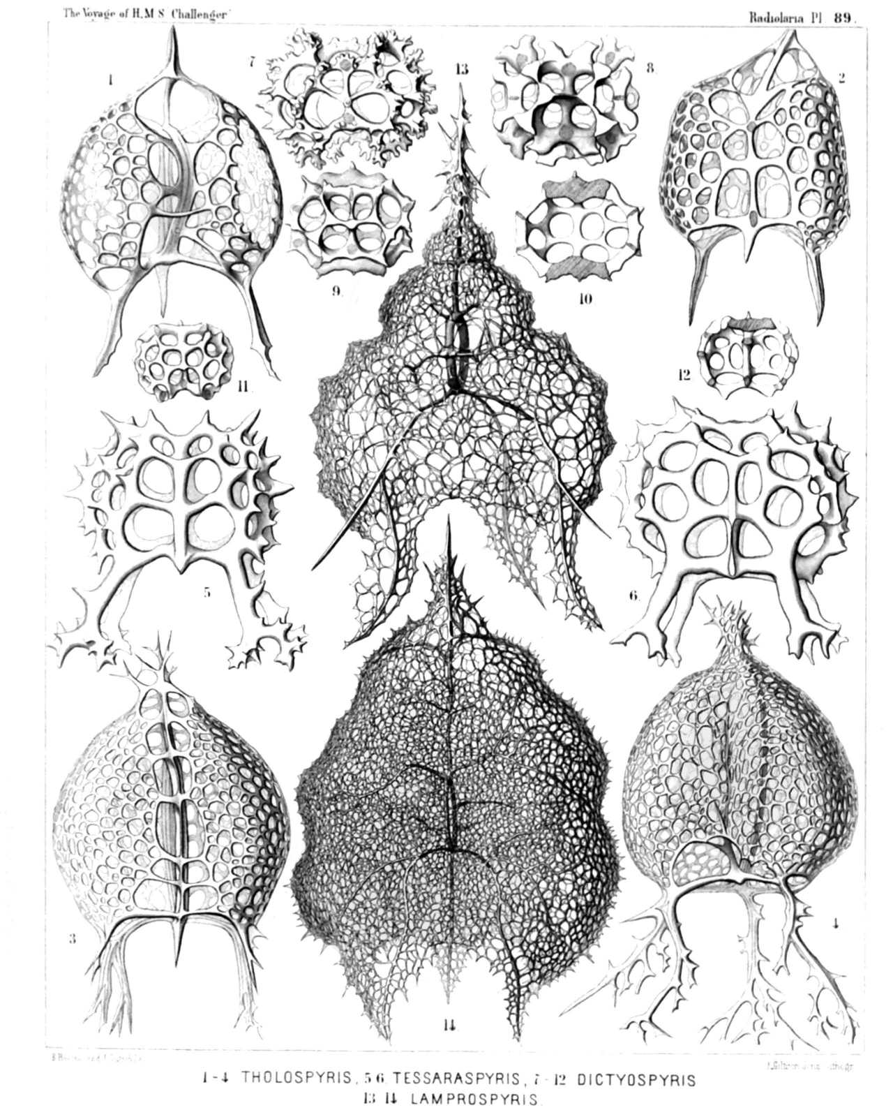 | ||||||||||||||||||||||||||||||||||||||||||||||||||||||||||||||||||||||||||||||||||
| PLATE 89. | |||
| Zygospyrida, Tholospyrida et Androspyrida. | |||
| Diam. | Page. | ||
Fig. 1. Tholospyris tripodiscus, n. sp., | × | 400 | 1079 |
| Ventral side. | |||
| Fig. 2. Tholospyris fenestrata, n. sp., | × | 400 | 1079 |
| Dorsal side. | |||
| Fig. 3. Tholospyris ramosa, n. sp., | × | 400 | 1079 |
| Dorsal side. | |||
| Fig. 4. Tholospyris cupola, n. sp., | × | 400 | 1080 |
| Ventral side. | |||
| Fig. 5. Therospyris leo, n. sp., | × | 400 | 1059 |
| Ventral side. | |||
| Fig. 6. Therospyris felis, n. sp., | × | 400 | 1059 |
| Dorsal side. | |||
| Fig. 7. Dictyospyris stalactites, n. sp., | × | 400 | 1073 |
| Ventral side. | |||
| Fig. 8. Dictyospyris anthophora, n. sp., | × | 400 | 1076 |
| Ventral side. | |||
| Fig. 9. Dictyospyris mammillaris, n. sp., | × | 400 | 1076 |
| Ventral side. | |||
| Fig. 10. Dictyospyris mammillaris, n. sp., | × | 400 | 1076 |
| Frontal section. | |||
| Fig. 11. Dictyospyris distoma, n. sp., | × | 300 | 1073 |
| Ventral side. | |||
| Fig. 12. Dictyospyris distoma, n. sp., | × | 300 | 1073 |
| Frontal section. | |||
| Fig. 13. Lamprospyris darwinii, n. sp., | × | 300 | 1094 |
| Ventral side. | |||
| Fig. 14. Lamprospyris huxleyi, n. sp., | × | 300 | 1094 |
| Ventral side. | |||
PLATE 90.
Legion NASSELLARIA.
Order SPYROIDEA.
Family Androspyrida.
| 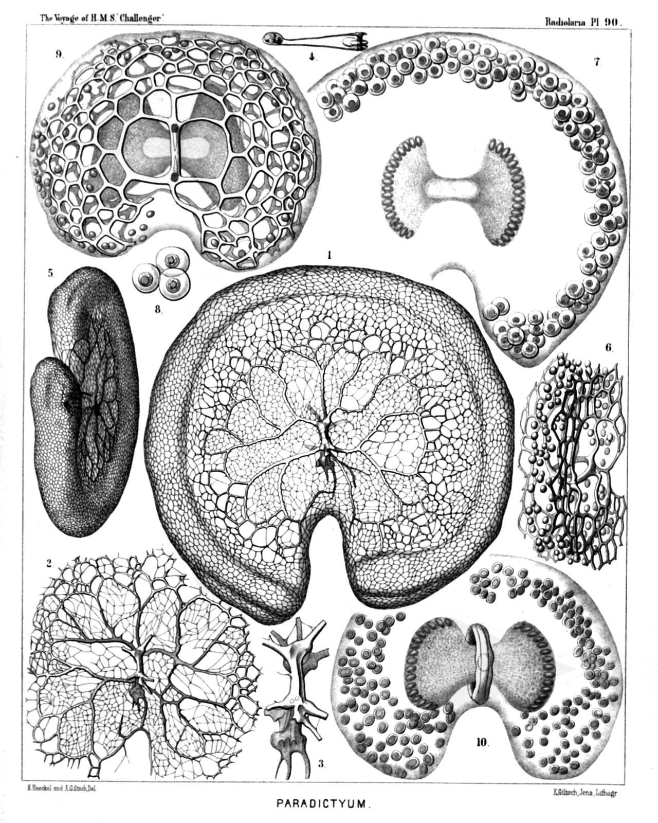 | ||||||||||||||||||||||||||||||||||||||||||||||||||||||||||||||
| PLATE 90. | |||
| Androspyrida. | |||
| Diam. | Page. | ||
Fig. 1. Nephrospyris paradictyum, n. sp. (vel Paradictyum paradoxum), | × | 250 | 1102 |
| The complete shell, seen from the frontal side. | |||
| Fig. 2. Nephrospyris paradictyum, n. sp., | × | 250 | 1102 |
| The incomplete shell, seen from the dorsal side. | |||
| Fig. 3. Nephrospyris paradictyum, n. sp., | × | 500 | 1102 |
| The sagittal ring, isolated, from the dorsal side; more enlarged. | |||
| Fig. 4. Nephrospyris paradictyum, n. sp., | × | 120 | 1102 |
| Vertical section through half the shell, exhibiting the thickened margin with the included symbiontes (compare page 1101). | |||
| Fig. 5. Nephrospyris paradictyum, n. sp., | × | 200 | 1102 |
| Oblique marginal view of the shell. | |||
| Fig. 6. Nephrospyris paradictyum, n. sp., | × | 250 | 1102 |
| Marginal view of a young specimen, with open fissure between the two parallel net-plates. | |||
| Fig. 7. Nephrospyris paradictyum, n. sp., | × | 250 | 1102 |
| The soft body alone, without the skeleton. The bilobed central capsule exhibits a central transverse nucleus, and on each lobe a stratum of oil-globules. The kidney-shaped calymma contains on the margin numerous symbiontes (Xanthellæ or Vorticellinæ? Compare page 1102). | |||
| Fig. 8. Nephrospyris paradictyum, n. sp., | × | 500 | 1102 |
| Three single unicellular symbiontes (Zooxanthellæ?). | |||
| Fig. 9. Nephrospyris renilla, n. sp. (vel Nephrodictyum renilla), | × | 250 | 1101 |
| The bilobed central capsule is enclosed by the discoidal shell and in the middle constricted by the sagittal ring; it contains a transverse nucleus. The kidney-shaped calymma contains in the peripheral part numerous symbiontes (Xanthellæ or Vorticellinæ? Compare page 1101). | |||
| Fig. 10. Nephrospyris renilla, n. sp., | × | 250 | 1101 |
| A singular abnormality (occurring not rarely), in which the reduced skeleton has nearly disappeared and the sagittal ring alone remained. The kidney-shaped calymma, however, which encloses numerous symbiontes, has preserved the form of the skeleton. The bilobed central capsule is similar to that in figs. 7 and 9, and is encircled by the thickened sagittal ring. | |||
PLATE 91.
Legion NASSELLARIA.
Orders NASSOIDEA et PLECTOIDEA.
Families Nassellida, Plagonida et Plectanida.
| 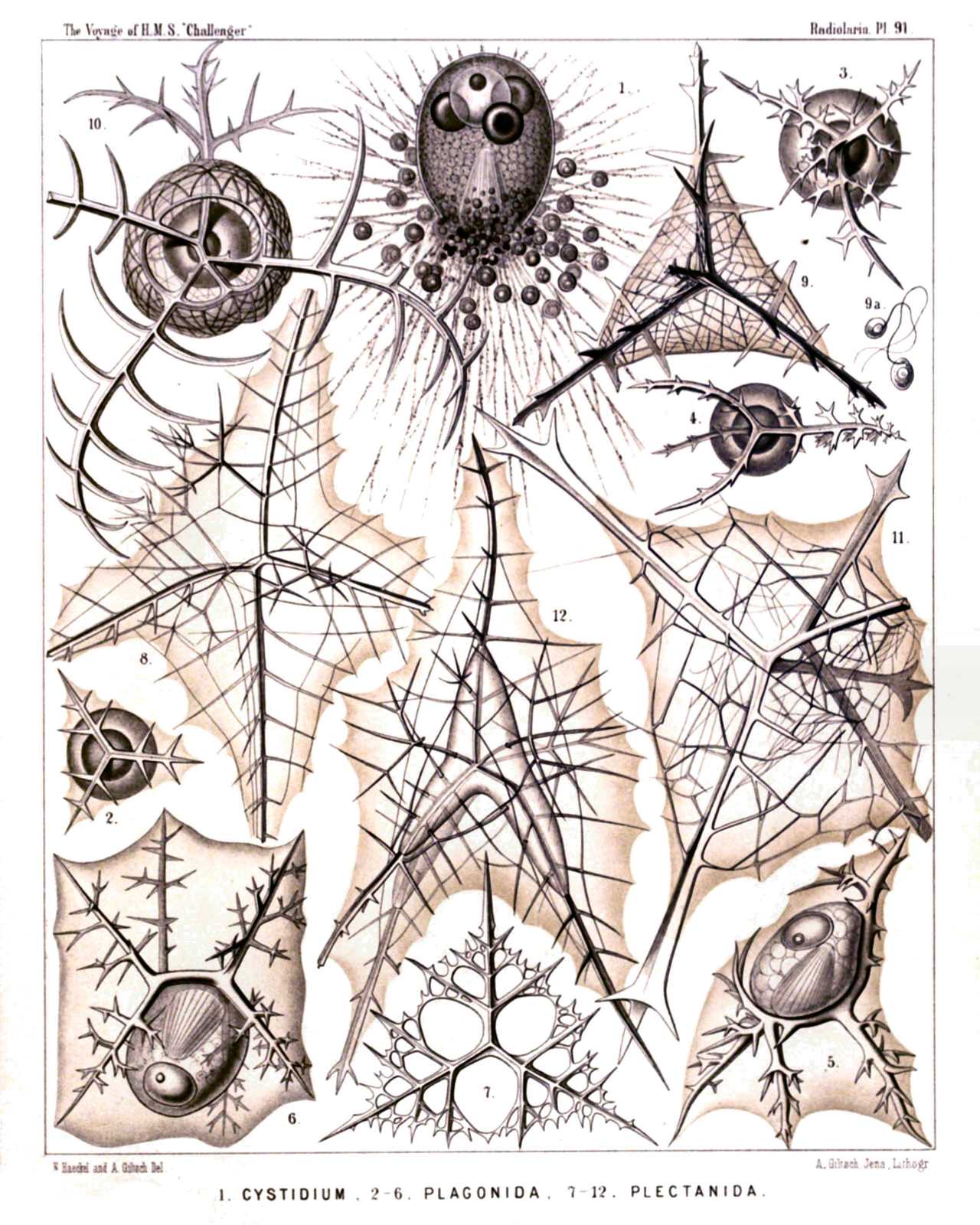 | ||||||||||||||||||||||||||||||||||||||||||||||||||||||||||||
| PLATE 91. | |||
| Nassellida, Plagonida et Plectanida. | |||
| Diam. | Page. | ||
Fig. 1. Cystidium princeps, n. sp., | × | 400 | 897 |
| Fig. 2. Triplagia primordialis, n. sp., | × | 100 | 909 |
| Fig. 3. Tetraplagia phænaxonia, n. sp., | × | 200 | 911 |
| Fig. 4. Plagoniscus tripodiscus, n. sp., | × | 200 | 912 |
| Fig. 5. Plagiocarpa procortina, n. sp., | × | 300 | 914 |
| Fig. 6. Plagonium sphærozoum, n. sp., | × | 300 | 916 |
| Fig. 7. Triplecta triactis, n. sp., | × | 300 | 922 |
| Fig. 8. Tetraplecta pinigera, n. sp., | × | 300 | 924 |
| Fig. 9. Plectaniscus cortiniscus, n. sp., | × | 300 | 925 |
| Fig. 10. Periplecta cortina, n. sp., | × | 400 | 926 |
| Fig. 11. Plectanium trigeminum, n. sp., | × | 400 | 928 |
| Fig. 12. Polyplecta heptacantha, n. sp., | × | 300 | 929 |
PLATE 92.
Legion NASSELLARIA.
Order STEPHOIDEA.
Families Stephanida et Semantida.
| 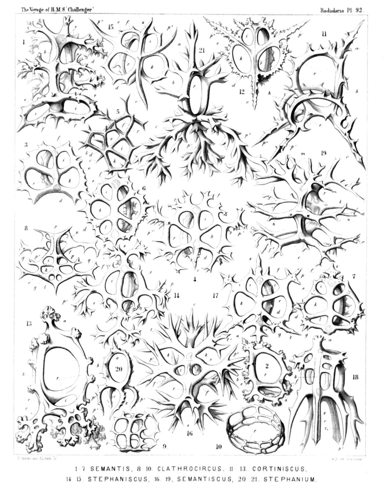 | ||||||||||||||||||||||||||||||||||||||||||||||||||||||||||||||||||||||||||||||||||||||||||||||||
| PLATE 92. | |||
| Stephanida et Semantida. | |||
| Diam. | Page. | ||
Fig. 1. Semantis sigillum, n. sp., | × | 400 | 957 |
| Fig. 2. Semantis biforis, n. sp., | × | 300 | 956 |
| Fig. 3. Semantrum tetrastoma, n. sp., | × | 300 | 959 |
| Fig. 4. Semantrum signarium, n. sp., | × | 400 | 960 |
| Fig. 5. Semantrum quadrifore, n. sp., | × | 400 | 958 |
| Fig. 6. Semantidium hexastoma, n. sp., | × | 400 | 960 |
| Fig. 7. Semantidium signatorium, n. sp., | × | 400 | 961 |
| Fig. 8. Clathrocircus stapedius, n. sp., | × | 400 | 962 |
| Fig. 9. Clathrocircus dictyospyris, n. sp., | × | 300 | 963 |
| Fig. 10. Clathrocircus multiforis, n. sp., | × | 300 | 963 |
| Fig. 11. Cortiniscus tripodiscus, n. sp., | × | 400 | 963 |
| Fig. 12. Cortiniscus typicus, n. sp., | × | 300 | 964 |
| Fig. 13. Cortiniscus dipylaris, n. sp., | × | 400 | 964 |
| Fig. 14. Stephaniscus quadrifurcus, n. sp., | × | 300 | 965 |
| Fig. 15. Stephaniscus quadrigatus, n. sp., | × | 400 | 965 |
| Fig. 16. Semantiscus hexapodius, n. sp., | × | 400 | 966 |
| Fig. 17. Semantiscus hexapylus, n. sp., | × | 400 | 967 |
| Fig. 18. Semantiscus hexaspyris, n. sp., | × | 400 | 966 |
| Fig. 19. Lithocircus tarandus, n. sp., | × | 400 | 944 |
| Fig. 20. Stephanium quadrupes, n. sp., | × | 200 | 952 |
| Fig. 21. Cortina cervina, n. sp., | × | 300 | 952 |
PLATE 93.
Legion NASSELLARIA.
Order STEPHOIDEA.
Families Coronida et Tympanida..
| 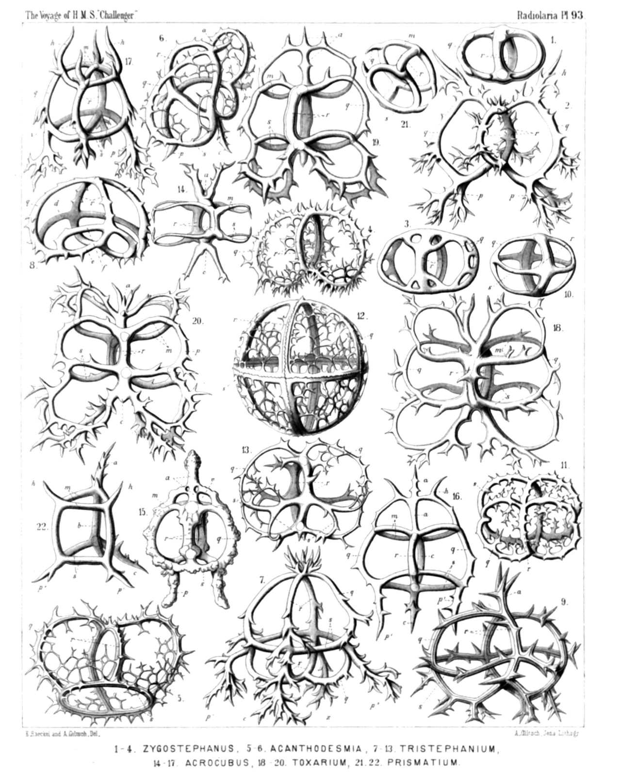 | ||||||||||||||||||||||||||||||||||||||||||||||||||||||||||||||||||||||||||||||||||||||||||||||||||||
| PLATE 93. | |||
| Coronida et Tympanida.. | |||
| Diam. | Page. | ||
Fig. 1. Zygostephanus dissocircus, n. sp., | × | 300 | 971 |
| Fig. 2. Zygostephanus bicornis, n. sp., | × | 300 | 972 |
| Fig. 3. Zygostephanium dizonium, n. sp., | × | 300 | 973 |
| Fig. 4. Zygostephanium paradictyum, n. sp., | × | 300 | 973 |
| Fig. 5. Acanthodesmia corona, n. sp., | × | 400 | 976 |
| Fig. 6. Plectocoronis pentacantha, n. sp., | × | 300 | 979 |
| Fig. 7. Tristephanium quadricorne, n. sp., | × | 300 | 984 |
| Fig. 8. Tristephanium octopyle, n. sp., | × | 300 | 983 |
| Fig. 9. Tristephanium dimensivum, n. sp., | × | 400 | 983 |
| Fig. 10. Trissocircus lentellipsis, n. sp., | × | 300 | 985 |
| Fig. 11. Trissocircus octostoma, n. sp., | × | 300 | 986 |
| Fig. 12. Trissocyclus sphæridium, n. sp., | × | 300 | 987 |
| Fig. 13. Tricyclidium dictyospyris, n. sp., | × | 300 | 984 |
| Fig. 14. Protympanium amphipodium, n. sp., | × | 300 | 992 |
| Fig. 15. Acrocubus arcuatus, n. sp., | × | 300 | 993 |
| Fig. 16. Acrocubus cortina, n. sp., | × | 300 | 994 |
| Fig. 17. Acrocubus amphithectus, n. sp., | × | 300 | 995 |
| Fig. 18. Toxarium thorax, n. sp., | × | 300 | 996 |
| Fig. 19. Toxarium cordatum, n. sp., | × | 300 | 996 |
| Fig. 20. Toxarium bifurcum, n. sp., | × | 300 | 997 |
| Fig. 21. Parastephanus quadrispinus, n. sp., | × | 300 | 1008 |
| Fig. 22. Prismatium tripodium, n. sp., | × | 300 | 1009 |
PLATE 94.
Legion NASSELLARIA.
Order STEPHOIDEA.
Family Tympanida.
| 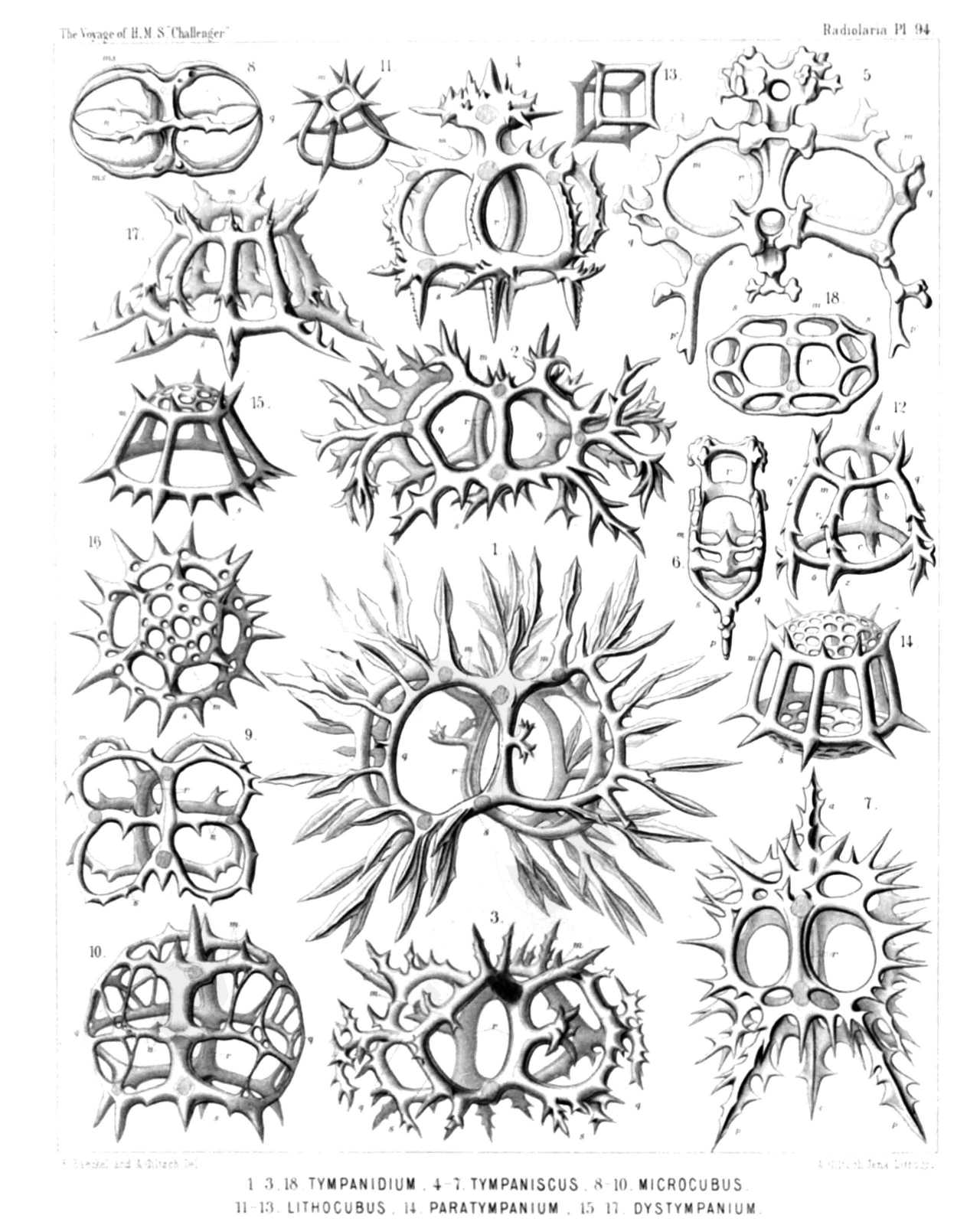 | |||||||||||||||||||||||||||||||||||||||||||||||||||||||||||||||||||||||||||||||||||||||||
| PLATE 94. | |||
| Tympanida. | |||
| Diam. | Page. | ||
Fig. 1. Tympanidium foliosum, n. sp., | × | 400 | 1003 |
| Fig. 2. Octotympanum cervicorne, n. sp., | × | 400 | 1000 |
| Fig. 3. Octotympanum octonarium, n. sp., | × | 400 | 1000 |
| Fig. 4. Tympaniscus quadrupes, n. sp., | × | 400 | 1002 |
| Fig. 5. Tympaniscus dipodiscus, n. sp., | × | 400 | 1001 |
| Frontal view. | |||
| Fig. 6. Tympaniscus dipodiscus, n. sp., | × | 400 | 1001 |
| Lateral view. | |||
| Fig. 7. Tympaniscus tripodiscus, n. sp., | × | 400 | 1002 |
| Frontal view. | |||
| Fig. 8. Microcubus zonarius, n. sp., | × | 300 | 998 |
| Fig. 9. Microcubus dodecastoma, n. sp., | × | 300 | 998 |
| Fig. 10. Microcubus amphispyris, n. sp., | × | 400 | 999 |
| Fig. 11. Pseudocubus obeliscus, n. sp., | × | 400 | 1010 |
| Fig. 12. Pseudocubus hexapylus, n. sp., | × | 300 | 1011 |
| Fig. 13. Lithocubus geometricus, n. sp., | × | 200 | 1011 |
| Fig. 14. Paratympanum octostylum, n. sp., | × | 400 | 1005 |
| Fig. 15. Dystympanium dictyocha, n. sp., | × | 400 | 1007 |
| Lateral view. | |||
| Fig. 16. Dystympanium dictyocha, n. sp., | × | 400 | 1007 |
| Apical view. | |||
| Fig. 17. Circotympanum octogonium, n. sp., | × | 500 | 1013 |
| Fig. 18. Tympanidium binoctonum, n. sp., | × | 400 | 1004 |
PLATE 95.
Legion NASSELLARIA.
Order SPYROIDEA.
Families Zygospyrida, Tholospyrida, Phormospyrida et Androspyrida.
| 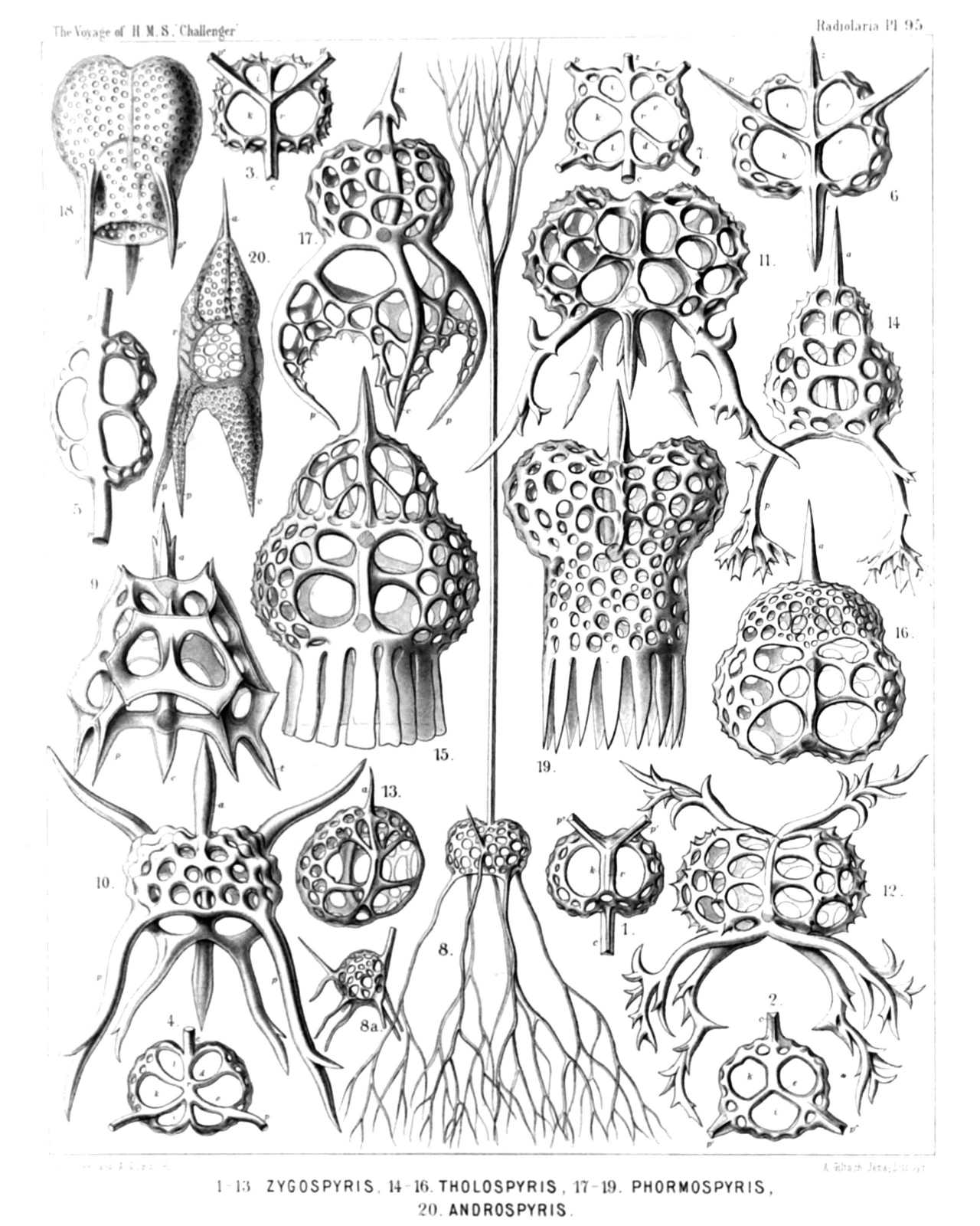 | ||||||||||||||||||||||||||||||||||||||||||||||||||||||||||||||||||||||||||||||||||||||||||||||||||||||||||||||||
| PLATE 95. | |||
| Zygospyrida, Tholospyrida, Phormospyrida et Androspyrida. | |||
| Diam. | Page. | ||
Fig. 1. Tripospyris cortina, n. sp., | × | 300 | 1025 |
| Basal view. | |||
| Fig. 2. Tripospyris triplecta, n. sp., | × | 300 | 1027 |
| Basal view. | |||
| Fig. 3. Tripospyris semantrum, n. sp., | × | 400 | 1027 |
| Basal view. | |||
| Fig. 4. Tripospyris hexomma, n. sp., | × | 300 | 1028 |
| Basal view. | |||
| Fig. 5. Brachiospyris diacantha, n. sp., | × | 400 | 1038 |
| Basal view. | |||
| Fig. 6. Tetraspyris stephanium, n. sp., | × | 300 | 1044 |
| Basal view. | |||
| Fig. 7. Liriospyris amphithecta, n. sp., | × | 300 | 1050 |
| Basal view. | |||
| Fig. 8. Hexaspyris hexacorethra, n. sp., | × | 300 | 1048 |
| Frontal view. | |||
| Fig. 9. Clathrospyris pyramidalis, n. sp., | × | 500 | 1052 |
| Frontal view. | |||
| Fig. 10. Aegospyris aegoceras, n. sp., | × | 400 | 1054 |
| Frontal view. | |||
| Fig. 11. Pentaspyris pentacantha, n. sp., | × | 400 | 1054 |
| Dorsal view. | |||
| Fig. 12. Taurospyris cervina, n. sp., | × | 400 | 1058 |
| Frontal view. | |||
| Fig. 13. Circospyris nucula, n. sp., | × | 300 | 1072 |
| Dorsal view. | |||
| Fig. 14. Lophospyris dipodiscus, n. sp., | × | 400 | 1080 |
| Frontal view. | |||
| Fig. 15. Sepalospyris platyphylla, n. sp., | × | 400 | 1081 |
| Dorsal view. | |||
| Fig. 16. Pylospyris canariensis, n. sp., | × | 400 | 1084 |
| Frontal view. | |||
| Fig. 17. Acrospyris clathrocanium, n. sp., | × | 300 | 1085 |
| Dorsal view. | |||
| Fig. 18. Phormospyris tridentata, n. sp., | × | 400 | 1087 |
| Frontal view. | |||
| Fig. 19. Patagospyris anthocyrtis, n. sp., | × | 500 | 1088 |
| Dorsal view. | |||
| Fig. 20. Androspyris pithecus, n. sp., | × | 400 | 1093 |
| Lateral view. | |||
PLATE 96.
Legion NASSELLARIA.
Order BOTRYODEA.
Families Cannobotryida, Lithobotryida et Pylobotryida.
| 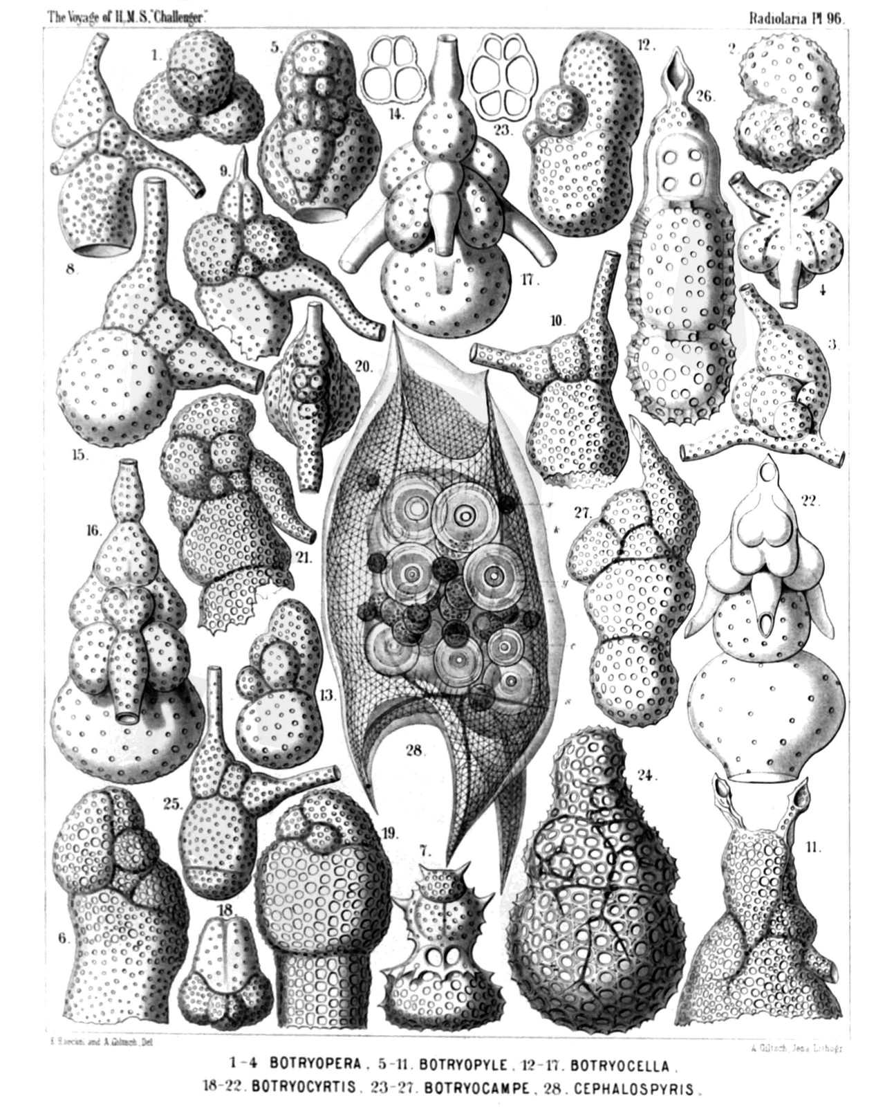 | ||||||||||||||||||||||||||||||||||||||||||||||||||||||||||||||||||||||||||||||||||||||||||||||||||||||||||||||||||||||||||||||||||||||||||||||||||||||||
| PLATE 96. | |||
| Cannobotryida, Lithobotryida et Pylobotryida | |||
| Diam. | Page. | ||
Fig. 1. Botryopera cyrtoloba, n. sp., | × | 500 | 1108 |
| Apical view. | |||
| Fig. 2. Botryopera quinqueloba, n. sp., | × | 500 | 1109 |
| Half lateral, half frontal view. | |||
| Fig. 3. Cannobotrys tricanna, n. sp., | × | 400 | 1110 |
| View half from the frontal, half from the left side. | |||
| Fig. 4. Cannobotrys cortina, n. sp., | × | 400 | 1110 |
| Basal view. | |||
| Fig. 5. Botryopyle inclusa, n. sp., | × | 500 | 1113 |
| Frontal view. | |||
| Fig. 6. Botryopyle dictyocephalus, n. sp., | × | 500 | 1113 |
| Lateral view (right side). | |||
| Fig. 7. Botryopyle sethocorys, n. sp., | × | 400 | 1112 |
| Frontal view. | |||
| Fig. 8. Acrobotrys trisolenia, n. sp., | × | 400 | 1115 |
| Lateral view (right side). | |||
| Fig. 9. Acrobotrys acuminata, n. sp., | × | 400 | 1115 |
| Lateral view (right side). | |||
| Fig. 10. Acrobotrys disolenia, n. sp., | × | 400 | 1114 |
| Lateral view (left side). | |||
| Fig. 11. Acrobotrys auriculata, n. sp., | × | 500 | 1115 |
| Lateral view (right side). | |||
| Fig. 12. Botryocella multicellaris, n. sp., | × | 500 | 1117 |
| Lateral view (left side). | |||
| Fig. 13. Botryocella quadricellaris, n. sp., | × | 400 | 1117 |
| Lateral view (left side). | |||
| Fig. 14. Botryocella quadrigemina, n. sp., | × | 400 | 1117 |
| Collar septum, between cephalis and thorax. | |||
| Fig. 15. Lithobotrys sphærothorax, n. sp., | × | 500 | 1119 |
| Lateral view (right side). | |||
| Fig. 16. Lithobotrys mascula, n. sp., | × | 500 | 1119 |
| Frontal view. | |||
| Fig. 17. Lithobotrys orchidea, n. sp., | × | 500 | 1119 |
| Frontal view. | |||
| Fig. 18. Botryocyrtis cerebellum, n. sp., | × | 400 | 1121 |
| Apical view. | |||
| Fig. 19. Botryocyrtis theocampe, n. sp., | × | 500 | 1121 |
| Lateral view (left side). | |||
| Fig. 20. Pylobotrys fontinalis, n. sp., | × | 400 | 1122 |
| Apical view. | |||
| Fig. 21. Pylobotrys putealis, n. sp., | × | 500 | 1121 |
| Lateral view (right side). | |||
| Fig. 22. Pylobotrys cerebralis, n. sp., | × | 500 | 1122 |
| Dorsal view. | |||
| Fig. 23. Botryocampe rotalia, n. sp., | × | 400 | 1123 |
| Collar septum. | |||
| Fig. 24. Botryocampe camerata, n. sp., | × | 500 | 1124 |
| Lateral view (left side). | |||
| Fig. 25. Phormobotrys cannothalamia, n. sp., | × | 400 | 1125 |
| Lateral view (right side). | |||
| Fig. 26. Phormobotrys trithalamia, n. sp., | × | 500 | 1124 |
| Frontal section. The dorsal wall is visible, in the cephalis the cruciform frontal septum. | |||
| Fig. 27. Phormobotrys pentathalamia, n. sp., | × | 400 | 1124 |
| Lateral view (left side). | |||
| Fig. 28. Cephalospyris triangulata, n. sp., | × | 400 | 1035 |
| The central capsule encloses numerous spherical concrements. | |||
PLATE 97.
Legion NASSELLARIA.
Orders STEPHOIDEA et CYRTOIDEA.
Families Stephanida, Coronida, Tripocalpida, Phænocalpida, Tripocyrtida, Podocyrtida et Podocampida.
| 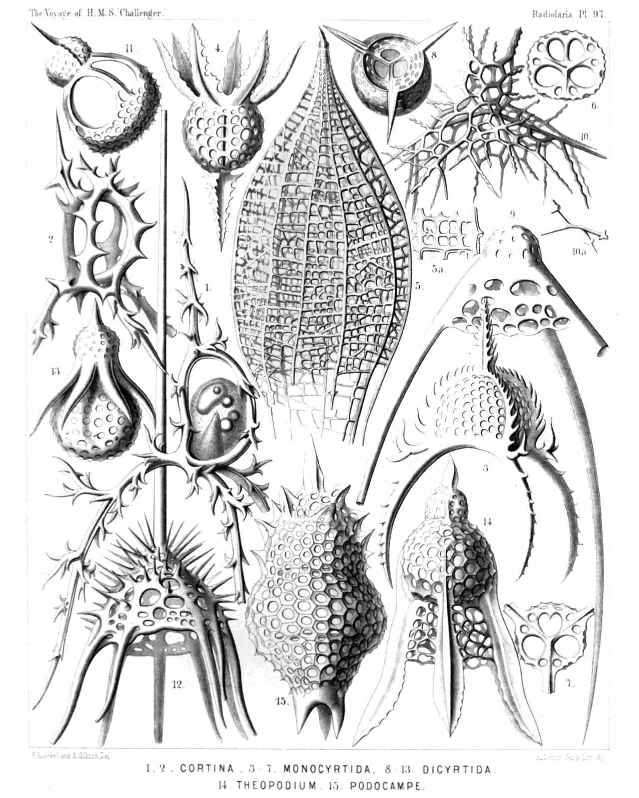 | ||||||||||||||||||||||||||||||||||||||||||||||||||||||||||||||||||||||||||||||
| PLATE 97. | |||
| Stephanida, Coronida, Tripocalpida, Phænocalpida, Tripocyrtida, Podocyrtida et Podocampida. | |||
| Diam. | Page. | ||
Fig. 1. Cortina typus, n. sp., | × | 300 | 951 |
| View from the right side. The upper part of the central capsule includes the nucleus, the lower part the podoconus, besides some oil-globules. The two pectoral feet are partly broken off. | |||
| Fig. 2. Podocoronis cortiniscus, n. sp., | × | 400 | 981 |
| View from the right anterior side. | |||
| Fig. 3. Tripocalpis cortinaris, n. sp., | × | 400 | 1137 |
| Fig. 4. Phænocalpis petalospyris, n. sp., | × | 400 | 1173 |
| Lateral view (inverted). | |||
| Fig. 5. Haliphormis lagena, n. sp., | × | 200 | 1167 |
| Fig. 6. Halicapsa lithapium, n. sp., | × | 300 | 1190 |
| Basal view. | |||
| Fig. 7. Peridium alatum, n. sp., | × | 300 | 1155 |
| Basal view. | |||
| Fig. 8. Sethopilium orthopus, n. sp., | × | 300 | 1202 |
| Basal view. | |||
| Fig. 9. Sethopilium macropus, n. sp., | × | 400 | 1203 |
| Fig. 10. Amphiplecta acrostoma, n. sp., | × | 400 | 1223 |
| Fig. 11. Sethopera tricostata, n. sp., | × | 400 | 1232 |
| Fig. 12. Acanthocorys macroceras, n. sp., | × | 200 | 1264 |
| Fig. 13. Sethophæna hexaptera, n. sp., | × | 400 | 1286 |
| Fig. 14. Theopodium tricostatum, n. sp., | × | 400 | 1328 |
| Fig. 15. Podocampe trictenota, n. sp., | × | 500 | 1446 |
PLATE 98.
Legion NASSELLARIA.
Order CYRTOIDEA.
Families Tripocalpida et Phænocalpida.
| 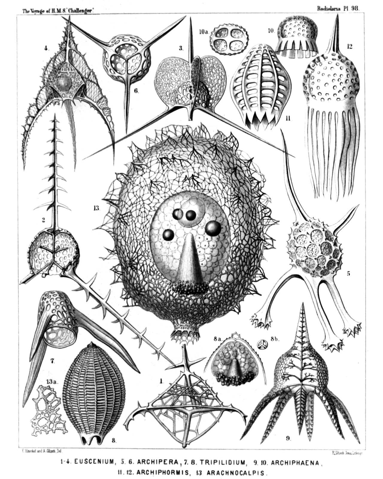 | ||||||||||||||||||||||||||||||||||||||||||||||||||||||||||||||||||||||||||||||
| PLATE 98. | |||
| Tripocalpida et Phænocalpida. | |||
| Diam. | Page. | ||
Fig. 1. Euscenium plectaniscus, n. sp., | × | 300 | 1146 |
| Half frontal, half basal view. | |||
| Fig. 2. Cladoscenium pectinatum, n. sp., | × | 400 | 1150 |
| Shell opened by a vertical section. | |||
| Fig. 3. Archiscenium cyclopterum, n. sp., | × | 400 | 1151 |
| View from the dorsal side. | |||
| Fig. 4. Pteroscenium arcuatum, n. sp., | × | 400 | 1152 |
| The central capsule contains a large spherical nucleus with a nucleolus. | |||
| Fig. 5. Archipera cortiniscus, n. sp., | × | 400 | 1155 |
| Fig. 6. Archibursa tripodiscus, n. sp., | × | 400 | 1157 |
| Basal view. | |||
| Fig. 7. Archipilium orthopterum, n. sp., | × | 400 | 1139 |
| Fig. 8. Tripilidium costatum, n. sp., | × | 300 | 1141 |
| Fig. 8a. Central capsule in the upper part of the shell, | |||
| Fig. 8b. Cortinar septum, | |||
| Fig. 9. Phænoscenium hexapodium, n. sp., | × | 300 | 1175 |
| Fig. 10. Archiphæna gorgospyris, n. sp., | × | 300 | 1178 |
| Fig. 10a. Cortinar septum with four collar pores, | × | 300 | |
| Fig. 11. Archiphormis urceolata, n. sp., | × | 300 | 1168 |
| Fig. 12. Halicalyptra petalospyris, n. sp., | × | 400 | 1169 |
| Fig. 13. Arachnocalpis ellipsoides, n. sp., | × | 300 | 1172 |
| The central capsule is filled up by clear vacuoles and exhibits in the upper half the ellipsoidal nucleus and four oil-globules, in the lower half the slender striated podoconus. | |||
| Fig. 13a. A piece of the network, more enlarged, | × | 900 | |
PLATE 99.
Legion PHÆODARIA.
Order PHÆOGROMIA.
Family Challengerida.
| 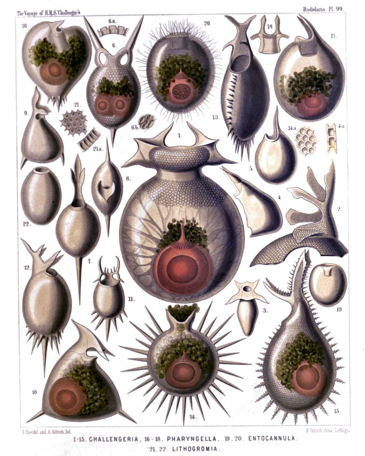 | |||||||||||||||||||||||||||||||||||||||||||||||||||||||||||||||||||||||||||||||||||||||||||||||||||||||||||||||||||||||
| PLATE 99. | |||
| Challengerida. | |||
| Diam. | Page. | ||
(The central capsule is coloured red and the phæodium green in Figs. 1, 6, 10, 14-17, 20). | |||
| Fig. 1. Challengeria murrayi, n. sp., | × | 50 | 1653 |
| From the dorsal side. Numerous streams of sarcode arise from the central capsule and pierce the calymma inside the shell. | |||
| Fig. 2. Challengeria wildi, n. sp., | × | 400 | 1653 |
| The peristome from the left side. | |||
| Fig. 3. Challengeria bromleyi, n. sp., | × | 400 | 1652 |
| From the dorsal side. | |||
| Fig. 4. Challengeria sloggettii, John Murray | × | 150 | 1649 |
| The ventral corner broken off. From the left side. | |||
| Fig. 4a. Vertical section through the shell-wall. | |||
| Fig. 5. Challengeria tritonis, n. sp., | × | 150 | 1649 |
| Fig. 6. Challengeron diodon, n. sp., | × | 400 | 1654 |
| From the dorsal side. The shell contains two central capsules. | |||
| Fig. 7. Challengeron pearceyi, n. sp., | × | 300 | 1654 |
| From the dorsal side. | |||
| Fig. 8. Challengeron richardsii, n. sp., | × | 100 | 1655 |
| From the oral margin | |||
| Fig. 9. Challengeron fergusoni, n. sp., | × | 100 | 1656 |
| From the right side. | |||
| Fig. 10. Challengeron triangulum, n. sp., | × | 200 | 1656 |
| From the right side. | |||
| Fig. 11. Challengeron crosbiei, n. sp., | × | 300 | 1657 |
| From the ventral side. | |||
| Fig. 12. Challengeron buchanani, n. sp., | × | 300 | 1657 |
| From the right side. | |||
| Fig. 13. Challengeron willemoesii, n. sp., | × | 400 | 1659 |
| From the ventral side. | |||
| Fig. 14. Challengeron moseleyi, n. sp., | × | 300 | 1658 |
| From the right side. | |||
| Fig. 15. Challengeron wyvillei, n. sp., | × | 300 | 1660 |
| From the left side. | |||
| Fig. 16. Porcupinia cordiformis, n. sp., | × | 200 | 1663 |
| From the right side. | |||
| Fig. 17. Pharyngella gastræa, n. sp., | × | 150 | 1662 |
| Fig. 18. Pharyngella gastrula, n. sp., | × | 150 | 1662 |
| Fig. 19. Entocannula infundibulum, n. sp., | × | 100 | 1661 |
| Fig. 20. Entocannula hirsuta, n. sp., | × | 150 | 1661 |
| Fig. 21. Lithogromia diatomacea, n. sp., | × | 400 | 1647 |
| A piece of the shell with diatomaceous structure. | |||
| Fig. 21a. Vertical section through the shell-wall. | |||
| Fig. 22. Lithogromia silicea, n. sp., | × | 150 | 1647 |
PLATE 100.
Legion PHÆODARIA.
Order PHÆOGROMIA.
Family Tuscarorida.
| 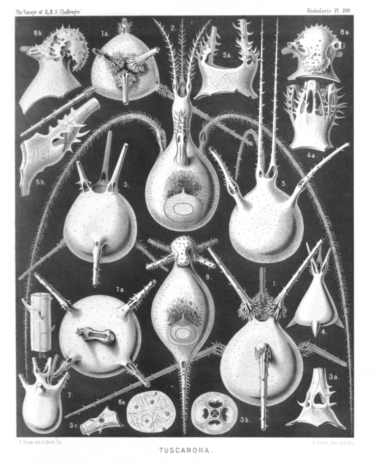 | |||||||||||||||||||||||||||||||||||||||||||||||||||||||||||||||||||||||||||
| PLATE 100. | |||
| Tuscarorida. | |||
| Diam. | Page. | ||
Fig. 1. Tuscarora bisternaria, John Murray, | × | 30 | 1706 |
| View from the dorsal side. | |||
| Fig. 1a. View from the mouth pole | × | 25 | |
| Fig. 2. Tuscarora murrayi, n. sp., | × | 30 | 1706 |
| View from the dorsal side. The central capsule (in the aboral half), and the phæodium (in the middle of the shell-cavity) are visible. A fine network of pseudopodia pierces the calymma, which fills up the shell-cavity. | |||
| Fig. 3. Tuscarora wyvillei, n. sp., | × | 30 | 1707 |
| View from the dorsal side. | |||
| Fig. 3a. Base of a tooth, | × | 100 | |
| Fig. 3b. Transverse section through the base of a tooth. | |||
| Fig. 3c. Base of a foot. | |||
| Fig. 4. Tuscarora tetrahedra, John Murray, | × | 15 | 1707 |
| View from the dorsal side. | |||
| Fig. 4a. Mouth with the three teeth, | × | 50 | |
| Fig. 5. Tuscarora tubulosa, John Murray, | × | 40 | 1707 |
| View from the ventral side. | |||
| Fig. 5a. Mouth with the two teeth, | × | 100 | |
| Fig. 5b. Basal part of a single tooth, | × | 150 | |
| Fig. 6. Tuscarora porcellana, John Murray, | × | 600 | 1708 |
| Fig. 6a. A piece of the shell, with five pores. | |||
| Fig. 6b. A piece of a tooth, with the internal axial rod and its transverse branches. | |||
| Fig. 7. Tuscarusa medusa, n. sp., | × | 25 | 1709 |
| View from the side. | |||
| Fig. 7a. View from the mouth, | × | 50 | |
| Fig. 8. Tuscaridium lithornithium, n. sp., | × | 20 | 1710 |
| View from the ventral side. Central capsule and calymma as in fig. 2. | |||
| Fig. 8a. Peristome from the ventral side. | |||
| Fig. 8b. Peristome from the right side. | |||
PLATE 101.
Legion PHÆODARIA.
Order PHÆOCYSTINA.
Families Phæodinida, Cannorrhaphida et Aulacanthida.
| 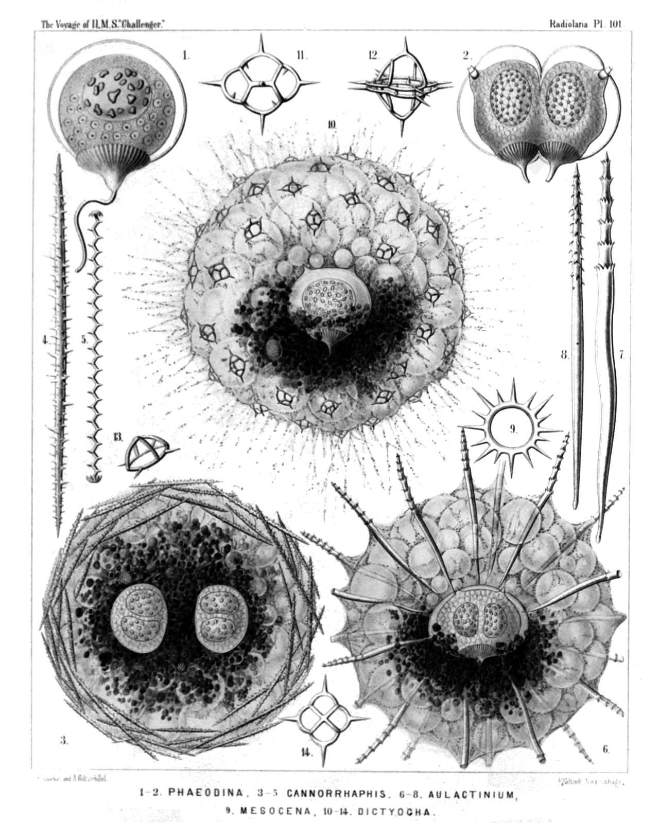 | ||||||||||||||||||||||||||||||||||||||||||||||||||||||||||||||||||||||||||||||||||
| PLATE 101. | |||
| Phæodinida, Cannorrhaphida et Aulacanthida. | |||
| Diam. | Page. | ||
Fig. 1. Phæocolla primordialis, n. sp., | × | 300 | 1544 |
| Central capsule, isolated. The double contoured outer membrane exhibits only one opening, with a radiate operculum and long proboscis. The granular protoplasm encloses clear spherical vacuoles. The sphæroidal nucleus contains irregular amœboid nucleoli. | |||
| Fig. 2. Phæodina tripylea, n. sp., | × | 300 | 1545 |
| A central capsule in self-division, with two elliptical nuclei. The astropyle is already bisected and has two proboscides. | |||
| Fig. 3. Cannorrhaphis spinulosa, n. sp., | × | 300 | 1552 |
| A complete specimen with two central capsules, each of which contains two nuclei. The alveolate calymma contains a dark phæodium and is surrounded by tangential tubular needles. | |||
| Fig. 4. Cannorrhaphis spinulosa, n. sp., | × | 300 | 1552 |
| A single tangential tube. | |||
| Fig. 5. Cannorrhaphis spathillata, n. sp., | × | 300 | 1552 |
| A single tangential tube. | |||
| Fig. 6. Aulactinium actinastrum, n. sp., | × | 100 | 1574 |
| A complete specimen, seen in optical meridional section. In the centre the spheroidal central capsule, with its double membrane and three openings (above two lateral parapylæ, below the large astropyle with its radiate operculum). The capsule encloses numerous spherical vacuoles and two hemispherical nuclei, each with numerous nucleoli. The anterior half of the capsule is surrounded by the blackish phæodium. The spherical calymma contains numerous globular alveoles and is pierced by the radial tubes, the proximal ends of which are in contact with the surface of the central capsule (compare Pl. 103, fig. 1). | |||
| Fig. 7. Aulactinium actinastrum, n. sp., | × | 300 | 1574 |
| A single radial tube. | |||
| Fig. 8. Aulactinium actinelium, n. sp., | × | 200 | 1574 |
| A single radial tube. | |||
| Fig. 9. Mesocena stellata, n. sp., | × | 600 | 1557 |
| A single annular piece of the skeleton | |||
| Fig. 10. Dictyocha stapedia, n. sp., | × | 300 | 1561 |
| A complete specimen, observed living at Ceylon. In the centre is visible the large, spheroidal, tripylean central capsule, with its three openings, containing a large nucleus with numerous nucleoli. Its oral half is covered with the dark phæodium. The voluminous spherical calymma contains numerous globular alveoles and its surface is covered with scattered, stirrup-shaped pieces of the skeleton. Numerous free pseudopodia arise from the surface. | |||
| Fig. 11. Dictyocha stapedia, n. sp., | × | 800 | 1561 |
| A single piece of the skeleton, from above. | |||
| Fig. 12. Dictyocha stapedia, n. sp., | × | 800 | 1561 |
| A twin piece of the skeleton. | |||
| Fig. 13. Dictyocha medusa, n. sp., | × | 800 | 1560 |
| A single piece of the skeleton, from the side. | |||
| Fig. 14. Dictyocha medusa, n. sp., | × | 800 | 1560 |
| A single piece of the skeleton, from above. | |||
PLATE 102.
Legion PHÆODARIA.
Order PHÆOCYSTINA.
Family Aulacanthida.
| 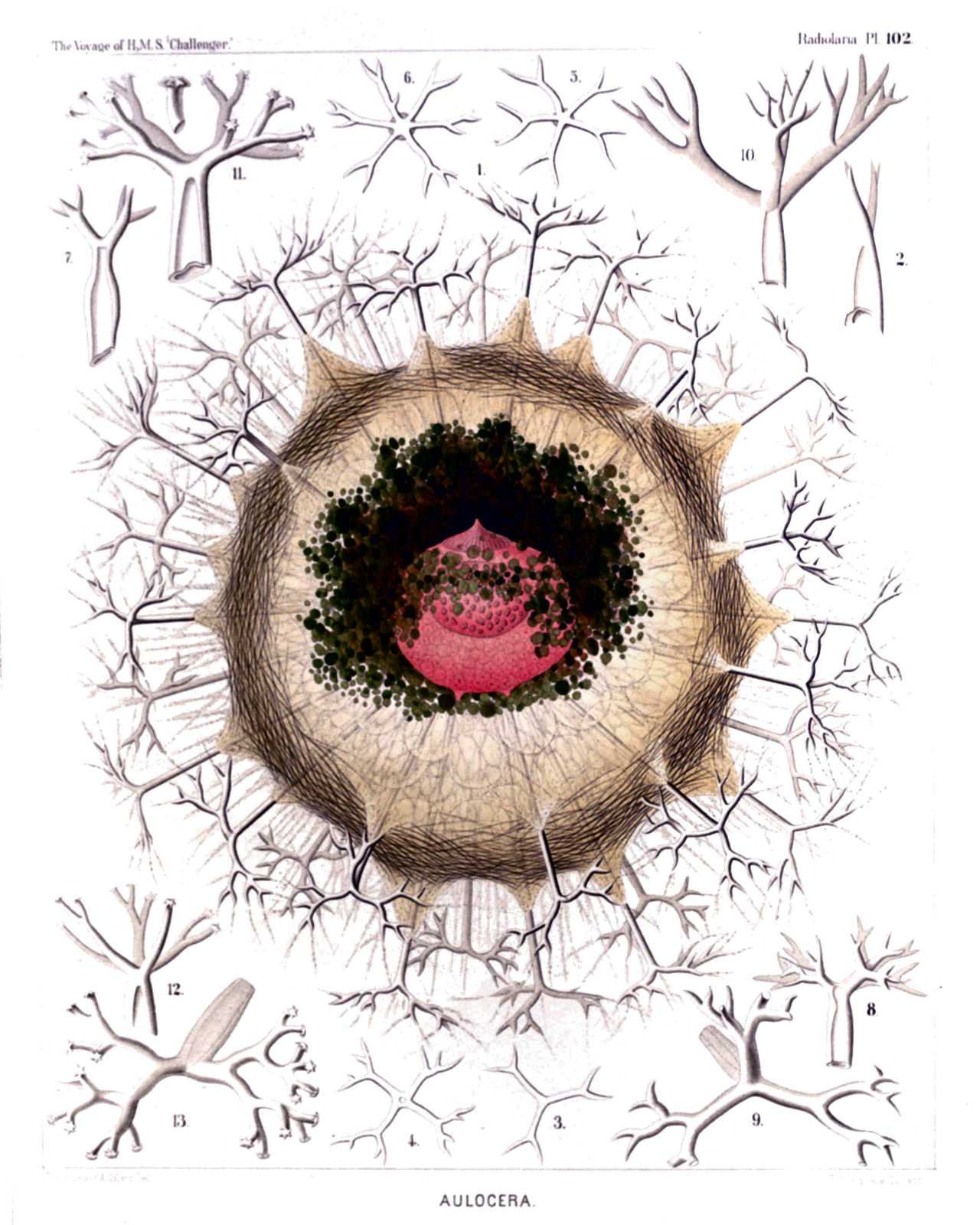 | |||||||||||||||||||||||||||||||||||||||||||||||
| PLATE 102. | |||
| Aulacanthida. | |||
| Diam. | Page. | ||
Fig. 1. Auloceros elegans, n. sp., | × | 80 | 1584 |
| A complete specimen, observed living at Ceylon. In the centre is visible the red central capsule with its three openings, containing a large nucleus of half the size, with numerous nucleoli. The alveolate calymma encloses a green excentric phæodium, is surrounded by a veil of interwoven tangential needles, and forms conical elevations, which enclose the piercing radial tubes. Between these radiate numerous pseudopodia (compare for the single parts, Pl. 103, fig. 1 and Pl. 104, figs. 1-3, and their explanation). | |||
| Figs. 2-6. Auloceros furcosus, n. sp., | × | 100 | 1583 |
| Distal ends of different radial tubes, exhibiting the great variability of this species. | |||
| Fig. 7. Auloceros trigeminus, n. sp., | × | 300 | 1584 |
| Distal end of a single tube. | |||
| Fig. 8. Auloceros capreolus, n. sp., | × | 200 | 1584 |
| Distal End of a Single Tube. | |||
| Figs. 9, 10. Auloceros cervinus, n. sp., | × | 300 | 1584 |
| Distal ends of two single tubes. | |||
| Fig. 12. Auloceros spathillaster, n. sp., | × | 300 | 1585 |
| Distal end of a single tube. | |||
| Figs. 11, 13. Auloceros arborescens, n. sp., | × | 300 | 1585 |
| Distal ends of two single tubes. | |||
PLATE 103.
Legion PHÆODARIA.
Order PHÆOCYSTINA.
Family Aulacanthida.
| 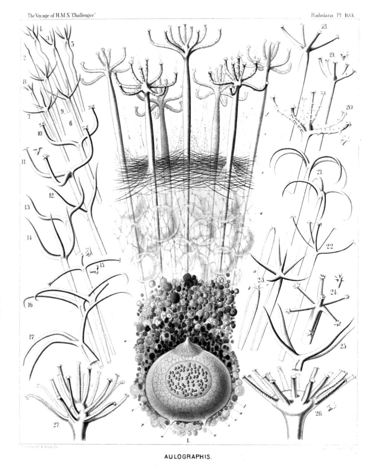 | ||||||||||||||||||||||||||||||||||||||||||||||||||||||||||||||||||||||||||||||||||||||||||||||||||||||
| PLATE 103. | |||
| Aulacanthida. | |||
| Diam. | Page. | ||
Fig. 1. Aulographis candelabrum, n. sp., | × | 100 | 1583 |
| p, The dark phæodium surrounding the central capsule on its oral part; a, a part of the surrounding alveolate calymma, also surrounding the central capsule; s, the veil of tangential needles covering the surface of the alveolate calymma; r, the big radial tubes, seven of which are visible, with an elegant verticil of terminal branches; f, the numerous pseudopodia radiating between the branches. The central capsule exhibits the following parts:—o, Astropyle; u, parapylæ; e, outer membrane; i, inner membrane; v, vacuoles; n, nucleus; l, nucleoli. | |||
| Figs. 2-9. Aulographis pandor, n. sp., | × | 100 | 1577 |
| Distal ends of various radial tubes of a single specimen, exhibiting the extraordinary variability of this species. | |||
| Fig. 10. Aulographis furcula, n. sp., | × | 400 | 1580 |
| A two-branched tube. | |||
| Fig. 11. Aulographis furcula, n. sp., | × | 400 | 1580 |
| A three-branched tube. | |||
| Figs. 12, 13. Aulographis bovicornis, n. sp., | × | 200 | 1577 |
| Two tubes with two branches. | |||
| Fig. 14. Aulographis bovicornis, n. sp., | × | 200 | 1577 |
| A tube with three branches. | |||
| Fig. 15. Aulographis triangulum, n. sp., | × | 200 | 1580 |
| A single tube. | |||
| Fig. 16. Aulographis taumorpha, n. sp., | × | 300 | 1577 |
| Two tubes, each with two branches. | |||
| Fig. 17. Aulographis triglochin, n. sp., | × | 300 | 1578 |
| A tube with three branches. | |||
| Figs. 18, 19. Aulographis hexancistra, n. sp., | × | 300 | 1581 |
| Distal end of two tubes (one with four, the other with five terminal branches). | |||
| Fig. 20. Aulographis dentata, n. sp., | × | 200 | 1582 |
| Distal end of a single tube. | |||
| Fig. 21. Aulographis ancorata, n. sp., | × | 300 | 1578 |
| Two tubes, each with four recurved branches. | |||
| Fig. 22. Aulographis tetrancistra, n. sp., | × | 300 | 1581 |
| A single tube. | |||
| Fig. 23. Aulographis stellata, n. sp., | × | 300 | 1578 |
| a and b, Two rudimentary or incompletely developed tubes; c, a well-developed tube of the usual form. | |||
| Fig. 24. Aulographis asteriscus, n. sp., | × | 300 | 1581 |
| Terminal verticil of a single tube. | |||
| Fig. 25. Aulographis cruciata, n. sp., | × | 300 | 1578 |
| Distal end of a single tube. | |||
| Fig. 26. Aulographis pulvinata, n. sp., | × | 400 | 1582 |
| Distal end of a single tube. | |||
| Fig. 27. Aulographis serrulata, n. sp., | × | 400 | 1582 |
| Distal end of a single tube. | |||
PLATE 104.
Legion PHÆODARIA.
Order PHÆOCYSTINA.
Family Aulacanthida.
| 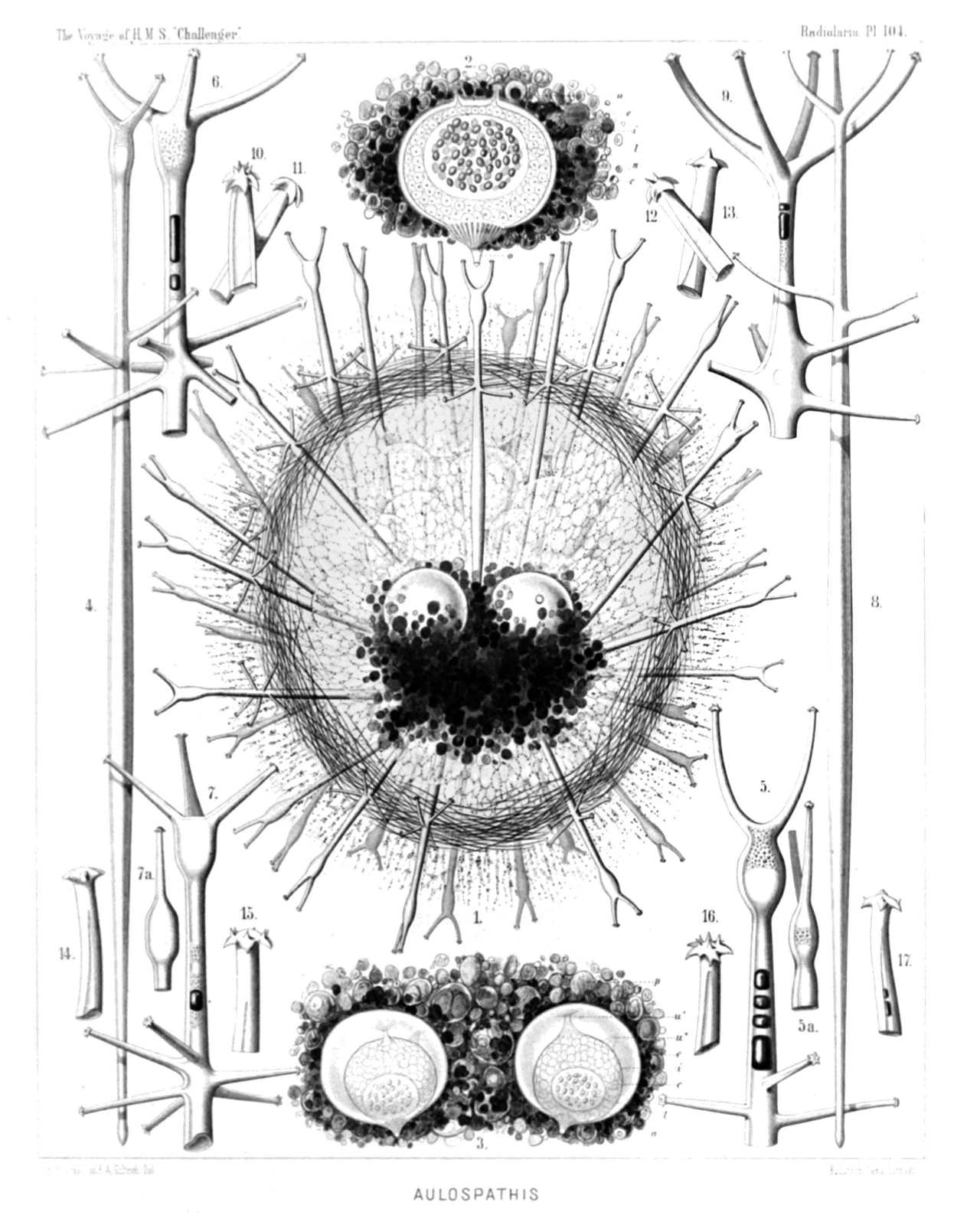 | ||||||||||||||||||||||||||||||||||||||||||||||||||||||||||||||||||||
| PLATE 104. | |||
| Aulacanthida. | |||
| Diam. | Page. | ||
Aulacanthida. | |||
| Fig. 1. Aulospathis bifurca, n. sp., | × | 50 | 1586 |
| A complete specimen, excellently preserved, with an ovate alveolate calymma and two central capsules. The surface of the calymma is covered with tangential needles. | |||
| Fig. 2. Aulospathis bifurca, n. sp., | × | 100 | 1586 |
| An isolated central capsule of another specimen, surrounded by granules of the phæodium. o, Radiate operculum of the astropyle; u, the two lateral parapylæ; e, external membrane of the capsule; i, internal membrane; c, vacuoles in the protoplasm; n, nucleus; l, numerous nucleoli. | |||
| Fig. 3. Aulospathis bifurca, n. sp., | × | 80 | 1586 |
| Two central capsules of another specimen, surrounded by the phæodium (Self-division). Characters as in fig. 2. | |||
| Fig. 4. Aulospathis bifurca, n. sp., | × | 100 | 1586 |
| A single radial tube. | |||
| Fig. 5. Aulospathis bifurca, n. sp., | × | 200 | 1586 |
| Distal part of another radial tube, partly filled up by air-bubbles. | |||
| Fig. 6. Aulospathis trifurca, n. sp., | × | 200 | 1586 |
| Distal part of a single radial tube. | |||
| Fig. 7. Aulospathis trifurca, n. sp., | × | 200 | 1586 |
| Distal part of another radial tube. | |||
| Fig. 8. Aulospathis triodon, n. sp., | × | 100 | 1587 |
| A single radial tube. | |||
| Fig. 9. Aulospathis tetrodon, n. sp., | × | 200 | 1588 |
| Distal end of single tube. | |||
| Figs. 10-13. Aulospathis polymorpha, n. sp., | × | 400 | 1587 |
| Four single terminal branches with very different forms of spathillæ. | |||
| Figs. 14-17. Aulospathis variabilis, n. sp., | × | 400 | 1588 |
| Four single terminal branches with very different forms of spathillæ. | |||
PLATE 105.
Legion PHÆODARIA.
Order PHÆOCYSTINA.
Family Aulacanthida.
| 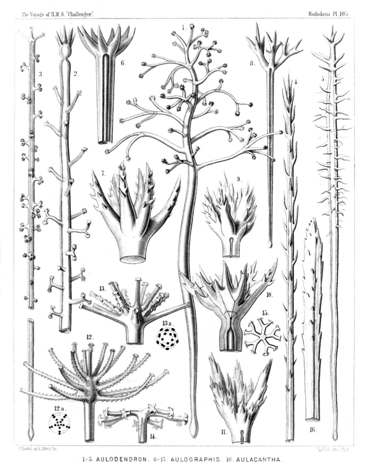 | |||||||||||||||||||||||||||||||||||||||||||||||||||||||||||||||||||||||||||||||||||||||||||||||
| PLATE 105. | |||
| Aulacanthida. | |||
| Diam. | Page. | ||
AULACANTHIDA. | |||
| Fig. 1. Aulodendron indicum, n. sp., | × | 200 | 1590 |
| A single tube. | |||
| Fig. 2. Aulodendron pacificum, n. sp., | × | 400 | 1589 |
| Distal half of a tube. | |||
| Fig. 3. Aulodendron australe, n. sp., | × | 300 | 1589 |
| A single tube. | |||
| Fig. 4. Aulacantha spinosa, n. sp., | × | 300 | 1575 |
| Distal half of a tube. | |||
| Fig. 5. Aulodendron antarcticum, n. sp., | × | 300 | 1589 |
| A single tube. | |||
| Fig. 6. Aulographis pistillum, n. sp., | × | 300 | 1579 |
| A single tube. | |||
| Fig. 7. Aulographis martagon, n. sp., | × | 300 | 1579 |
| Distal end of a single tube. | |||
| Fig. 8. Aulographis triæna, n. sp., | × | 80 | 1579 |
| A single tube. | |||
| Fig. 9. Aulographis flammabunda, n. sp., | × | 100 | 1579 |
| Distal end of a tube. | |||
| Fig. 10. Aulographis flosculus, n. sp., | × | 300 | 1580 |
| Distal end of a tube. | |||
| Fig. 11. Aulographis gemmascens, n. sp., | × | 100 | 1580 |
| Distal end of a tube. | |||
| Fig. 12. Aulographis verticillata, n. sp., | × | 400 | 1582 |
| Distal end of a tube. | |||
| Fig. 12a. Apical view, with four verticils of five branches. | |||
| Fig. 13. Aulographis tripentas, n. sp., | × | 300 | 1582 |
| Distal end of a tube. | |||
| Fig. 13a. Apical view, with three verticils of five branches. | |||
| Fig. 14. Auloceros dicranaster, n. sp., | × | 400 | 1585 |
| Distal end of a tube, seen from the side. | |||
| Fig. 15. Auloceros dicranaster, n. sp., | × | 200 | 1585 |
| Distal end of a tube, seen from the terminal face. | |||
| Fig. 16. Aulacantha cannulata, n. sp., | × | 300 | 1576 |
| Distal end of a tube. | |||
PLATE 106.
Legion PHÆODARIA.
Orders PHÆOSPHÆRIA.
Family Orosphærida.
| 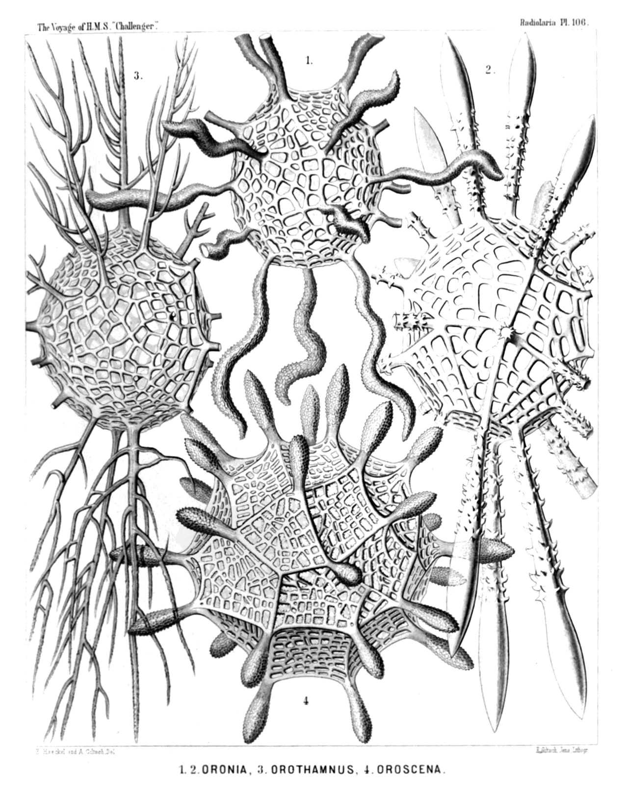 | ||||||||||||||||||||||||||||||||||
| PLATE 106. | |||
| Orosphærida. | |||
| Diam. | Page. | ||
Orosphærida. | |||
| Fig. 1. Orosphæra serpentina, n. sp., | × | 50 | 1595 |
| The entire shell. | |||
| Fig. 2. Orosphæra horrida, n. sp., | × | 50 | 1596 |
| The entire shell. | |||
| Fig. 3. Orosphæra arborescens, n. sp. (vel Orothamnus arborescens), | × | 50 | 1597 |
| The entire shell. | |||
| Fig. 4. Oroscena gegenbauri, n. sp., | × | 50 | 1597 |
| The entire shell. | |||
| (Compare Pl. 12, fig. 1.) | |||
PLATE 107.
Legion PHÆODARIA.
Order PHÆOSPHÆRIA.
Family Orosphærida.
| 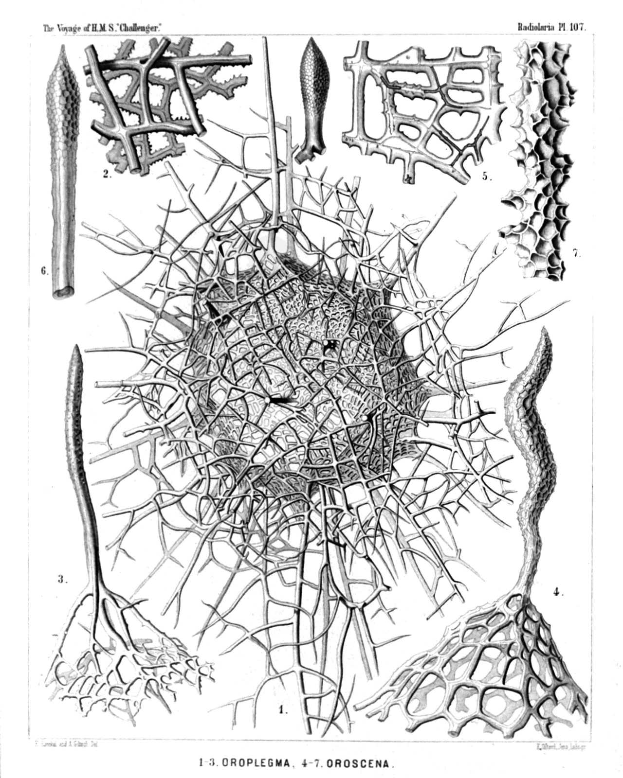 | |||||||||||||||||||||||||||||||||||||||||||||||||||||
| PLATE 107. | |||
| Orosphærida. | |||
| Diam. | Page. | ||
(Fig. 8 of this Plate has no number, by mistake; it is at the top in the middle.) | |||
| Fig. 1. Oroplegma diplosphæra, n. sp., | × | 50 | 1600 |
| The entire shell, enveloped by an outer mantle of spongy framework. | |||
| Fig. 2. Oroplegma giganteum, n. sp., | × | 200 | 1601 |
| A small piece of the spongy framework. | |||
| Fig. 3. Oroplegma spongiosum, n. sp., | × | 50 | 1601 |
| A pyramidal elevation of the inner shell, with its spongy framework, and a radial spine on the top. | |||
| Fig. 4. Oroscena bærii, n. sp., | × | 100 | 1598 |
| A pyramidal elevation of the shell, with a radial spine on its top. | |||
| Fig. 5. Orona maxima, n. sp., | × | 300 | 1594 |
| A small piece of the network; the central canals of the bars are partly filled by air. | |||
| Fig. 6. Oroscena cuvieri, n. sp., | × | 50 | 1598 |
| A single radial spine. | |||
| Fig. 7. Orona crassissima, n. sp., | × | 300 | 1594 |
| A single bar of the coarse network, with dimpled surface. | |||
| Fig. 8. Oroscena mülleri, n. sp., | × | 50 | 1598 |
| A single radial spine. | |||
PLATE 108.
Legion PHÆODARIA.
Order PHÆOSPHÆRIA.
Family Sagosphærida.
| 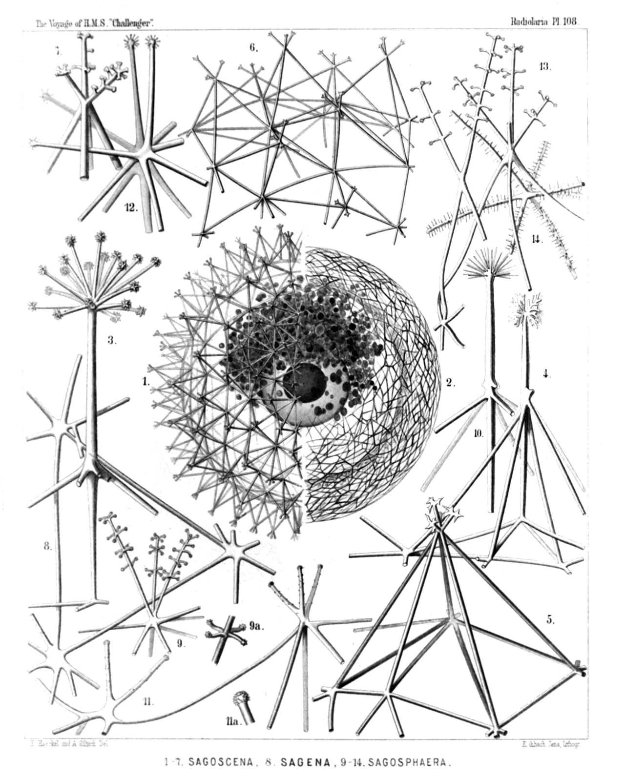 | |||||||||||||||||||||||||||||||||||||||||||||||||||||||||||||||||||||||||||||||||||||
| PLATE 108. | |||
| Sagosphærida. | |||
| Diam. | Page. | ||
Sagosphærida. | |||
| Fig. 1. Sagoscena castra, n. sp., | × | 50 | 1608 |
| Half the shell, with the enclosed central capsule and the phæodium, stained by carmine. (The central nucleus dark.) | |||
| Fig. 2. Sagmarium spongodictyum, n. sp., | × | 50 | 1612 |
| Half the shell, with its delicate spongy framework. | |||
| Fig. 3. Sagenoscena stellata, n. sp., | × | 300 | 1610 |
| Top and axial rod of a pyramid, prolonged into a crowned radial spine. | |||
| Fig. 4. Sagenoscena ornata, n. sp., | × | 300 | 1610 |
| A single pyramid with its axial rod, prolonged into a crowned radial spine. | |||
| Fig. 5. Sagoscena pellorium, n. sp., | × | 300 | 1609 |
| A single pyramid of the shell-surface. | |||
| Fig. 6. Sagoscena tentorium, n. sp., | × | 100 | 1608 |
| A piece of the shell with eight pyramids. | |||
| Fig. 7. Sagoscena prætorium, n. sp., | × | 400 | 1609 |
| Top of a pyramid. | |||
| Fig. 8. Sagena ternaria, n. sp., | × | 400 | 1606 |
| A single triangular mesh of the lattice sphere. | |||
| Fig. 9. Sagmidium crucicorne, n. sp., | × | 400 | 1613 |
| A single nodal point with three radial spines. | |||
| Fig. 9a. A portion of a spine, more highly magnified. | |||
| Fig. 10. Sagosphæra penicilla, n. sp., | × | 400 | 1607 |
| One nodal point and its radial spine. | |||
| Fig. 11. Sagosphæra furcilla, n. sp., | × | 300 | 1607 |
| Two nodal points of the network. | |||
| Fig. 11a. Extremity of a spine. | |||
| Fig. 12. Sagmidium quadricorne, n. sp., | × | 400 | 1614 |
| A nodal point of the shell surface, with four divergent spines. | |||
| Fig. 13. Sagoplegma scenophora, n. sp., | × | 300 | 1615 |
| Tops of two pyramids. | |||
| Fig. 14. Sagmarium plegmosphærium, n. sp., | × | 300 | 1612 |
| A nodal point of the spongy framework. | |||
PLATE 109.
Legion PHÆODARIA.
Order PHÆOSPHÆRIA.
Family Aulosphærida.
| 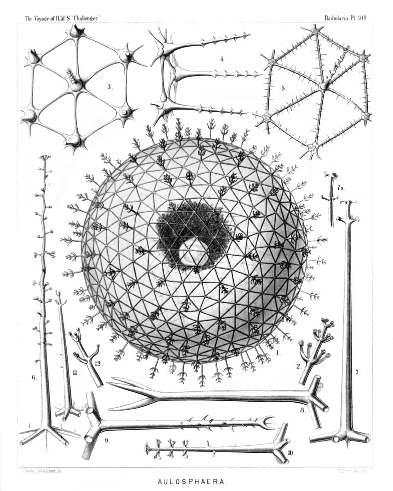 | |||||||||||||||||||||||||||||||||||||||||||||||||||||||||||||||||||||||||||
| PLATE 109. | |||
| Aulosphærida. | |||
| Diam. | Page. | ||
Fig. 1. Aulosphæra dendrophora, n. sp., | × | 50 | 1625 |
| The entire shell, with the central capsule and its nucleus, enveloped by the dark granular phæodium. | |||
| Fig. 2. Aulosphæra dendrophora, n. sp., | × | 300 | 1625 |
| A single radial tube. | |||
| Fig. 3. Aulosphæra sceptrophora, n. sp., | × | 300 | 1625 |
| A hexagonal group of six triangular meshes. | |||
| Fig. 4. Aulosphæra sceptrophora, n. sp., | × | 300 | 1625 |
| A similar group, seen from the side, with three radial tubes. | |||
| Fig. 5. Aulosphæra spinosa, n. sp., | × | 300 | 1627 |
| A hexagonal group of six triangular meshes. | |||
| Fig. 6. Aulosphæra undulata, n. sp., | × | 400 | 1627 |
| A single radial tube. | |||
| Fig. 7. Aulosphæra spathillata, n. sp., | × | 400 | 1624 |
| A single radial tube. | |||
| Fig. 7a. An abnormal variety, | × | 400 | |
| Fig. 8. Aulosphæra triodon, n. sp., | × | 400 | 1623 |
| A single radial tube. | |||
| Fig. 9. Aulosphæra trifurca, n. sp., | × | 400 | 1626 |
| A single radial tube. | |||
| Fig. 10. Aulosphæra cruciata, n. sp., | × | 300 | 1624 |
| A single radial tube. | |||
| Fig. 11. Aulosphæra bisternaria, n. sp., | × | 300 | 1624 |
| A single radial tube. | |||
| Fig. 12. Aulosphæra bisternaria, n. sp., | × | 600 | 1624 |
| Distal end of a single radial tube. | |||
PLATE 110.
Legion PHÆODARIA.
Order PHÆOSPHÆRIA.
Family Aulosphærida.
| 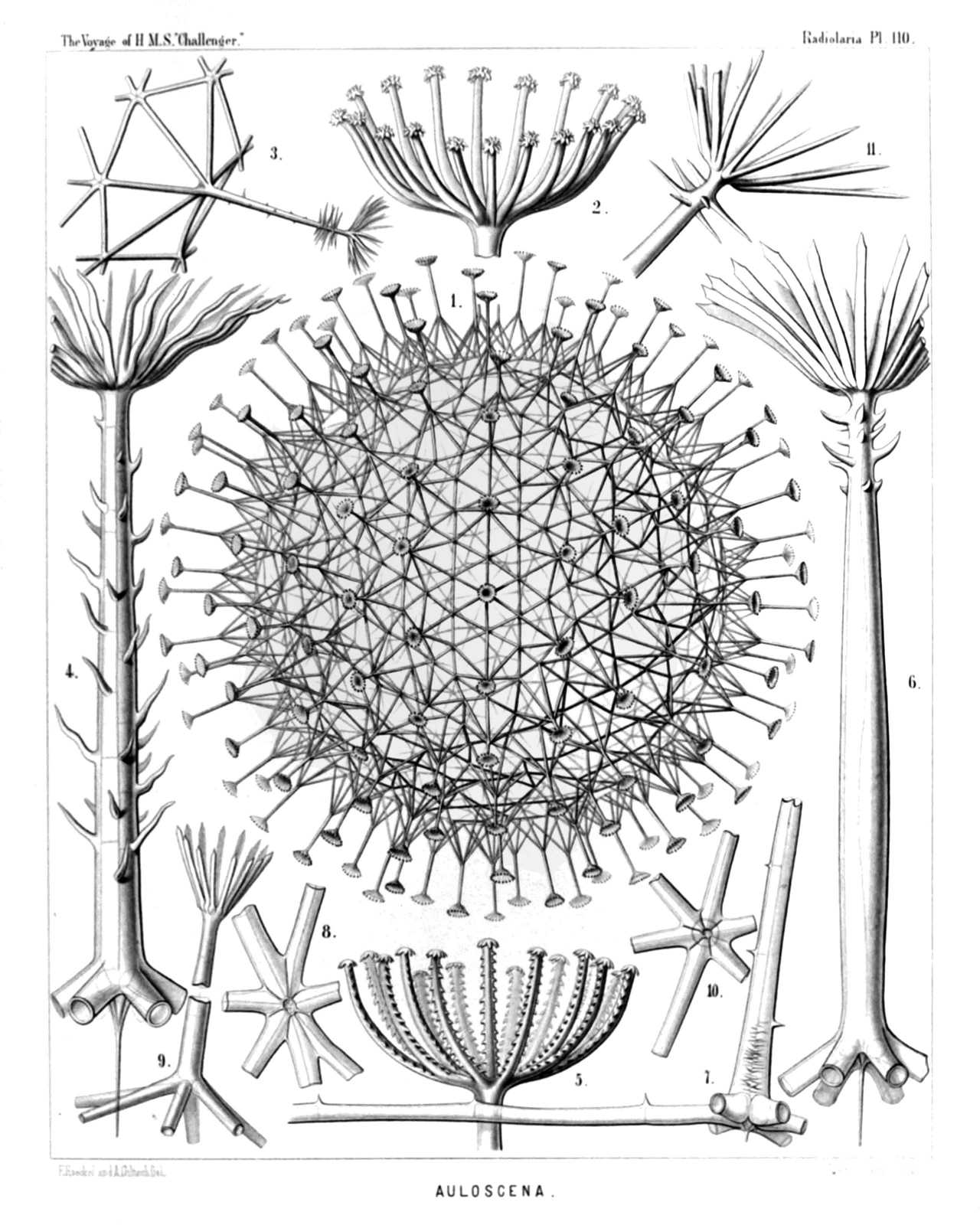 | |||||||||||||||||||||||||||||||||||||||||||||||||||||||||||||||||||
| PLATE 110. | |||
| Aulosphærida. | |||
| Diam. | Page. | ||
Fig. 1. Auloscena mirabilis, n. sp., | × | 50 | 1628 |
| The complete shell, representing a regular latticed sphere, which is composed of equal hexagonal pyramids; the top of each pyramid bears a radial tube with a terminal corona. | |||
| Fig. 2. Auloscena mirabilis, n. sp., | × | 600 | 1628 |
| Terminal corona of a single radial tube. | |||
| Fig. 3. Auloscena penicillus, n. sp., | × | 200 | 1629 |
| A single tent-shaped elevation or six-sided pyramid, bearing on the top a brush-shaped radial tube. | |||
| Fig. 4. Auloscena flammabunda, n. sp., | × | 400 | 1629 |
| A single radial tube, with a centripetal free prolongation at the base and a verticil of undulate terminal branches at the distal end. | |||
| Fig. 5. Auloscena serrata, n. sp., | × | 600 | 1630 |
| Terminal corona of a single radial tube. | |||
| Fig. 6. Auloscena tentorium, n. sp., | × | 400 | 1628 |
| A single radial tube, with a centripetal prolongation at the base and a terminal corona at the distal end. | |||
| Fig. 7. Auloscena gigantea, n. sp., | × | 400 | 1629 |
| Basal part of a radial tube, exhibiting the internal axial thread and its connection with the six tubes, which form the edges of a flat six-sided pyramid (usually more elevated than the figure exhibits). | |||
| Fig. 8. Auloscena spectabilis, n. sp., | × | 400 | 1628 |
| Apex of an abnormal pyramid (sometimes occurring), in which seven radial tubes are united, instead of six. | |||
| Fig. 9. Auloscena spectabilis, n. sp., | × | 800 | 1628 |
| Basal part of a radial tube, in the top of a flat six-sided pyramid; above it the distal part of the same tube with its terminal corona (middle part of the tube wanting). | |||
| Fig. 10. Auloscena verticillus, n. sp., | × | 300 | 1629 |
| Apex of a six-sided pyramid, seen from the inside. | |||
| Fig. 11. Auloscena verticillus, n. sp., | × | 400 | 1629 |
| Distal part of a single radial tube, with the terminal corona. | |||
PLATE 111.
Legion PHÆODARIA.
Order PHÆOSPHÆRIA.
Family Aulosphærida.
| 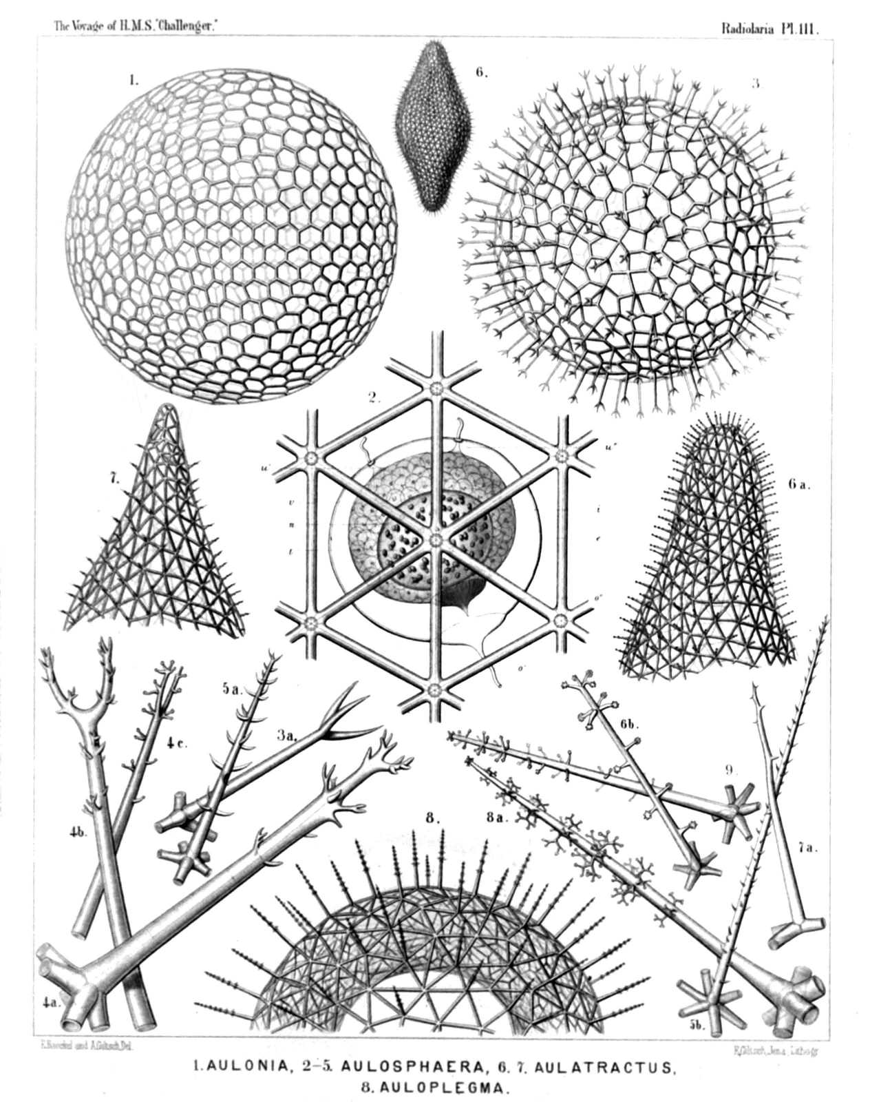 | |||||||||||||||||||||||||||||||||||||||||||||||||||||||||||||||||||||||||||||||||||||||
| PLATE 111. | |||
| Aulosphærida. | |||
| Diam. | Page. | ||
Fig. 1. Aulonia hexagonia, n. sp., | × | 30 | 1634 |
| The complete spherical shell. | |||
| Fig. 2. Aularia ternaria, n. sp., | × | 300 | 1621 |
| A group of six triangular meshes, with seven nodal points of radial tubes. Behind the central capsule, with its double membrane (e, outer; i, inner) and radiate operculum (o); u, the two outer parapylæ; v, vacuoles in the protoplasm. The ellipsoidal nucleus (n) contains numerous nucleoli (l). | |||
| Fig. 3. Aulastrum triceros, n. sp., | × | 50 | 1635 |
| The complete shell. | |||
| Fig. 3a. Aulastrum triceros, n. sp., | × | 300 | 1635 |
| A single radial tube. | |||
| Figs. 4a, 4b, 4c. Aulastrum dendroceros, n. sp., | × | 400 | 1635 |
| Three single radial spines (taken from three different specimens). | |||
| Fig. 5a. Aulophacus lenticularis, n. sp., | × | 300 | 1631 |
| A single radial spine. | |||
| Fig. 5b. Aulophacus amphidiscus, n. sp., | × | 300 | 1631 |
| A single radial spine. | |||
| Fig. 6. Aulatractus fusiformis, n. sp., | × | 5 | 1632 |
| The complete shell, five times enlarged. | |||
| Fig. 6a. Aulatractus fusiformis, n. sp., | × | 20 | 1632 |
| Apical part of the shell. | |||
| Fig. 6b. Aulatractus fusiformis, n. sp., | × | 400 | 1632 |
| A single radial tube. | |||
| Fig. 7. Aulatractus diploconus, n. sp., | × | 20 | 1632 |
| Apical part of the shell. | |||
| Fig. 7a. Aulatractus diploconus, n. sp., | × | 400 | 1632 |
| A single radial tube. | |||
| Fig. 8. Auloplegma perplexum, n. sp., | × | 50 | 1630 |
| Half the shell. | |||
| Fig. 8a. Auloplegma perplexum, n. sp., | × | 400 | 1630 |
| A single radial tube. | |||
| Fig. 9. Auloplegma spongiosum, n. sp., | × | 300 | 1631 |
| A single radial tube. | |||
PLATE 112.
Legion PHÆODARIA.
Orders PHÆOSPHÆRIA.
Family Cannosphærida.
| 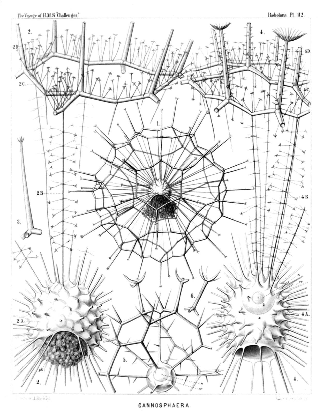 | |||||||||||||||||||||||||||||||||||||||||||
| PLATE 112. | |||
| Cannosphærida. | |||
| Diam. | Page. | ||
Cannosphærida. | |||
| Fig. 1. Cannosphæra antarctica, n. sp., | × | 50 | 1640 |
| The entire shell. The inner mammillate shell, from the month of which is prominent the phæodium, in connected by numerous radial beams with the outer shell. | |||
| Fig. 2. Cannosphæra antarctica, n. sp., | × | 200 | 1640 |
| The inner shell, from the mouth of which is prominent the phæodium, and a single hexagonal mesh of the outer shell, connected with the former by thin radial threads. | |||
| Fig. 3. Cannosphæra antarctica, n. sp., | × | 200 | 1640 |
| A single radial spine, with four terminal branches. | |||
| Fig. 4. Cannosphæra pacifica, n. sp., | × | 200 | 1641 |
| The inner shell, exhibiting on its base the widely open mouth, and in its upper half the transparent spherical central capsule with its nucleus. Of the outer shell (which is connected with the inner by thin radial threads), only a few polygonal meshes are visible. | |||
| Fig. 5. Cannosphæra atlantica, n. sp., | × | 200 | 1640 |
| The inner shell, connected by spiny radial beams with the outer shell, a quadrant only of which is visible. | |||
| Fig. 6. Cannosphæra atlantica, n. sp., | × | 200 | 1640 |
| A single radial spine, with five terminal branches. | |||
PLATE 113.
Legion PHÆODARIA.
Order PHÆOGROMIA.
Family Castanellida.
| 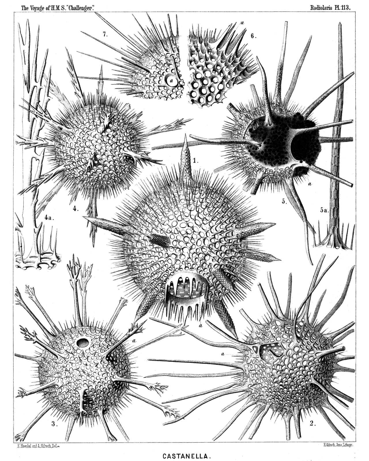 | ||||||||||||||||||||||||||||||||||||||||||||||||||||
| PLATE 113. | |||
| Castanellida. | |||
| Diam. | Page. | ||
Fig. 1. Castanissa challengeri, n. sp., | × | 100 | 1686 |
| In the lower part of the figure is visible the large corona of teeth around the mouth (a). | |||
| Fig. 2. Castanidium moseleyi, n. sp., | × | 80 | 1686 |
| In the upper part of the figure, at left, is visible the irregular polygonal mouth (a). | |||
| Fig. 3. Castanopsis naresi, n. sp., | × | 80 | 1688 |
| In the upper part of the figure is visible the smooth circular mouth (a). | |||
| Fig. 4. Castanura tizardi, n. sp., | × | 80 | 1689 |
| Fig. 4a. A single main-spine of the same, | × | 400 | |
| Fig. 5. Castanidium murrayi, n. sp., | × | 100 | 1685 |
| With a large phæodium, partly protruded through the circular mouth. | |||
| Fig. 5a. A single main-spine of the same, hexagonally dimpled, | × | 400 | |
| Fig. 6. Castanella wyvillei, n. sp., | × | 100 | 1683 |
| A piece of the shell with the mouth, armed with six large teeth (a). | |||
| Fig. 7. Castanidium buchanani, n. sp., | × | 100 | 1685 |
| A piece of the shell with the smooth roundish mouth (a). | |||
PLATE 114.
Legion PHÆODARIA.
Orders PHÆOCYSTINA et PHÆOGROMIA.
Families Cannorrhaphida et Circoporida.
| 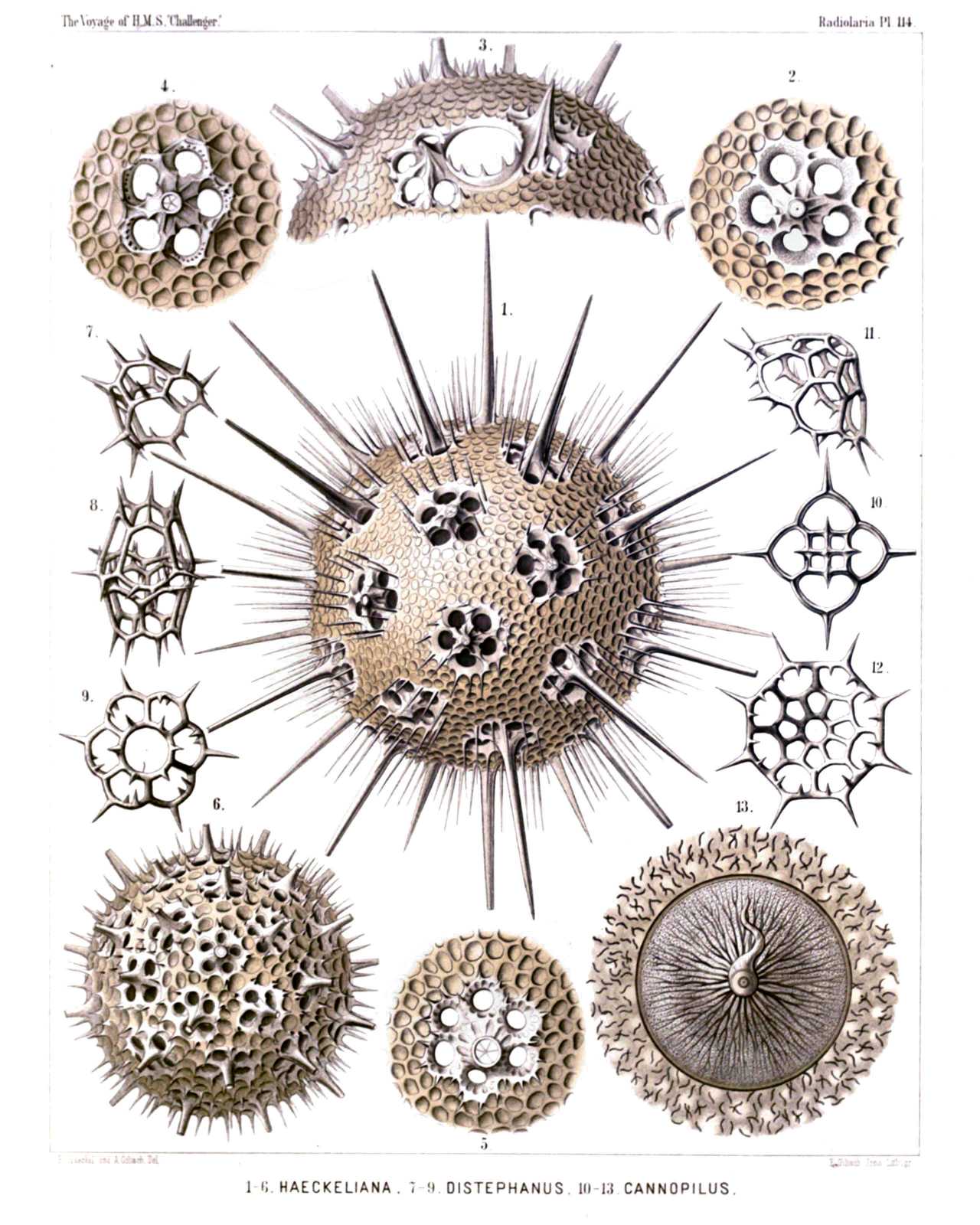 | |||||||||||||||||||||||||||||||||||||||||||||||||||||||||||||||||||||||||||||
| PLATE 114. | |||
| Cannorrhaphida et Circoporida. | |||
| Diam. | Page. | ||
Fig. 1. Haeckeliana darwiniana, n. sp., | × | 200 | 1702 |
| A complete shell. | |||
| Fig. 2. Haeckeliana darwiniana, n. sp., | × | 400 | 1702 |
| A single coronet of pores. | |||
| Fig. 3. Haeckeliana gœtheana, n. sp., | × | 300 | 1702 |
| The oral part of the shell with the mouth. | |||
| Fig. 4. Haeckeliana lamarckiana, n. sp., | × | 400 | 1701 |
| A single coronet of pores. | |||
| Fig. 5. Haeckeliana maxima, n. sp., | × | 300 | 1701 |
| A single coronet of pores. | |||
| Fig. 6. Haeckeliana porcellana, John Murray, | × | 200 | 1701 |
| A complete shell. | |||
| Fig. 7. Distephanus corona, n. sp., | × | 800 | 1566 |
| A single pileated piece (half from the side, half from below). | |||
| Fig. 8. Distephanus corona, n. sp., | × | 800 | 1566 |
| Two coupled pileated pieces caught into one another (twin-piece). | |||
| Fig. 9. Distephanus corona, n. sp., | × | 800 | 1566 |
| A single pileated piece, seen from above. | |||
| Fig. 10. Cannopilus diplostaurus, n. sp., | × | 800 | 1568 |
| A single pileated piece, seen from above. | |||
| Fig. 11. Cannopilus cyrtoides, n. sp., | × | 800 | 1569 |
| A single pileated piece, seen obliquely from the side. | |||
| Fig. 12. Cannopilus cyrtoides, n. sp., | × | 800 | 1569 |
| A single pileated piece, seen from below. | |||
| Fig. 13. Haeckeliana porcellana, John Murray, | × | 600 | 1526 |
| The radiate operculum of the central capsule. | |||
PLATE 115.
Legion PHÆODARIA.
Order PHÆOGROMIA.
Family Circoporida.
| 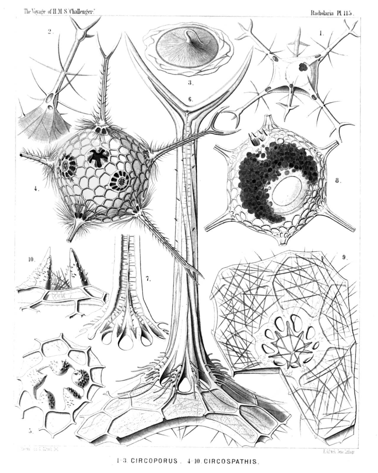 | ||||||||||||||||||||||||||||||||||||||||||||||||||||||||||||||
| PLATE 115. | |||
| Circoporida. | |||
| Diam. | Page. | ||
Fig. 1. Circoporus sexfuscinus, n. sp., | × | 100 | 1695 |
| The cruciform mouth is visible in the upper part of the figure, to the right. | |||
| Fig. 2. Circoporus sexfuscinus, n. sp., | × | 200 | 1695 |
| A single radial spine, with four cruciate pores at the base. | |||
| Fig. 3. Circoporus sexfuscinus, n. sp., | × | 600 | 1695 |
| The radiate operculum of the central capsule, with the proboscis. | |||
| Fig. 4. Circospathis furcata, n. sp., | × | 100 | 1696 |
| Five of the nine spines are visible, two others (on the upper face) broken off. Between the latter the pentagonal mouth (with five teeth). | |||
| Fig. 5. Circospathis furcata, n. sp., | × | 300 | 1696 |
| The mouth with its five teeth. | |||
| Fig. 6. Circospathis furcata, n. sp., | × | 400 | 1696 |
| A piece of the shell with a radial spine. | |||
| Fig. 7. Circospathis furcata, n. sp., | × | 400 | 1696 |
| Vertical section through the base of a radial spine, to show the central funicle. | |||
| Fig. 8. Circogonia dodecacantha, n. sp., | × | 100 | 1698 |
| The central capsule with the elliptical nucleus (to the right) and the dark phæodium (to the left) are visible, in the upper part (to the left) the mouth of the shell, with six teeth. | |||
| Fig. 9. Circogonia dodecacantha, n. sp., | × | 400 | 1698 |
| A fragment of the shell, exhibiting its peculiar structure (needles tangentially scattered in the cement of the porcellanous substance), and a circle of nine pores around the base of a broken spine. | |||
| Fig. 10. Circospathis tetrodonta, n. sp., | × | 400 | 1697 |
| The mouth with four teeth, in profile view. | |||
PLATE 116.
Legion PHÆODARIA.
Order PHÆOGROMIA.
Families Medusettida et Circoporida.
| 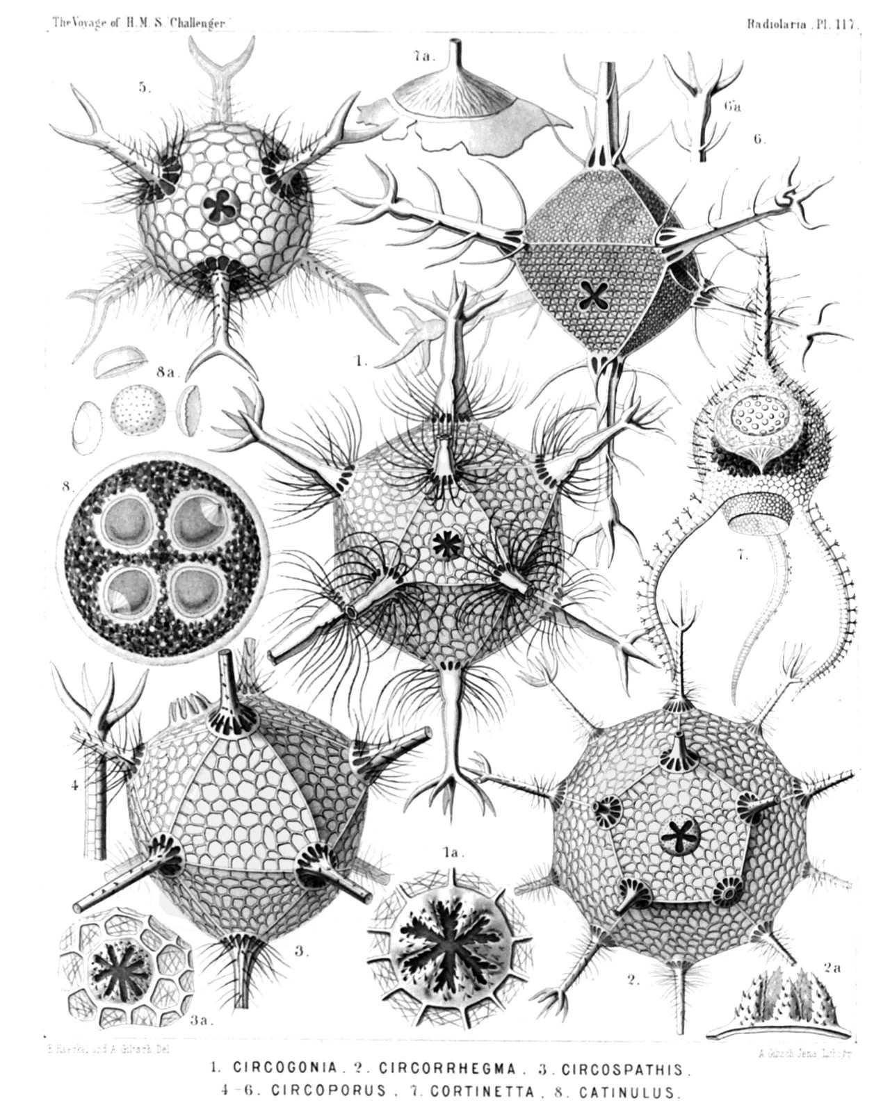 | ||||||||||||||||||||||||||||||||||||||||||
| PLATE 116. | |||
| Medusettida et Circoporida. | |||
| Diam. | Page. | ||
Fig. 1. Polypetta mammillata, n. sp., | × | 500 | 1677 |
| In the upper part of the figure the dentate proboscis. | |||
| Fig. 1a. Vertical section through the shell-wall, showing two of the hollow alveoles, opening on its inside, | × | 1000 | |
| Fig. 2. Polypetta tabulata, n. sp., | × | 500 | 1677 |
| In the upper part of the figure the dentate proboscis. | |||
| Fig. 2a. A piece of the shell, seen from the surface, with the triangular plates, | × | 1000 | |
| Fig. 2b. Vertical section through the shell-wall, with an alveole, | × | 1000 | |
| Fig. 3. Circostephanus coronarius, n. sp., | × | 150 | 1699 |
| The polyhedral shell exhibits in its wall the small tangential needles. The radial spines are partly broken off. The mouth of the shell, surrounded by eight short conical teeth, is visible on the left side of the figure. | |||
| Fig. 3a. The mouth of the shell, seen in profile, with eight conical spinulate teeth, | × | 400 | |
| Fig. 3b. The base of a radial spine broken off, to show the corona of (five or six) basal pores, | × | 400 | |
PLATE 117.
Legion PHÆODARIA.
Orders PHÆOCYSTINA ET PHÆOGROMIA.
Families Cannorrhaphida, Medusettida et Circoporida.
| 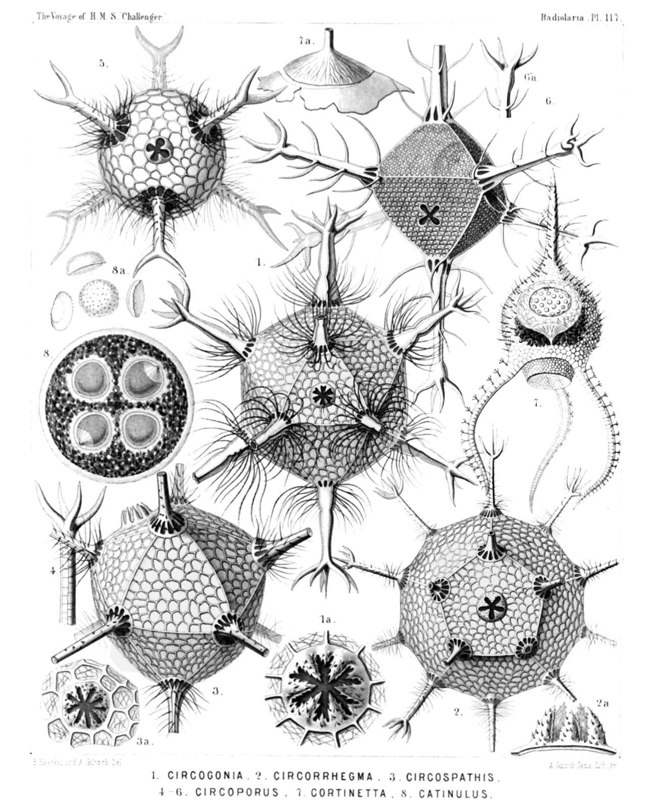 | |||||||||||||||||||||||||||||||||||||||||||||||||||||||||||||||||||
| PLATE 117. | |||
| Cannorrhaphida, Medusettida et Circoporida. | |||
| Diam. | Page. | ||
Fig. 1. Circogonia icosahedra, n. sp., | × | 80 | 1698 |
| The entire shell, with twelve radial tubes and twenty triangular faces. In the centre of one face is the mouth, with six teeth. | |||
| Fig. 1a. The mouth alone, with its six spinulate teeth, | × | 400 | |
| Fig. 2. Circorrhegma dodecahedra, n. sp., | × | 80 | 1699 |
| The entire shell, with twenty radial tubes and twelve pentagonal faces. In the centre of one face is the mouth, with five teeth. | |||
| Fig. 2a. The mouth alone, with its five spinulate teeth, seen in profile, | × | 200 | |
| Fig. 3. Circospathis novena, n. sp., | × | 100 | 1696 |
| The entire shell, with nine radial tubes and fourteen triangular faces. In one face (to the left above) is the mouth with nine teeth. | |||
| Fig. 3a. The mouth alone, with its nine spinulate teeth, | × | 150 | |
| Fig. 4. Circoporus hexastylus, n. sp., | × | 80 | 1695 |
| A single radial spine. | |||
| Fig. 5. Circoporus sexfurcus, n. sp., | × | 80 | 1694 |
| The entire spherical shell with six forked and ciliated radial tubes. In the centre the cruciform mouth with four teeth. | |||
| Fig. 6. Circoporus octahedrus, n. sp., | × | 300 | 1695 |
| The entire shell, with six verticillate radial tubes and eight triangular faces. In the centre of one face is the mouth, with four teeth. | |||
| Fig. 7. Cortinetta tripodiscus, n. sp., | × | 300 | 1667 |
| The entire shell with the enclosed central capsule, and the phæodium around the astropyle. | |||
| Fig. 7a. The astropyle, partly detached from the wall of the central capsule, seen in profile, | × | 800 | |
| Fig. 8. Catinulus quadrifidus, n. sp., | × | 80 | 1553 |
| A complete specimen, with four equal central capsules, united in a single spherical calymma. | |||
| Fig. 8a. Some single pieces of the skeleton, | × | 400 | |
PLATE 118.
Legion PHÆODARIA.
Order PHÆOGROMIA.
Family Medusettida.
| 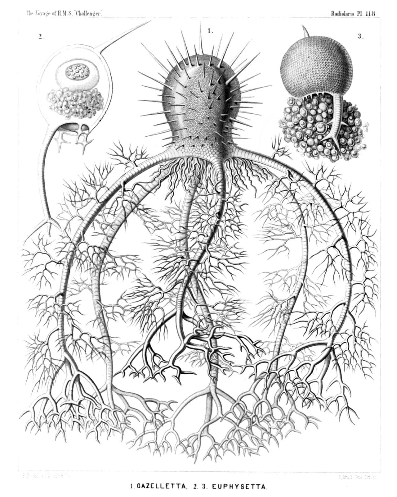 | |||||||||||||||||||||||||||
| PLATE 118. | |||
| Medusettida. | |||
| Diam. | Page. | ||
Fig. 1. Gazelletta melusina, n. sp., | × | 300 | 1674 |
| From the peristome of the thorny campanulate shell arise six large descending feet, which are studded with arborescent fragile lateral branches, and armed at the distal end with stouter dichotomous terminal branches. | |||
| Fig. 2. Euphysetta staurocodon, n. sp., | × | 300 | 1670 |
| The peristome of the ovate shell bears an odd large foot with three terminal branches and three cruciate rudimentary feet. In the upper part of the shell-cavity is visible the sphæroidal central capsule (containing a nucleus of half the size, with numerous nucleoli); in the lower half the dark pigment-masses of the green phæodium. | |||
| Fig. 3. Euphysetta amphicodon, n. sp., | × | 300 | 1670 |
| The shell-wall exhibits the regular alveolate structure. From the mouth are prominent large masses of the phæodium, which is more voluminous than the shell-cavity, and seems to contain nucleated cells. | |||
PLATE 119.
Legion PHÆODARIA.
Order PHÆOGROMIA.
Family Medusettida.
| 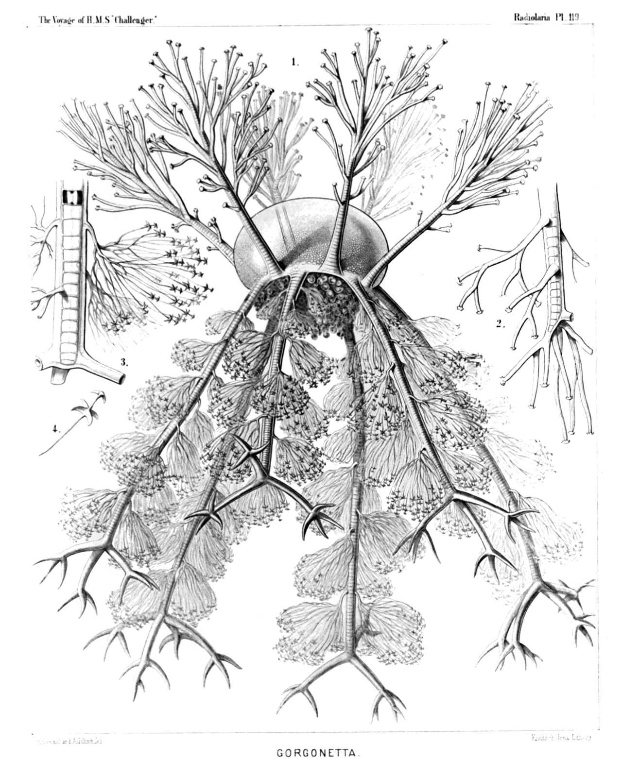 | ||||||||||||||||||||||||||||||||
| PLATE 119. | |||
| Medusettida. | |||
| Diam. | Page. | ||
Fig. 1. Gorgonetta mirabilis, n. sp., | × | 100 | 1674 |
| The entire body. From the margin of the cap-shaped shell arise six ascending arborescent feet and six alternating descending feet, which are covered with anchor-pencils and branched at the distal end. From the mouth of the delicately alveolate shell depend prominent parts of the dark voluminous phæodium. | |||
| Fig. 2. Gorgonetta mirabilis, n. sp., | × | 300 | 1674 |
| The distal end of an ascending foot; the branches bear a terminal spathilla with small recurved teeth. | |||
| Fig. 3. Gorgonetta mirabilis, n. sp., | × | 300 | 1674 |
| The distal end of a descending foot, with three lateral anchor-pencils and three terminal branches (broken off). Odd alveole contains an air-bubble. | |||
| Fig. 4. Gorgonetta mirabilis, n. sp., | × | 600 | 1674 |
| A single thread of an anchor-pencil with two quadridentate spathillæ, a larger proximal and a smaller distal (terminal). | |||
PLATE 120.
Legion PHÆODARIA.
Order PHÆOGROMIA.
Family Medusettida.
| 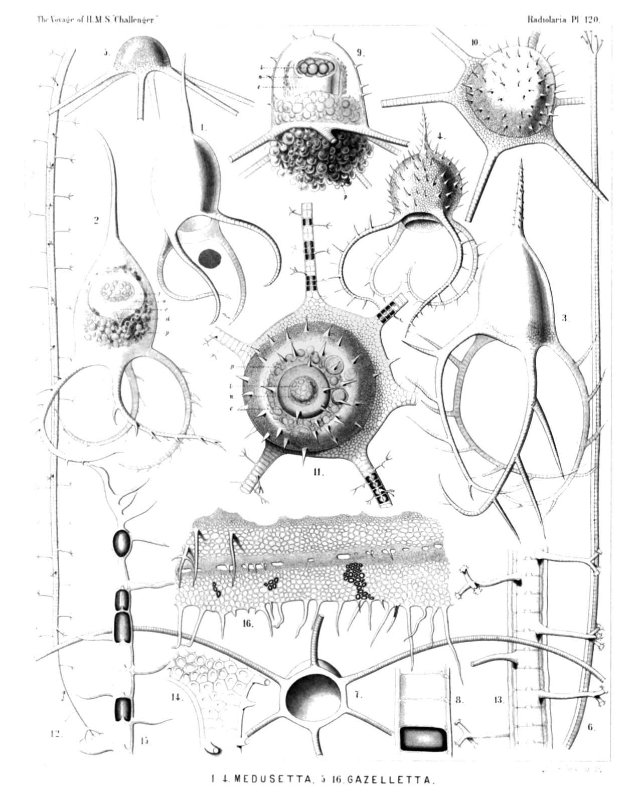 | ||||||||||||||||||||||||||||||||||||||||||||||||||||||||||||||||||||||||||||||||||||||||
| PLATE 120. | |||
| Medusettida. | |||
| Diam. | Page. | ||
Fig. 1. Medusetta codonium, n. sp., | × | 400 | 1668 |
| Fig. 2. Medusetta quadrigata, n. sp., | × | 400 | 1668 |
| The central capsule is visible in the upper half, the phæodium in the lower half of the shell-cavity. | |||
| Fig. 3. Medusetta tetranema, n. sp., | × | 400 | 1669 |
| Fig. 4. Medusetta craspedota, n. sp., | × | 400 | 1669 |
| Fig. 5. Gazelletta hexanema, n. sp., | × | 300 | 1671 |
| Fig. 6. Gazelletta bifurca, n. sp., | × | 300 | 1672 |
| A single alveolate foot. | |||
| Fig. 7. Gazelletta macronema, n. sp., | × | 200 | 1671 |
| Oral view of the shell. | |||
| Fig. 8. Gazelletta macronema, n. sp., | × | 800 | 1671 |
| Three joints of an alveolate foot. | |||
| Fig. 9. Gazelletta cyrtonema, n. sp., | × | 300 | 1671 |
| The upper part of the shell encloses the central capsule with its nucleus. The voluminous phæodium is prominent over the mouth. | |||
| Fig. 10. Gazelletta orthonema, n. sp., | × | 200 | 1671 |
| The central capsule and its nucleus are visible in the shell-cavity. | |||
| Fig. 11. Gazelletta schleinitzii, n. sp., | × | 400 | 1673 |
| Oblique apical view, with the enclosed central capsule, the nucleus of which contains numerous nucleoli. | |||
| Fig. 12. Gazelletta schleinitzii, n. sp., | × | 300 | 1673 |
| A single alveolate foot. | |||
| Fig. 13. Gazelletta trispathilla, n. sp., | × | 400 | 1673 |
| The middle part of a foot. | |||
| Fig. 14. Gazellatta robusta, n. sp., | × | 300 | 1673 |
| The base of a foot, exhibiting the pores of the alveoli. | |||
| Fig. 15. Gazelletta studeri, n. sp., | × | 400 | 1673 |
| The distal end of a foot; four alveoli filled up by air-bubbles. | |||
| Fig. 16. Gazelletta dendronema, n. sp., | × | 300 | 1674 |
| A part of the velum, seen from the inside. The alveoles are partly filled by air. | |||
PLATE 121.
Legion PHÆODARIA.
Order PHÆOCONCHIA.
Family Cœlodendrida.
| 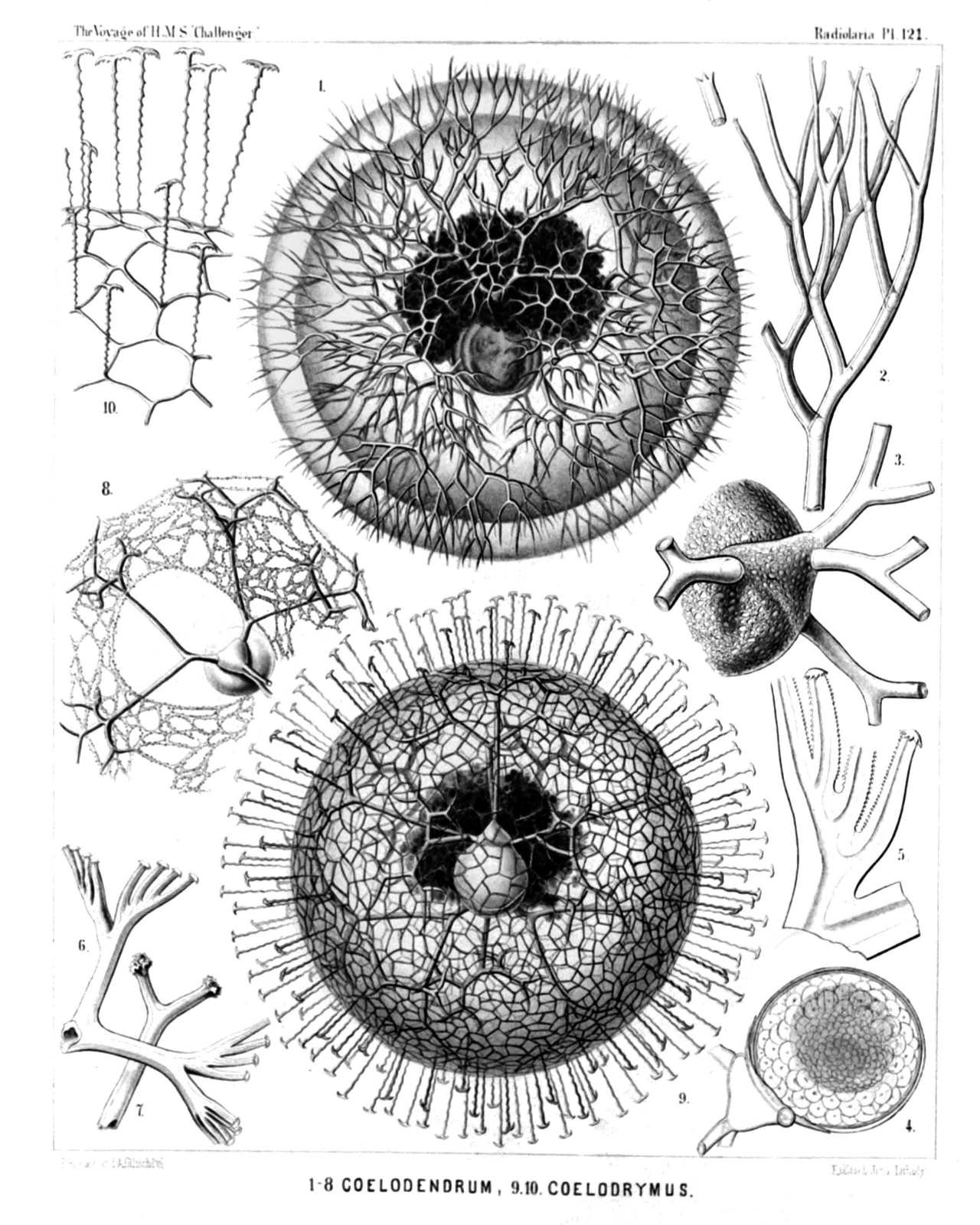 | ||||||||||||||||||||||||||||||||||||||||||||||||||||||||||||||
| PLATE 121. | |||
| Cœlodendrida. | |||
| Diam. | Page. | ||
Fig. 1. Cœlodendrum furcatissimum, n. sp., | × | 50 | 1735 |
| A complete specimen with the central capsule and the big phæodium. The spherical calymma envelops almost the entire skeleton. | |||
| Fig. 2. Cœlodendrum furcatissimum, n. sp., | × | 300 | 1735 |
| A distal branch with its terminal ramification. | |||
| Fig. 3. Cœlodendrum furcatissimum, n. sp., | × | 100 | 1735 |
| One valve of the shell, with its galea and the four hollow forked tubes arising from it. | |||
| Fig. 4. Cœlodendrum furcatissimum, n. sp., | × | 100 | 1735 |
| The central capsule with its nucleus; on the left side one valve of the closely enveloping shell (seen in vertical section), and its galea with the origin of the four tubes. | |||
| Fig. 5. Cœlodendrum serratum, n. sp., | × | 400 | 1737 |
| A flabellate terminal branch. | |||
| Fig. 6. Cœlodendrum flabellatum, n. sp., | × | 150 | 1737 |
| A flabellate terminal branch. | |||
| Fig. 7. Cœlodendrum spinosissimum, n. sp., | × | 300 | 1735 |
| Forked distal end of a terminal branch. | |||
| Fig. 8. Cœlodendrum cervicorne, n. sp., | × | 150 | 1736 |
| One valve of the shell, with its galea and the four tubes arising from it. A network of protoplasm connects the distal branches. | |||
| Fig. 9. Cœlodrymus ancoratus, n. sp., | × | 50 | 1738 |
| A complete specimen, with the central capsule and the enveloping phæodium. The surface of the spherical calymma is covered by a dense network, from which arise numerous, anchor-bearing, radial tubules. | |||
| Fig. 10. Cœlodrymus ancoratus, n. sp., | × | 150 | 1738 |
| A small piece of the superficial network of the skeleton, with the zigzag radial tubules arising from it, each of which bears an anchor with two recurved denticulate teeth on the distal end. | |||
PLATE 122.
Legion PHÆODARIA.
Order PHÆOCONCHIA.
Family Cœlographida.
| 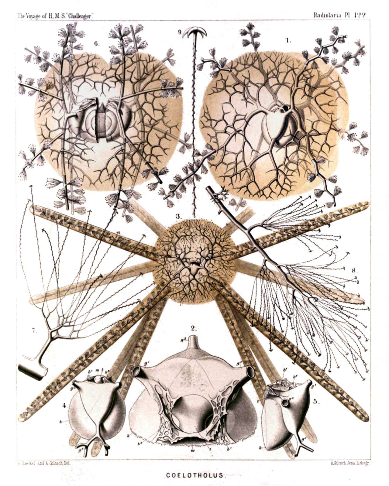 | |||||||||||||||||||||||||||||||||||||||||||||||||||||||||
| PLATE 122. | |||
| Cœlographida. | |||
| Diam. | Page. | ||
Fig. 1. Cœlotholus octonus, n. sp., | × | 30 | 1749 |
| The entire bivalved shell, seen obliquely from the dorsal and somewhat from the right side, enveloped by the yellowish calymma. | |||
| Fig. 2. Cœlotholus octonus, n. sp., | × | 100 | 1749 |
| One valve of the shell (h) with its large galea and the origin of the three styles. The base of the two lateral styles (g1, g2) is connected by two latticed lateral frenula (b1, b2) with the mouth (m) of the rhinocanna (t). The odd style (g3) is free. | |||
| Fig. 3. Cœlothauma duodenum, n. sp., | × | 20 | 1750 |
| The entire shell, seen from the dorsal side. The long styles are enveloped by the yellowish calymma. | |||
| Fig. 4. Cœlothauma duodenum, n. sp., | × | 80 | 1750 |
| One valve of the shell (h), seen from the apical side; t, rhinocanna; m, its mouth; b1, b2, the two lateral frenula; g1, g2, the two paired styles; g3, the odd style. | |||
| Fig. 5. Cœlothauma duodenum, n. sp., | × | 80 | 1750 |
| One valve of the shell, seen in profile. Characters as in fig. 4. | |||
| Fig. 6. Cœlothamnus bivalvis, n. sp., | × | 30 | 1751 |
| The entire shell, enveloped by the yellowish calymma, seen from the left side; between the two valves is the central capsule, with nucleus and astropyle. | |||
| Fig. 7. Cœlothamnus bivalvis, n. sp., | × | 100 | 1751 |
| A single lateral anchor-pencil. | |||
| Fig. 8. Cœlothamnus bivalvis, n. sp., | × | 200 | 1751 |
| Distal end of a style, with its anchor-pencils. | |||
| Fig. 9. Cœlothamnus bivalvis, n. sp., | × | 400 | 1751 |
| A single anchor-thread, with its quadridentate terminal spathilla. | |||
PLATE 123.
Legion PHÆODARIA.
Order PHÆOCONCHIA.
Family Concharida.
| 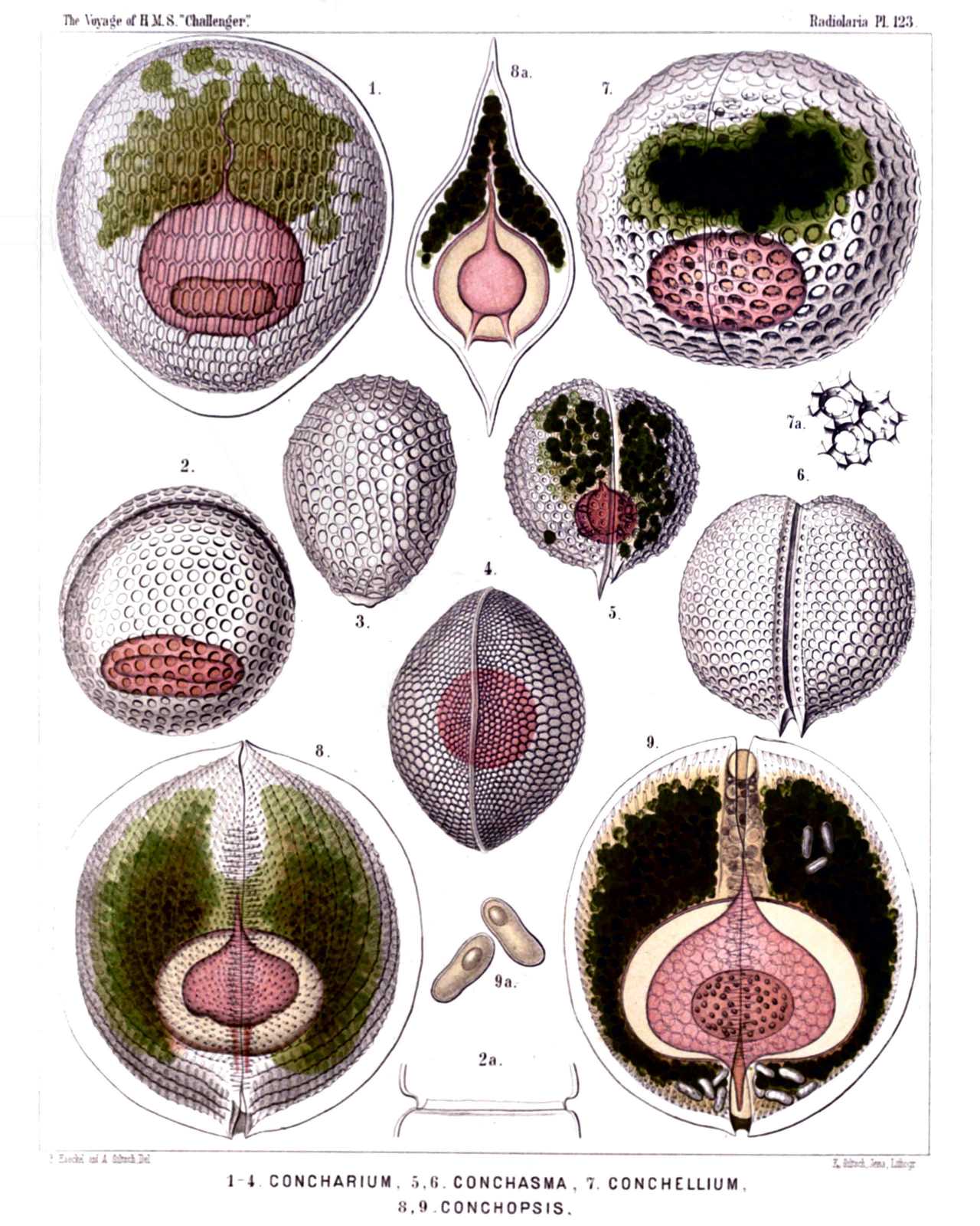 | |||||||||||||||||||||||||||||||||||||||||||||||||||||||||||
| PLATE 123. | |||
| Concharida. | |||
| Diam. | Page. | ||
(The central capsule is coloured red in the figures of this plate, the phæodium green). | |||
| Fig. 1. Concharium diatomeum, n. sp., | 1717 | ||
| Dorsal view. The central capsule (red) exhibits above the anterior tubular main-opening (astropyle), and below the two small posterior lateral openings (right and left parapylæ). | |||
| Fig. 2. Concharium bivalvum, n. sp., | × | 150 | 1717 |
| Dorsal view. The central capsule is visible in the lower part, the margin of the two valves in the upper part of the figure. | |||
| Fig. 2a exhibits the two smooth lateral margins of the valves, catching into one another. (Lateral view). | |||
| Fig. 3. Concharium nucula, n. sp., | 1717 | ||
| The dorsal valve alone, seen from the outside. | |||
| Fig. 4. Concharium bacillarium, n. sp., | 1718 | ||
| Lateral view from the smooth margin, by which the two valves are united. | |||
| Fig. 5. Conchasma radiolites, n. sp., | × | 300 | 1719 |
| Lateral view. In the aboral half of the shell-cavity lies the red central capsule, in the oral half the green phæodium. | |||
| Fig. 6. Conchasma sphærulites, n. sp., | × | 300 | 1719 |
| Lateral view. On the aboral pole the two horns of the hinge. | |||
| Fig. 7. Conchellium tridacna, n. sp., | × | 200 | 1720 |
| Oblique lateral view (from the right and ventral side). | |||
| Fig. 7a. Three pores of the same, with their hexagonal frames and six internal denticles, | × | 400 | |
| Fig. 8. Conchopsis carinata, n. sp., | × | 150 | 1725 |
| Lateral view, from the left side. | |||
| Fig. 9. Conchopsis lenticula, n. sp., | × | 150 | 1726 |
| Lateral view, from the right side. The two membranes of the central capsule are separated by a wide interval in this and the preceding figure. The nucleus contains numerous nucleoli. | |||
| Fig. 9a. Two of the peculiar cells, which are contained in the green phæodium in large numbers, | × | 400 | |
PLATE 124.
Legion PHÆODARIA.
Order PHÆOCONCHIA.
Family Concharida.
| 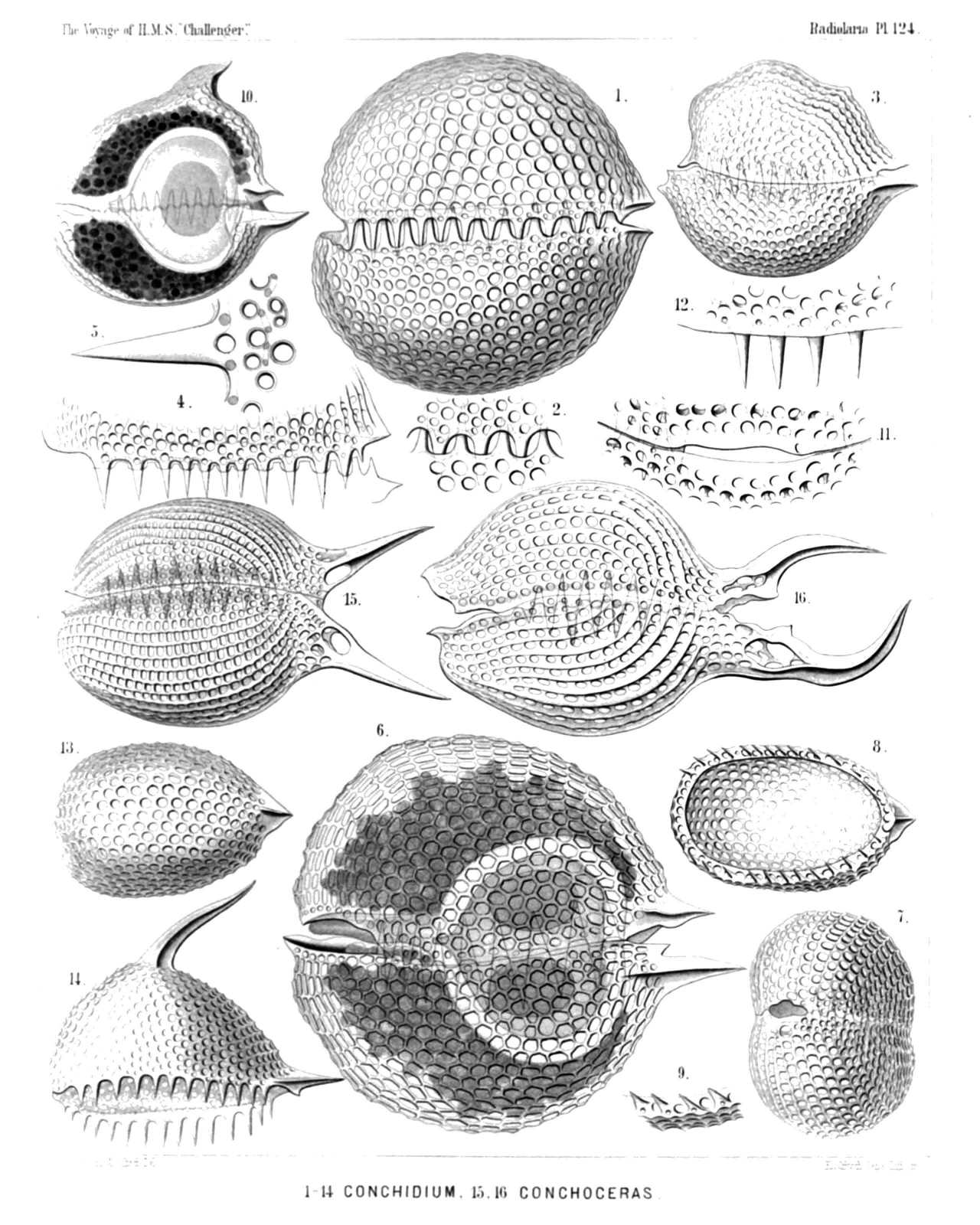 | ||||||||||||||||||||||||||||||||||||||||||||||||||||||||||||||||||||||||||||||||||||||||||||
| PLATE 124. | |||
| Concharida. | |||
| Diam. | Page. | ||
Fig. 1. Conchidium terebratula, n. sp., | × | 400 | 1721 |
| Lateral view, from the left side. | |||
| Fig. 2. Conchidium terebratula, n. sp., | × | 800 | 1721 |
| A piece of the frontal girdle-fissure, with the teeth of both valves catching into one another. | |||
| Fig. 3. Conchidium rhynchonella, n. sp., | × | 200 | 1722 |
| Lateral view, from the left side. | |||
| Fig. 4. Conchidium leptæna, n. sp., | × | 300 | 1722 |
| Girdle-fissure with the teeth, seen from the left side. | |||
| Fig. 5. Conchidium leptæna, n. sp., | × | 800 | 1722 |
| A single tooth with its base. | |||
| Fig. 6. Conchidium thecidium, n. sp., | × | 300 | 1721 |
| Lateral view, from the left side. In the oral part of the shell-cavity the dark phæodium, in the aboral part the central capsule with two nuclei (a dorsal and a ventral). | |||
| Fig. 7. Conchidium argiope, n. sp., | × | 300 | 1722 |
| Oblique oral view (half from the anterior, half from the left side). | |||
| Fig. 8. Conchidium argiope, n. sp., | × | 300 | 1722 |
| Dorsal valve, from below. | |||
| Fig. 9. Conchidium argiope, n. sp., | × | 600 | 1722 |
| A piece of the valve margin, with four teeth. | |||
| Fig. 10. Conchonia diodon, n. sp., | × | 200 | 1723 |
| Lateral view, from the left side. In the anterior part of the shell-cavity the dark phæodium, in the posterior part the central capsule with the nucleus. The two valves are connected at the posterior hinge by a ligament (to the right in the figure). | |||
| Fig. 11. Conchonia diodon, n. sp., | × | 400 | 1723 |
| Mouth of the shell, with its two lips, seen from the oral pole. | |||
| Fig. 12. Conchonia diodon, n. sp., | × | 400 | 1723 |
| A piece of the valve-margin, with four teeth. | |||
| Fig. 13. Conchonia triodon, n. sp., | × | 300 | 1724 |
| Ventral valve, seen from the lower face. | |||
| Fig. 14. Conchonia triodon, n. sp., | × | 300 | 1724 |
| Dorsal valve, seen from the left side. | |||
| Fig. 15. Conchoceras caudatum, n. sp., | × | 300 | 1727 |
| Lateral view, from the left side. | |||
| Fig. 16. Conchoceras cornutum, n. sp., | × | 200 | 1728 |
| Lateral view, from the left side. | |||
PLATE 125.
Legion PHÆODARIA.
Order PHÆOCONCHIA.
Family Concharida.
| 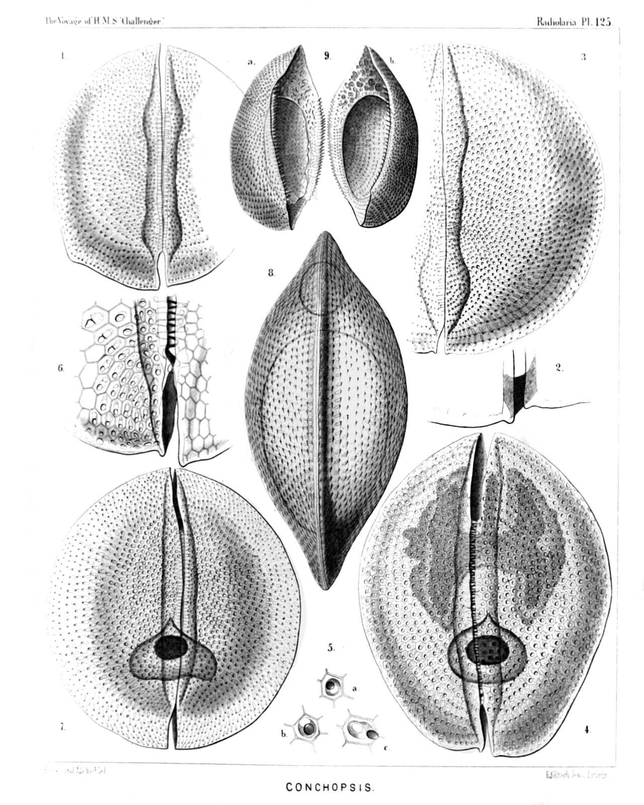 | |||||||||||||||||||||||||||||||||||||||||||||||||||||||||
| PLATE 125. | |||
| Concharida. | |||
| Diam. | Page. | ||
Fig. 1. Conchopsis aspidium, n. sp., | × | 150 | 1726 |
| Lateral view, from the left side. | |||
| Fig. 2. Conchopsis aspidium, n. sp., | × | 300 | 1726 |
| The hinge of another specimen, in which the two valves are connected by a ligament (as in figs. 8 and 9, Pl. 123). | |||
| Fig. 3. Conchopsis orbicularis, n. sp., | × | 200 | 1725 |
| Lateral view, from the left side. | |||
| Fig. 4. Conchopsis navicula, n. sp., | × | 150 | 1727 |
| Lateral view, from the right side. In the lower (posterior) half of the figure is visible the central capsule with its dark nucleus, in the upper (anterior) half the phæodium with two broad sagittal wings. | |||
| Fig. 5. Conchopsis navicula, n. sp., | × | 400 | 1727 |
| Three single pores with their hexagonal external frame and the dilated internal ovate or ampullaceous channel. | |||
| Fig. 6. Conchopsis navicula, n. sp., | × | 400 | 1727 |
| Hinge of the shell, from the right side. | |||
| Fig. 7. Conchopsis compressa, n. sp., | × | 150 | 1725 |
| Lateral view from the left side. The triangular central capsule with the dark nucleus is visible. | |||
| Fig. 8. Conchopsis compressa, n. sp., | × | 150 | 1725 |
| Dorsal view of the upper valve with its keel. | |||
| Fig. 9. Conchopsis pilidium, n. sp., | × | 80 | 1726 |
| The two valves separated and seen obliquely, half from the lateral, half from the internal side. The inner opening of each valve is bordered and partly closed by a broad horizontal velum or diaphragm like the deck of a boat. | |||
PLATE 126.
Legion PHÆODARIA.
Order PHÆOCONCHIA.
Family Cœlographida.
| 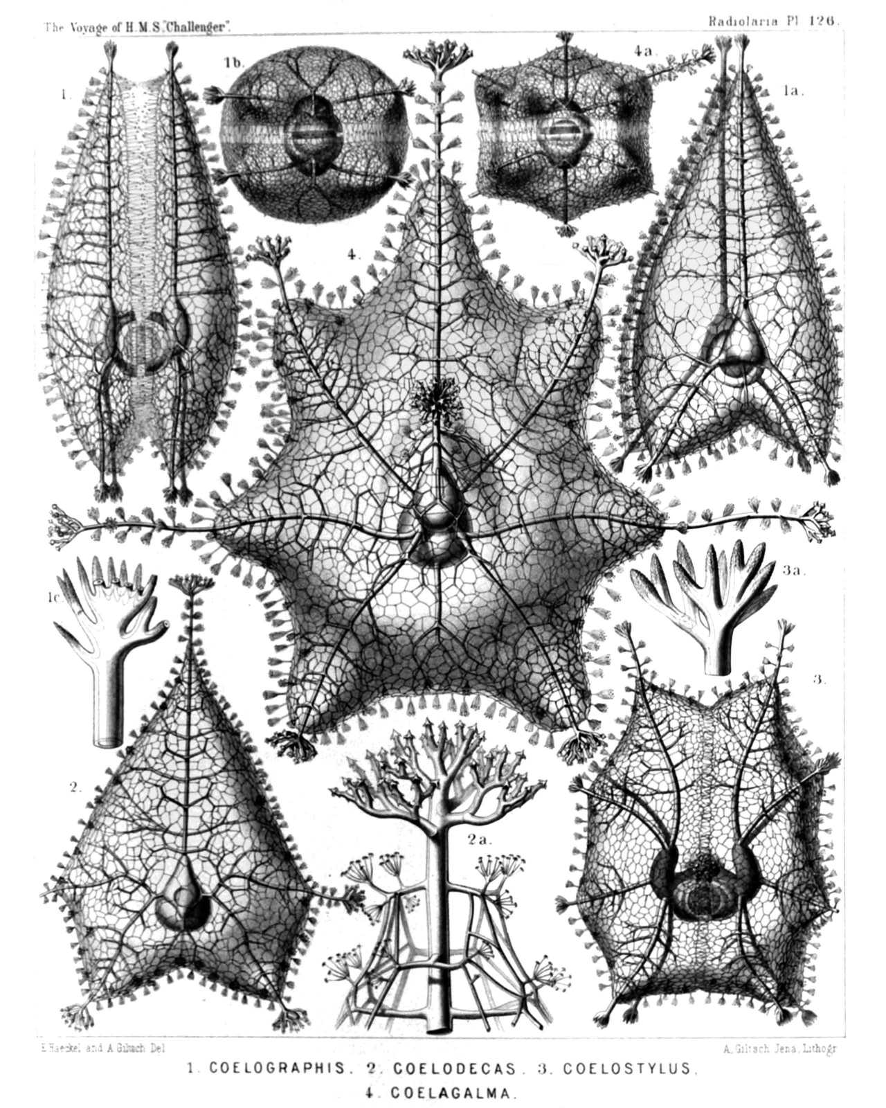 | ||||||||||||||||||||||||||||||||||||||||||||||||||
| PLATE 126. | |||
| Cœlographida. | |||
| Diam. | Page. | ||
Figs. 1-1c. Cœlographis regina, n. sp., | 1752 | ||
| Fig. 1. Lateral view. The central capsule it visible between the two valves of the inner shell, the galeæ of which are filled by the phæodium, | × | 20 | |
| Fig. 1a. Dorsal view (somewhat obliquely from the left side). The galeæ appear triangular, | × | 20 | |
| Fig. 1b. Basal view, | × | 20 | |
| Fig. 1c. Distal end of a style, | × | 300 | |
| Figs. 2-2b. Cœlodecas sagittaria, n. sp., | 1755 | ||
| Fig. 2. One valve of the shell, seen from the outside, | × | 30 | |
| Fig. 2a. Distal end of a style, | × | 300 | |
| Figs. 3-3a. Cœlostylus bisenarius, n. sp., | 1756 | ||
| Fig. 3. Lateral view of the bivalved shell. The central capsule is visible between the two valves of the inner shell, the galeæ of which are filled by the phæodium, | × | 20 | |
| Fig. 3a. Distal end of a style, | × | 300 | |
| Figs. 4-4a. Cœlagalma mirabile, n. sp., | 1759 | ||
| Fig. 4. Dorsal view of the bivalved shell, | × | 30 | |
| Fig. 4a. Basal view of the bivalved shell, | × | 10 | |
PLATE 127.
Legion PHÆODARIA.
Order PHÆOCONCHIA.
Family Cœlographida.
| 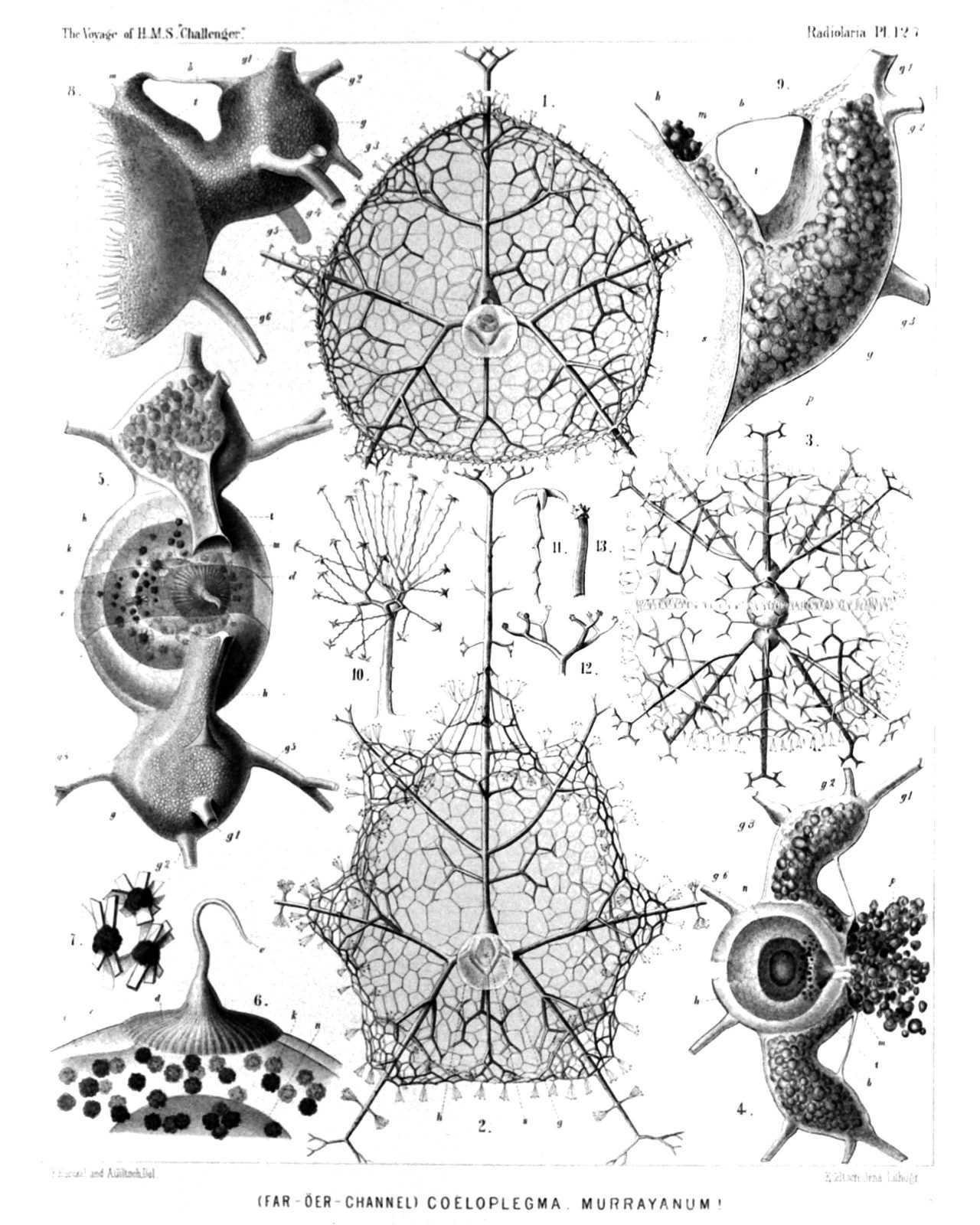 | |||||||||||||||||||||||||||||||||||||||||||||||||||||||||||||||||||||||||||||
| PLATE 127. | |||
| Cœlographida. | |||
| Diam. | Page. | ||
Fig. 1. Cœloplegma murrayanum, n. sp., | × | 40 | 1757 |
| One valve of the bivalved shell, seen from the inside, of the usual ovate form. | |||
| Fig. 2. Cœloplegma murrayanum, n. sp., | × | 40 | 1757 |
| One valve of the bivalved shell, seen from the inside, of the rarer polyhedral form, which may be distinguished as a different species (Cœloplegma tritonis, compare p. 1758). h, hemispherical inner valve; g, galea; s, its base. | |||
| Fig. 3. Cœloplegma murrayanum, n. sp., | × | 40 | 1757 |
| The entire shell, seen from the base of the aboral pole (dorsal and ventral valve connected by delicate teeth, catching into one another). | |||
| Fig. 4. Cœloplegma murrayanum, n. sp., | × | 100 | 1757 |
| h, The two hemispherical inner valves of the shell, seen from the right side; n, the central nucleus inside the central capsule; d, the astropyle; g, the galea; t, the nasal tube, arising from its base; m, its mouth; p, the phæodium, which is partly thrown out by the nasal openings, filling up the galea and nasal tube. | |||
| Fig. 5. Cœloplegma murrayanum, n. sp., | × | 200 | 1757 |
| h, The two hemispherical inner valves of the shell and the enclosed central capsule, seen from the oral side; d, the radiate operculum of the astropyle, seen in the frontal fissure between the two valves; n, the nucleus; k, the crystals; g, galea; g1-g5, the styles arising from the galea; t, nasal tube; m, mouth of it. | |||
| Fig. 6. Cœloplegma murrayanum, n. sp., | × | 600 | 1757 |
| Oral part of a central capsule, in profile. o, Opening of the proboscis; d, radiate operculum of the astropyle, which gives rise to the proboscis; e, the outer, i, the inner membrane of the capsule; k, groups of crystals; n, nucleus. | |||
| Fig. 7. Cœloplegma murrayanum, n. sp., | × | 1000 | 1757 |
| Three single groups of crystals, taken from the central capsule. | |||
| Fig. 8. Cœloplegma murrayanum, n. sp., | × | 300 | 1757 |
| One inner valve of the shell, in profile. h, hemispherical valve; g, galea; g1-g5, the tubes arising from it; t, rhinocanna or nasal tube; m, its mouth; b, frenulum. | |||
| Fig. 9. Cœloplegma murrayanum, n. sp., | × | 400 | 1757 |
| g, The galea; t, rhinocanna of one inner valve; m, its mouth; p, phæodella filling up both the galea and the mouth of the rhinocanna. g1-g5, the styles arising from the galea; s, sieve-plate, which separates the cavity of the galea from the hemispherical valve (h). View in profile. | |||
| Fig. 10. Cœloplegma murrayanum, n. sp., | × | 300 | 1757 |
| An anchor-pencil of the outer shell. | |||
| Fig. 11. Cœloplegma murrayanum, n. sp., | × | 1000 | 1757 |
| A single anchor-thread of a pencil. | |||
| Fig. 12. Cœloplegma murrayanum, n. sp., | × | 300 | 1757 |
| Terminal branches of a style. | |||
| Fig. 13. Cœloplegma murrayanum, n. sp., | × | 1000 | 1757 |
| A single terminal branch of a style. | |||
PLATE 128.
Legion PHÆODARIA.
Order PHÆOCONCHIA.
Family Cœlographida.
| 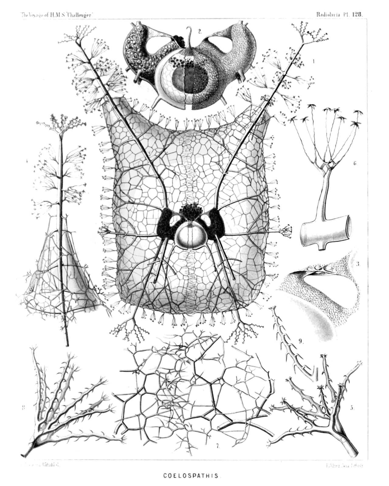 | |||||||||||||||||||||||||||||||||||||||||||||||||||||||||
| PLATE 128. | |||
| Cœlographida. | |||
| Diam. | Page. | ||
Fig. 1. Cœlospathis ancorata, n. sp., | × | 50 | 1754 |
| Lateral view of the entire shell. The central capsule is visible between the two valves of the inner shell. The galeæ and rhinocannæ of the two inner valves are filled up by the black phæodium. | |||
| Fig. 2. Cœlospathis ancorata, n. sp., | × | 100 | 1754 |
| The two valves of the inner shell; the galeæ and rhinocannæ of which are filled up by the black phæodium. Between the mouth of the two rhinocannæ is prominent the proboscis of the astropyle, arising from the radiate operculum of the central capsule. The latter contains numerous crystals and a big dark nucleus. Lateral view. | |||
| Fig. 3. Cœlospathis ancorata, n. sp., | × | 200 | 1754 |
| The rhinocanna or the nasal tube of one valve, and the latticed frenulum which connects its mouth with the top of the galea. | |||
| Fig. 4. Cœlospathis ancorata, n. sp., | × | 80 | 1754 |
| Distal end of a style. | |||
| Fig. 5. Cœlospathis ancorata, n. sp., | × | 200 | 1754 |
| Terminal branches of a style. | |||
| Fig. 6. Cœlospathis ancorata, n. sp., | × | 600 | 1754 |
| Lateral branch of a style, with an anchor-pencil. | |||
| Fig. 7. Cœlospathis ancorata, n. sp., | × | 300 | 1754 |
| The lateral margins of the latticed valves of the outer shell or mantle, catching into one another, without being connected directly. | |||
| Fig. 8. Cœlospathis octostyla, n. sp., | × | 300 | 1754 |
| Terminal branches of a style. | |||
| Fig. 9. Cœlospathis octodactyla, n. sp., | × | 400 | 1755 |
| A single terminal branch of a style. | |||
PLATE 129.
Legion ACANTHARIA.
Orders ACTINELLIDA et ACANTHONIDA.
Families Astrolophida, Litholophida, Chiastolida et Astrolonchida.
| 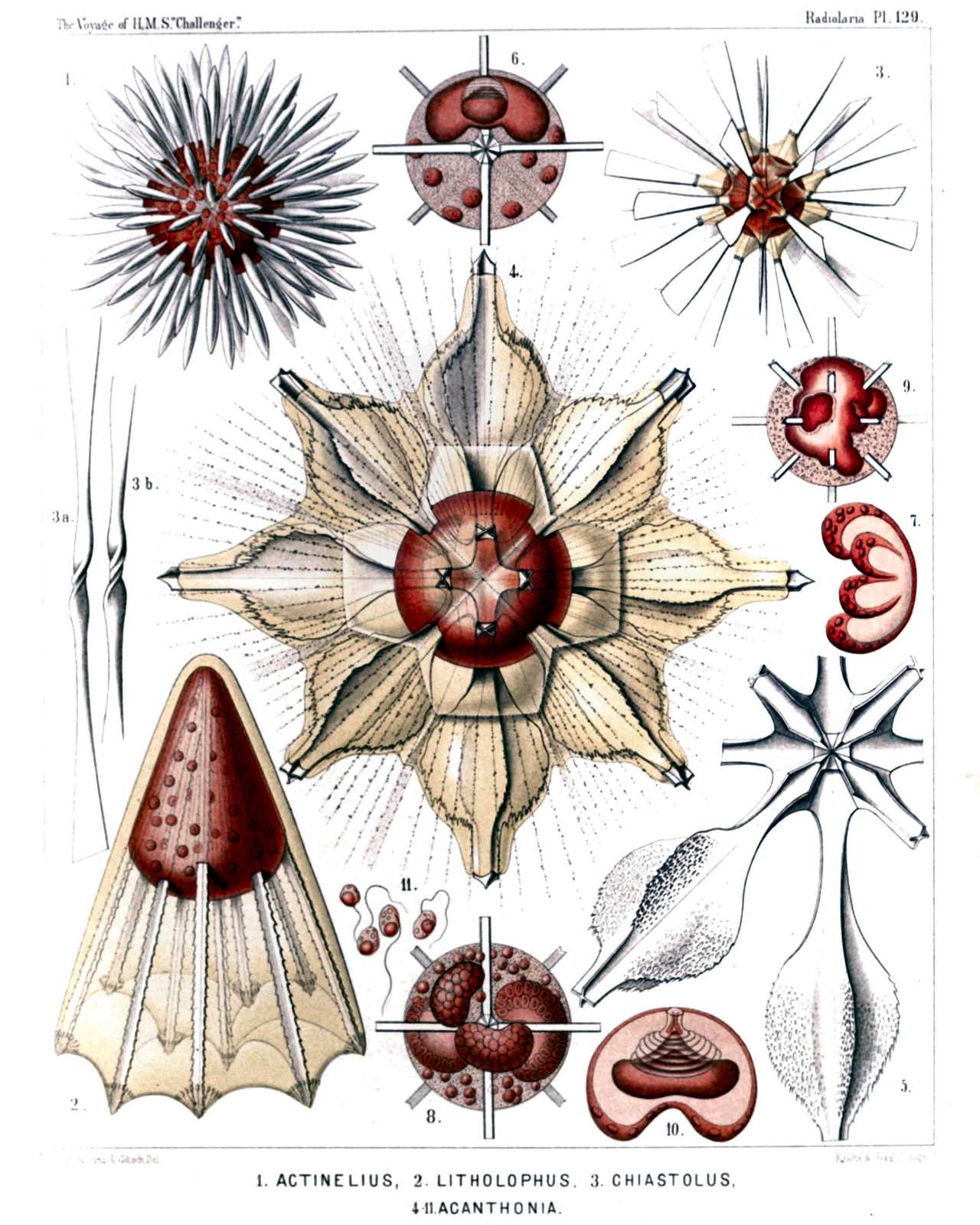 | ||||||||||||||||||||||||||||||||||||||||||||||||||||||||||||||||||||||
| PLATE 129. | |||
| Astrolophida, Litholophida, Chiastolida et Astrolonchida. | |||
| Diam. | Page. | ||
Fig. 1. Actinelius primordialis, n. sp., | × | 100 | 730 |
| The red central capsule, coloured by carmine, contains numerous intensely stained nuclei. | |||
| Fig. 2. Litholophus decapristis, n. sp., | × | 300 | 735 |
| The conical central capsule contains numerous nuclei. The calymma exhibits on the distal end of each spine a coronet of myophriscs. | |||
| Fig. 3. Chiastolus amphicopium, n. sp., | × | 150 | 738 |
| Sixteen diametral spines pierce the spherical, red coloured capsule. The conical sheets of the calymma bear myophriscs. | |||
| Figs. 3a, 3b. Two isolated diametral spines exhibiting the peculiar spiral revolution at their central part, | × | 300 | |
| Fig. 4. Xiphacantha ciliata, n. sp., | × | 300 | 761 |
| The spherical central capsule is coloured red. The yellowish calymma envelops the radial spines completely. The polygonal network of lines, in which the radiating pseudopodia are symmetrically arranged, is partly visible. | |||
| Fig. 5. Xiphacantha ciliata, n. sp., | × | 300 | 761 |
| The central part of the skeleton, exhibiting the central junction of the radial spines. | |||
| Fig. 6. Acanthometron dolichoscion, n. sp., | × | 300 | 743 |
| Central capsule of a young specimen; in its upper half the peculiar kidney-shaped nucleus is visible, with its invagination; in the lower half some nucleated yellow cells are visible (intracapsular xanthellæ). These and the nucleus are stained by carmine. | |||
| Fig. 7. Acanthometron dolichoscion, n. sp., | × | 300 | 743 |
| Cleavage of an isolated nucleus, with four buds. | |||
| Fig. 8. Acanthometron dolichoscion, n. sp., | × | 300 | 743 |
| A central capsule with four large budding nuclei; and numerous small spherical nuclei produced by gemmation. | |||
| Fig. 9. Acanthonia tetracopa, n. sp., | × | 400 | 749 |
| Central capsule of s young specimen, with a large, irregularly lobate nucleus. | |||
| Fig. 10. Acanthonia tetracopa, n. sp., | × | 400 | 749 |
| An isolated nucleus, exhibiting the peculiar invagination, with its circular folds, and the connection with the flatly conical nucleolus. | |||
| Fig. 11. Acanthonia tetracopa, n. sp., | × | 800 | 749 |
| Four flagellate spores. | |||
PLATE 130.
Legion ACANTHARIA.
Order ACANTHONIDA.
Family Astrolonchida.
| 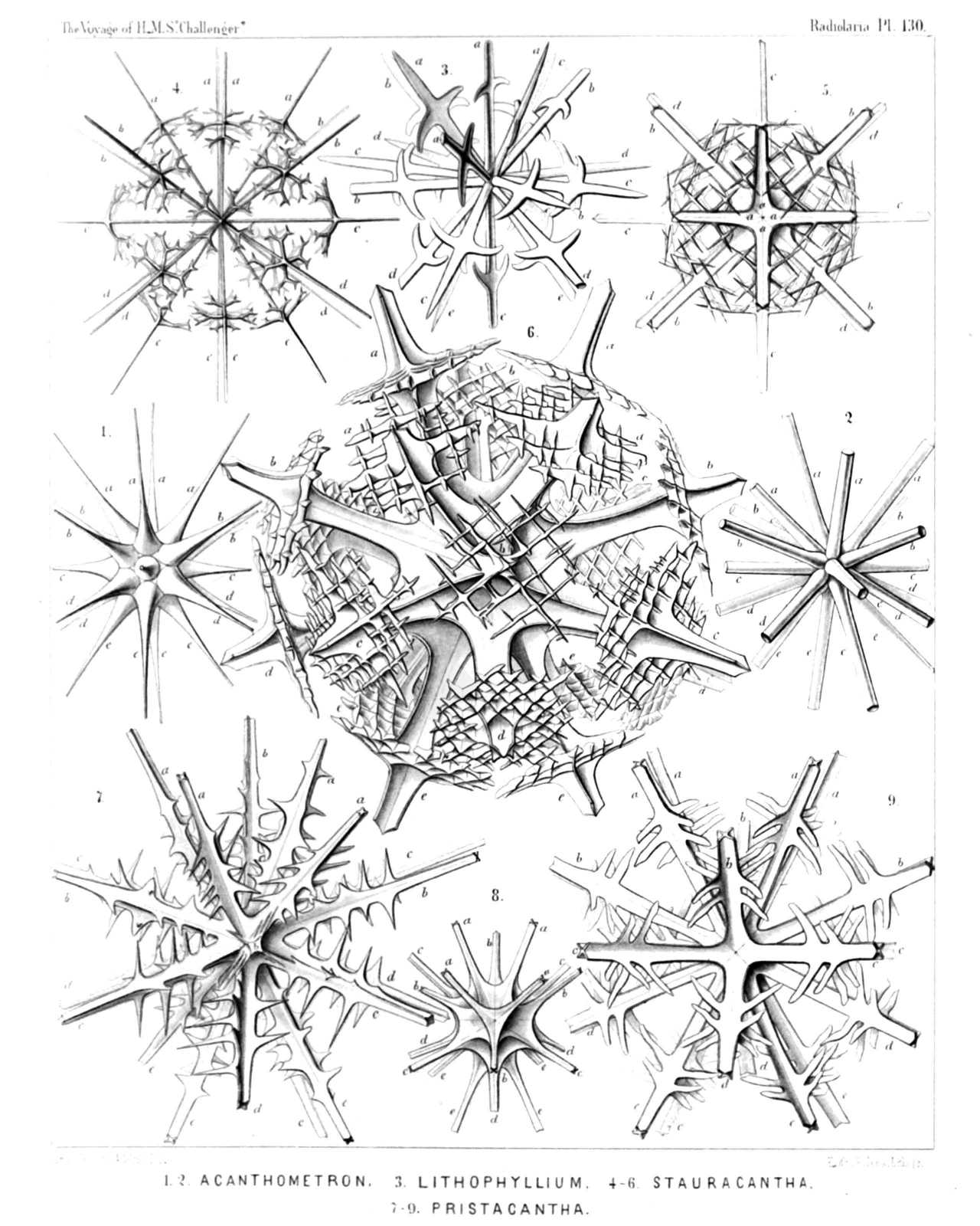 | ||||||||||||||||||||||||||||||||||||||||||||||||||||||||||||||||||
| PLATE 130. | |||
| Astrolonchida. | |||
| Diam. | Page. | ||
Fig. 1. Acanthometron bulbiferum, n. sp., | × | 300 | 745 |
| Fig. 2. Acanthometron cylindricum, n. sp., | × | 200 | 743 |
| Fig. 3. Lithophyllium gladiatum, n. sp., | × | 200 | 754 |
| Fig. 4. Stauracantha quadrifurca, n. sp., | × | 300 | 764 |
| Fig. 5. Stauracantha orthostaura, n. sp., | × | 200 | 762 |
| Fig. 6. Phatnacantha icosaspis, n. sp., | × | 400 | 765 |
| Fig. 7. Pristacantha polyodon, n. sp., | × | 300 | 766 |
| Fig. 8. Pristacantha dodecodon, n. sp., | × | 300 | 766 |
| Only the central parts and the leaf-cross. | |||
| Fig. 9. Pristacantha octodon, n. sp., | × | 200 | 765 |
PLATE 131.
Legion ACANTHARIA.
Order ACANTHONIDA.
Family Quadrilonchida.
| 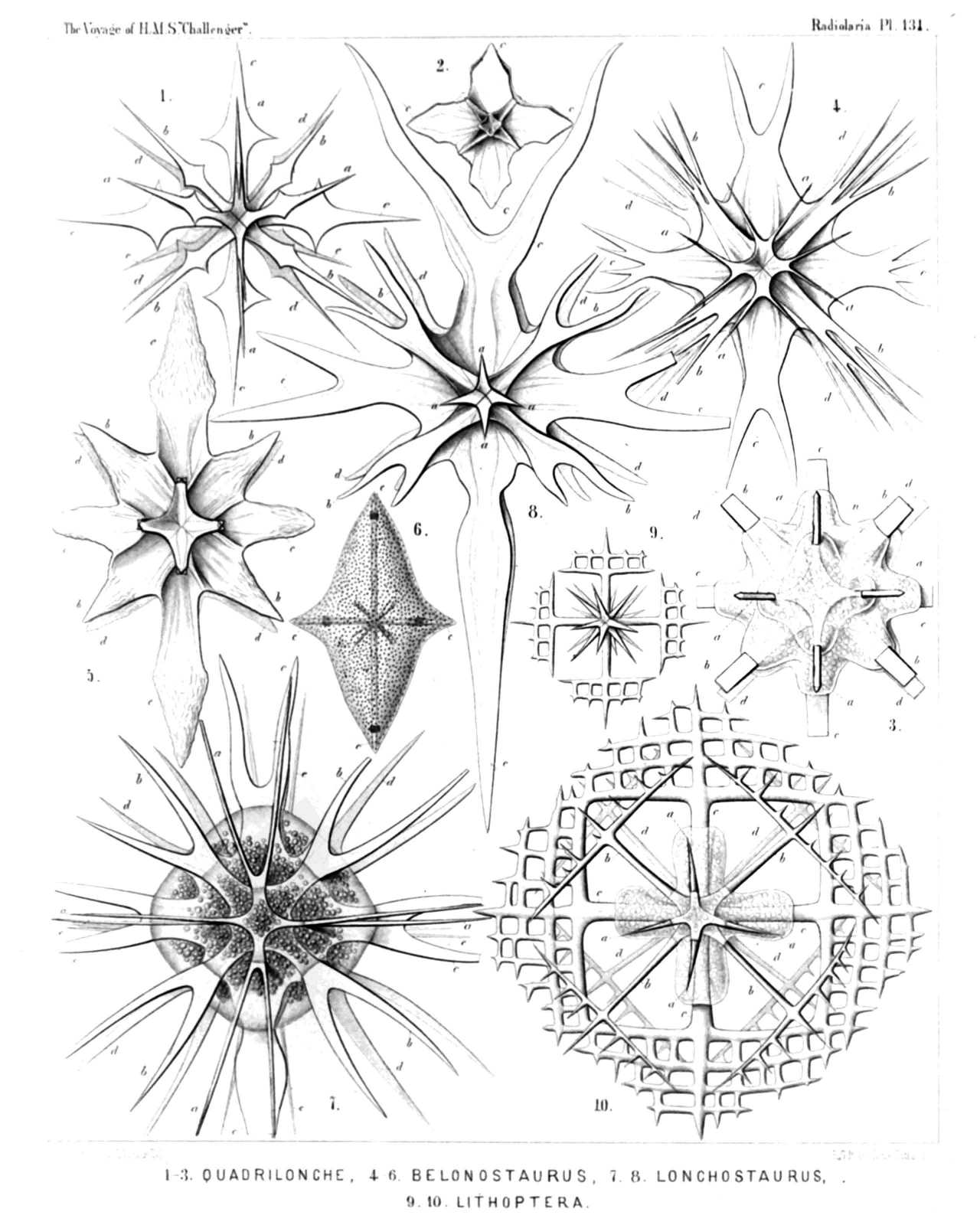 | |||||||||||||||||||||||||||||||||||||||||||||||||||||||||||||||||||||||||
| PLATE 131. | |||
| Quadrilonchida. | |||
| Diam. | Page. | ||
Fig. 1. Quadrilonche mesostaura, n. sp., | × | 300 | 777 |
| Fig. 2. Quadrilonche platystaura, n. sp., | × | 100 | 777 |
| Fig. 3. Xiphoptera dodecactena, n. sp., | × | 200 | 778 |
| The central capsule with the central part of the skeleton. | |||
| Fig. 4. Lonchostaurus bifurcus, n. sp., | × | 300 | 773 |
| Fig. 5. Lonchostaurus crystallinus, n. sp., | × | 400 | 773 |
| Fig. 6. Lonchostaurus rhomboides, n. sp., | × | 200 | 772 |
| The radial spines are completely enclosed in the rhombic calymma, the surface of which is covered with small plates, similar to those in the shell of the Sphærocapsida. | |||
| Fig. 7. Zygostaurus amphithectus, n. sp., | × | 300 | 774 |
| The square central capsule envelops the half skeleton. | |||
| Fig. 8. Zygostaurus sagittalis, n. sp., | × | 300 | 775 |
| Fig. 9. Lithoptera tetraptera, n. sp., | × | 300 | 779 |
| Fig. 10. Lithoptera quadrata, n. sp., | × | 300 | 780 |
| The central part of the skeleton is enclosed by the four-lobed central capsule. | |||
PLATE 132.
Legion ACANTHARIA.
Orders ACTINELLIDA et ACANTHONIDA.
Families Astrolophida, Astrolonchida et Amphilonchida.
| 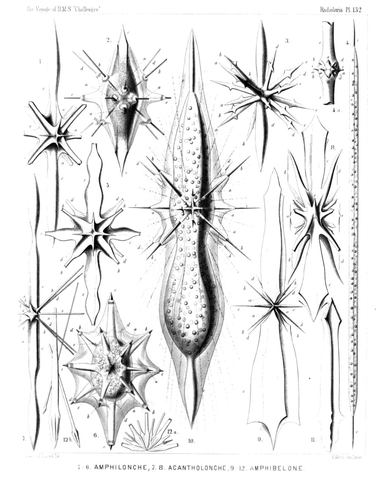 | |||||||||||||||||||||||||||||||||||||||||||||||||||||||||||||||||||||||||||||||||||||||||
| PLATE 132. | |||
| Astrolophida, Astrolonchida et Amphilonchida. | |||
| Diam. | Page. | ||
Fig. 1. Amphilonche lanceolata, n. sp., | × | 300 | 783 |
| Fig. 2. Amphilonche hydrotomica, n. sp., | × | 300 | 786 |
| The spindle-shaped central capsule is filled up with small granules. The clear calymma forms conical sheaths for the spines, with myophriscs. | |||
| Fig. 3. Amphilonche diodon, n. sp., | × | 300 | 783 |
| Fig. 4. Amphilonche concreta, n. sp., | × | 100 | 787 |
| A complete specimen with the cylindrical central capsule. | |||
| Fig. 4a. Central part of the skeleton, | × | 400 | |
| Fig. 5. Amphilonche violina, n. sp., | × | 300 | 787 |
| Fig. 6. Amphilonche conica, n. sp., | × | 300 | 785 |
| The ellipsoidal central capsule contains numerous nuclei and is enclosed by the calymma. The conical sheaths of the latter include the radial spines completely and exhibit coronets of myophriscs. | |||
| Fig. 7. Acantholonche amphipolaris, n. sp., | × | 200 | 790 |
| Fig. 8. Acantholonche peripolaris, n. sp., | × | 300 | 791 |
| Fig. 9. Amphibelone pyramidata, n. sp., | × | 300 | 789 |
| Fig. 10. Amphibelone cultellata, n. sp., | × | 400 | 789 |
| The central capsule contains numerous spherical nuclei and is enclosed by the hyaline calymma, which forms conical sheaths around the spines. | |||
| Fig. 11. Stauracantha johannis, n. sp., | × | 400 | 763 |
| Basal part of a radial spine, exhibiting the peculiar torsion of the basal leaf-cross and the central apex. | |||
| Fig. 12. Astrolophus solaris, n. sp., | × | 200 | 732 |
| Fig. 12a. A group of larger and smaller radial spines united in the centre. | |||
| Fig. 12b. Three isolated spines (one larger and two smaller), | × | 200 | |
PLATE 133.
Legion ACANTHARIA.
Order SPHÆROPHRACTA.
Families Sphærocapsida, Dorataspida et Phractopeltida.
| 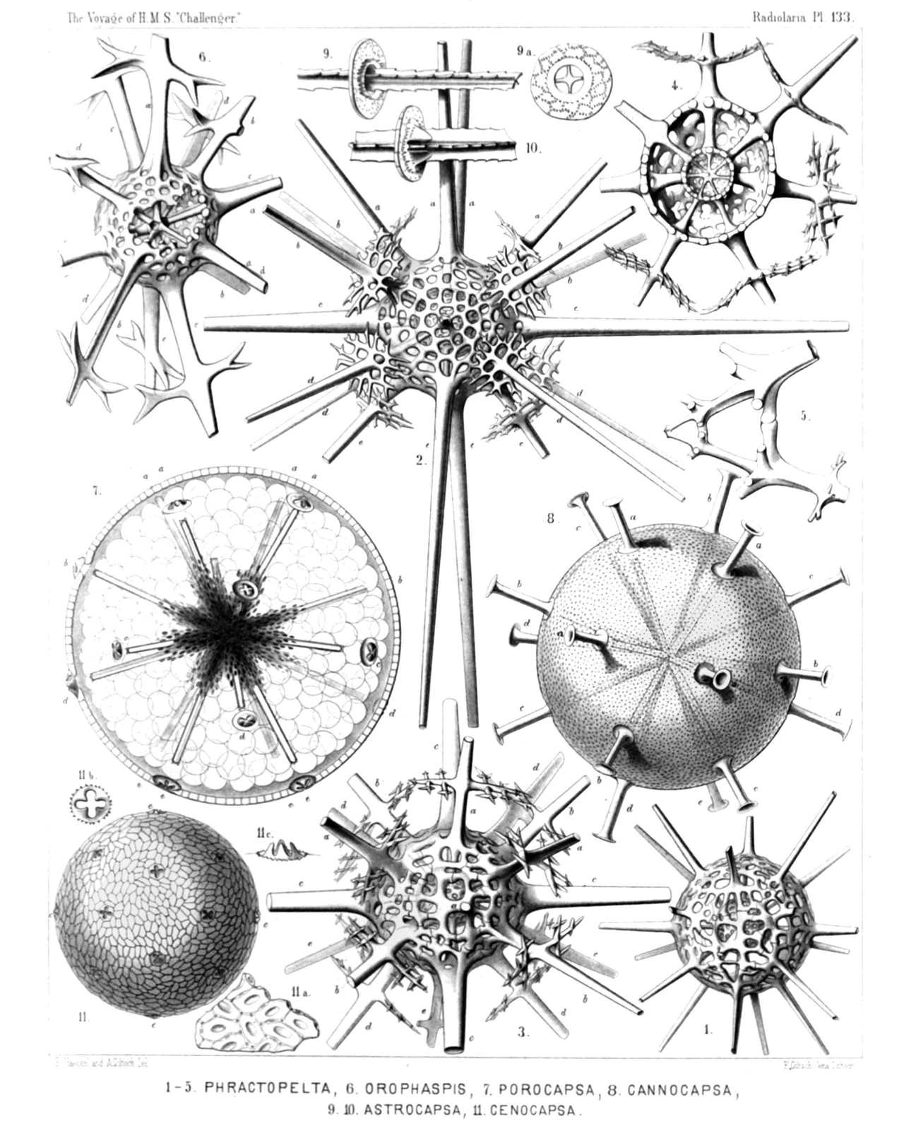 | ||||||||||||||||||||||||||||||||||||||||||||||||||||||||||||||||||||||||||||||||||||||||||||
| PLATE 133. | |||
| Sphærocapsida, Dorataspida et Phractopeltida. | |||
| Diam. | Page. | ||
Fig. 1. Phractopelta dorataspis, n. sp., | × | 300 | 852 |
| Fig. 2. Dorypelta tessaraspis, n. sp., | × | 300 | 858 |
| Fig. 3. Stauropelta cruciata, n. sp., | × | 400 | 859 |
| Fig. 4. Pantopelta icosaspis, n. sp., | × | 400 | 855 |
| Meridional section through the double shell. | |||
| Fig. 5. Octopelta scutella, n. sp., | × | 400 | 856 |
| Proximal part of two meeting spines, isolated. | |||
| Fig. 6. Orophaspis furcata, n. sp., | × | 400 | 818 |
| Fig. 7. Porocapsa murrayana, n. sp., | × | 300 | 800 |
| The central capsule is filled up by spherical vacuoles and enclosed by the porous shell; in the centre radii of small granules (nuclei ?) occur. | |||
| Fig. 8. Cannocapsa stethoscopium, n. sp., | × | 300 | 801 |
| The shell alone. | |||
| Fig. 9. Astrocapsa coronata, n. sp., | × | 400 | 799 |
| Middle part of one spine with the four aspinal holes. | |||
| Fig. 9a. Transverse section of a radial spine, with the four surrounding aspinal holes and the neighbouring part of the shell, | × | 400 | |
| Fig. 10. Astrocapsa stellata, n. sp., | × | 400 | 799 |
| Part of one spine, with the aspinal holes and their four triangular teeth. | |||
| Fig. 11. Cenocapsa nirvana, n. sp., | × | 200 | 802 |
| The entire shell, with its pavement of small plates and the twenty cruciform perspinal holes. | |||
| Fig. 11a. A group of small ovate plates which compose the shell; in each plate a dimple with a porule, | × | 400 | |
| Fig. 11b. A cruciform perspinal hole, seen from the face, | × | 400 | |
| Fig. 11c. A cruciform perspinal hole, with its four teeth, seen in profile, | × | 400 | |
PLATE 134.
Legion ACANTHARIA.
Order SPHÆROPHRACTA.
Family Dorataspida.
| 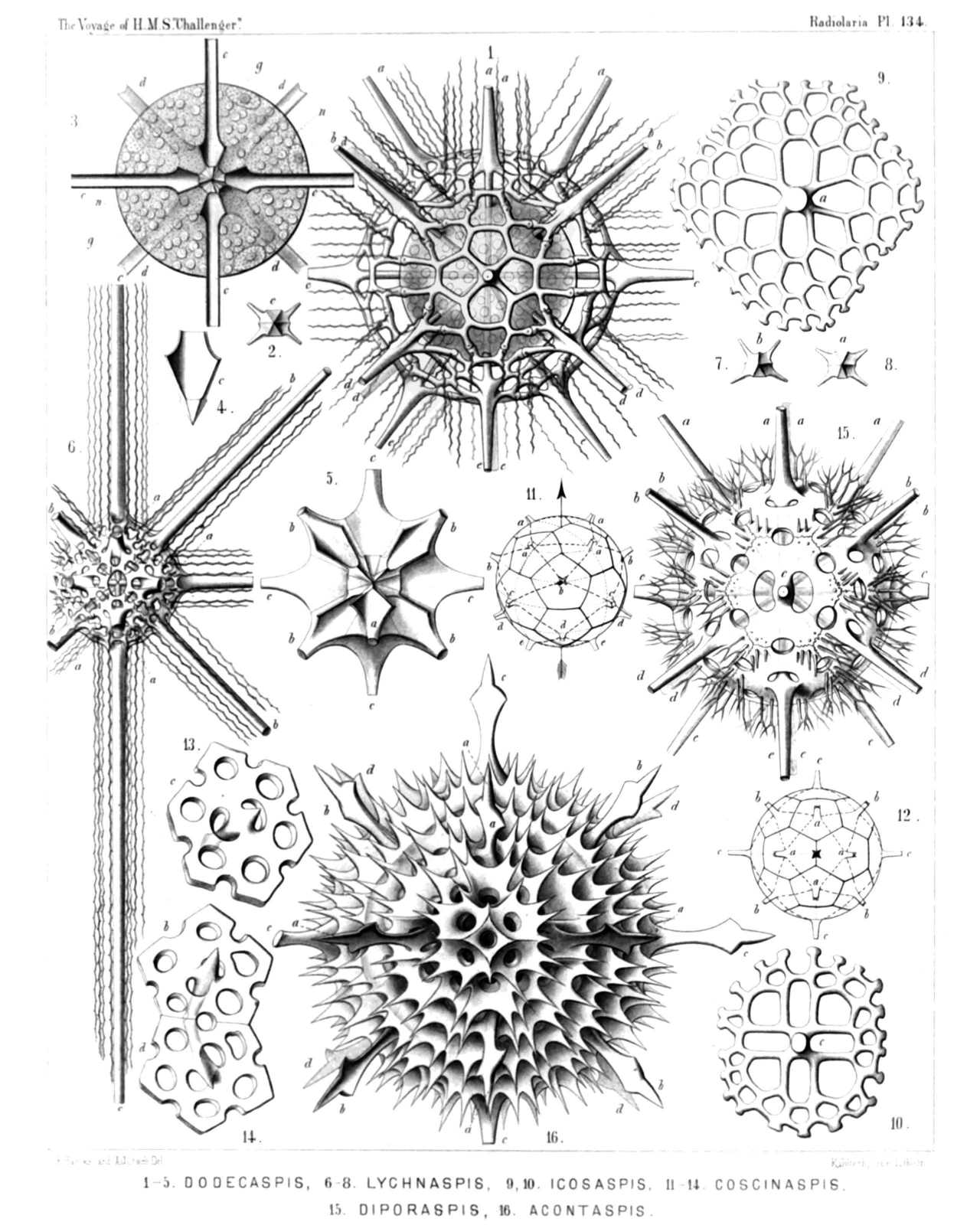 | ||||||||||||||||||||||||||||||||||||||||||||||||||||||||||||||||||||||||||||||||||||||||||||||||||||||
| PLATE 134. | |||
| Dorataspida. | |||
| Diam. | Page. | ||
Fig. 1. Dodecaspis tricincta, n. sp., | × | 400 | 834 |
| The enclosed central capsule contains numerous spherical nuclei. | |||
| Fig. 2. Lychnaspis minima, n. sp., | × | 400 | 841 |
| Six-sided basal pyramid of an equatorial spine, with the leaf-cross, seen from the centre. | |||
| Fig. 3. Zonaspis cingulata, n. sp., | × | 400 | 834 |
| Equatorial section through the central capsule. n, nuclei; g, yellow bodies (intracapsular xanthellæ). | |||
| Fig. 4. Zonaspis cingulata, n. sp., | × | 800 | 834 |
| Central pyramidal base of an equatorial spine, with the leaf-cross. | |||
| Fig. 5. Stauraspis cruciata, n. sp., | × | 400 | 831 |
| Central union of the radial spines, three polar spines being taken off. | |||
| Fig. 6. Lychnaspis longissima, n. sp., | × | 400 | 841 |
| Fig. 7. Lychnaspis minima, n. sp., | × | 400 | 841 |
| Five-sided basal pyramid of a tropical spine, with the leaf-cross, seen from the centre. | |||
| Fig. 8. Lychnaspis minima, n. sp., | × | 400 | 841 |
| Six-sided basal pyramid of a polar spine, with the leaf-cross, seen from the centre. | |||
| Fig. 9. Icosaspis elegans, n. sp., | × | 400 | 844 |
| An isolated polar plate. | |||
| Fig. 10. Icosaspis cruciata, n. sp., | × | 400 | 844 |
| An isolated equatorial plate. | |||
| Fig. 11, 12. Dorataspis species, | × | 100 | |
| Diagram of the composition of the shell of twenty plates (and also of the central union of the basal leaf-cross). | |||
| Fig. 11. Oblique equatorial aspect. | |||
| Fig. 12. Accurate polar aspect (compare p. 804, 805). | |||
| Fig. 13. Coscinaspis isopora, n. sp., | × | 400 | 828 |
| An isolated equatorial plate (with two aspinal and six coronal pores). | |||
| Fig. 14. Coscinaspis isopora, n. sp., | × | 400 | 828 |
| Two isolated tropical plates (b, northern; d, southern), each with two aspinal and five coronal pores. | |||
| Fig. 15. Diporaspis nephropora, n. sp., | × | 400 | 816 |
| Fig. 16. Acontaspis hastata, n. sp., | × | 400 | 829 |
PLATE 135.
Legion ACANTHARIA.
Order SPHÆROPHRACTA.
Families Sphærocapsida et Dorataspida.
| 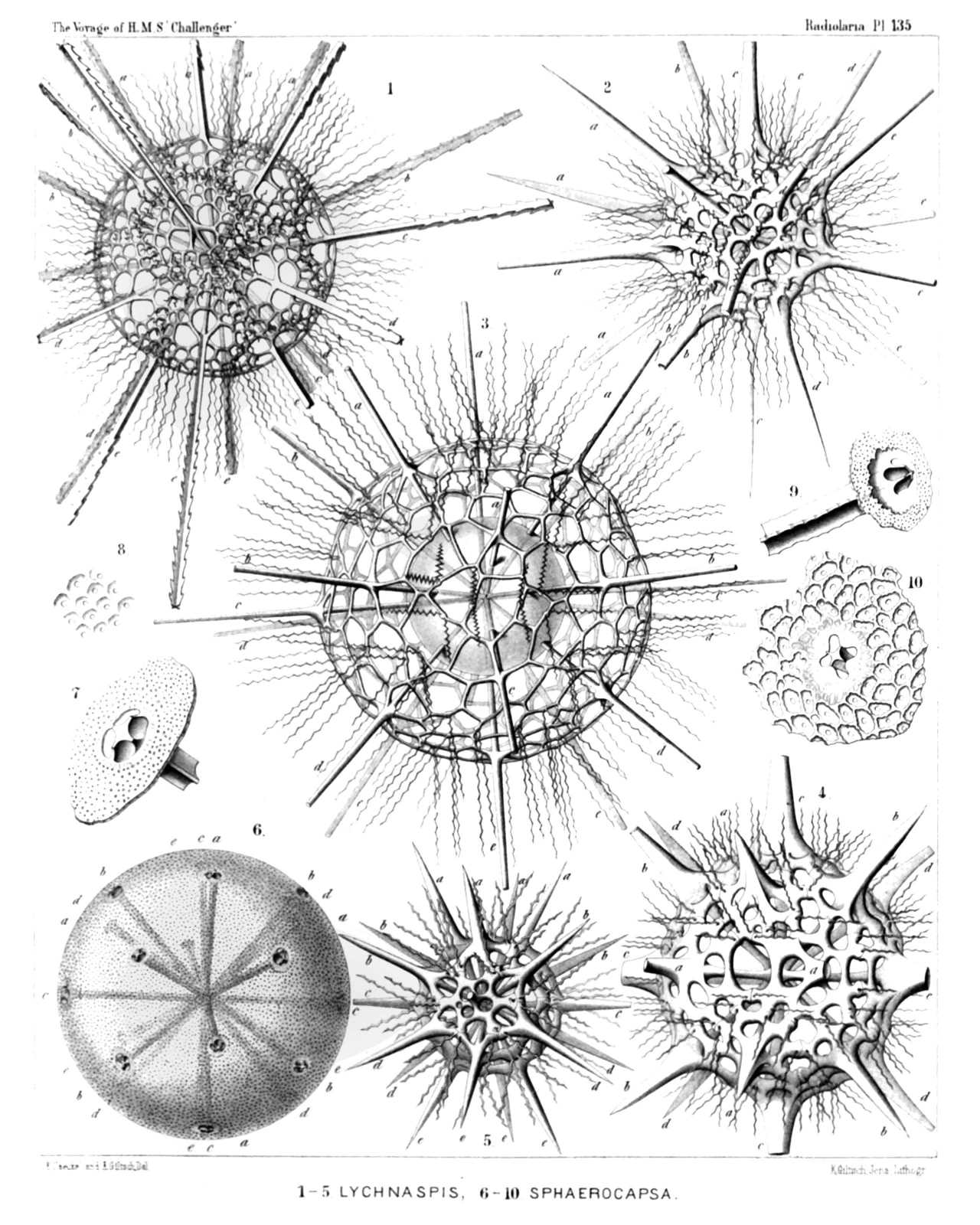 | |||||||||||||||||||||||||||||||||||||||||||||||||||||||||||||||||||||||||||
| PLATE 135. | |||
| Sphærocapsida et Dorataspida. | |||
| Diam. | Page. | ||
Fig. 1. Hylaspis serrulata, n. sp., | × | 300 | 846 |
| Fig. 2. Lychnaspis undulata, n. sp., | × | 400 | 841 |
| Fig. 3. Lychnaspis giltschii, n. sp., | × | 400 | 839 |
| The spherical central capsule is enclosed in the shell. | |||
| Fig. 4. Lychnaspis rottenburgii, n. sp., | × | 400 | 841 |
| Fig. 5. Zonaspis æquatorialis, n. sp., | × | 300 | 834 |
| Fig. 6. Sphærocapsa cruciata, n. sp., | × | 150 | 798 |
| The entire shell, with its twenty cruciate perspinal holes. | |||
| Fig. 7. Sphærocapsa cruciata, n. sp., | × | 800 | 798 |
| Insertion of one spine in the cruciate perspinal hole of the shell. | |||
| Fig. 8. Sphærocapsa quadrata, n. sp., | × | 800 | 798 |
| A group of pores and dimples in the shell surface. | |||
| Fig. 9. Sphærocapsa dentata, n. sp., | × | 800 | 798 |
| Insertion of one spine in the cruciate perspinal hole of the shell. | |||
| Fig. 10. Sphærocapsa pavimentata, n. sp., | × | 800 | 798 |
| Insertion of one spine in the perspinal hole of the shell, which is composed of four cruciate aspinal holes and surrounded by a group of dimples and pores. | |||
PLATE 136.
Legion ACANTHARIA.
Orders SPHÆROPHRACTA et PRUNOPHRACTA.
Families Dorataspida et Belonaspida.
| 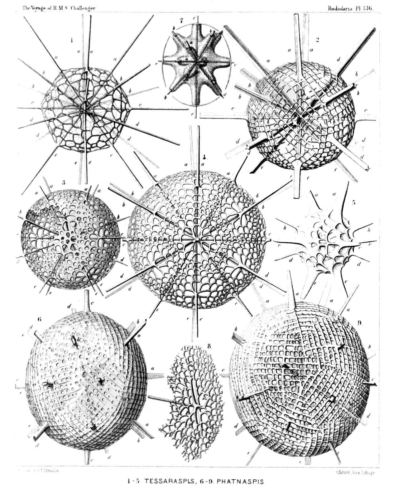 | |||||||||||||||||||||||||||||||||||||||||||||||||||||||||||||||||||
| PLATE 136. | |||
| Dorataspida et Belonaspida. | |||
| Diam. | Page. | ||
Fig. 1. Tessaraspis arachnoides, n. sp., | × | 300 | 836 |
| Fig. 2. Icosaspis tabulata, n. sp., | × | 200 | 843 |
| Fig. 3. Icosaspis icosastaura, n. sp., | × | 400 | 846 |
| Fig. 4. Icosaspis elegans, n. sp., | × | 300 | 844 |
| Fig. 5. Tessaraspis concreta, n. sp., | × | 400 | 838 |
| Fig. 6. Phatnaspis cristata, n. sp., | × | 400 | 869 |
| Fig. 7. Phatnaspis haliommidium, n. sp., | × | 200 | 871 |
| Central capsule within the shell—outline. | |||
| Fig. 8. Coscinaspis polypora, n. sp., | × | 300 | 827 |
| A single lattice-plate of the shell. | |||
| Fig. 9. Phatnaspis lacunaria, n. sp., | × | 400 | 869 |
PLATE 137.
Legion ACANTHARIA.
Order SPHÆROPHRACTA.
Family Dorataspida.
| 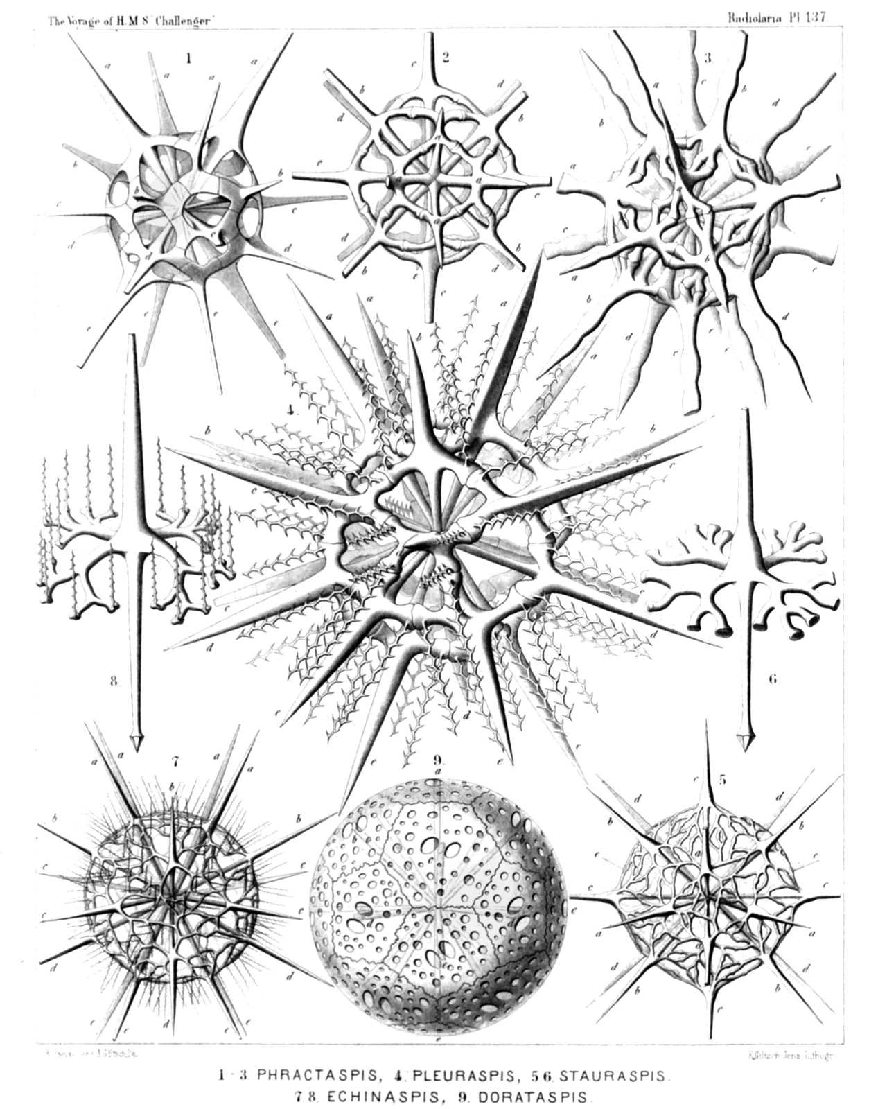 | |||||||||||||||||||||||||||||||||||||||||||||||||||||||||||||||||||
| PLATE 137. | |||
| Dorataspida. | |||
| Diam. | Page. | ||
Fig. 1. Phractaspis complanata, n. sp., | × | 400 | 809 |
| Fig. 2. Phractaspis prototypus, n. sp., | × | 400 | 809 |
| Fig. 3. Phractaspis constricta, n. sp., | × | 400 | 810 |
| Fig. 4. Pleuraspis horrida, n. sp., | × | 400 | 811 |
| Fig. 5. Stauruspis stauracantha, n. sp., | × | 300 | 832 |
| Fig. 6. Stauruspis stauracantha, n. sp., | × | 600 | 832 |
| A single spine. | |||
| Fig. 7. Echinaspis echinoides, n. sp., | × | 300 | 833 |
| Fig. 8. Echinaspis echinoides, n. sp., | × | 800 | 833 |
| A single spine. | |||
| Fig. 9. Coscinaspis parmipora, n. sp., | × | 400 | 827 |
PLATE 138.
Legion ACANTHARIA.
Order SPHÆROPHRACTA.
Family Dorataspida.
| 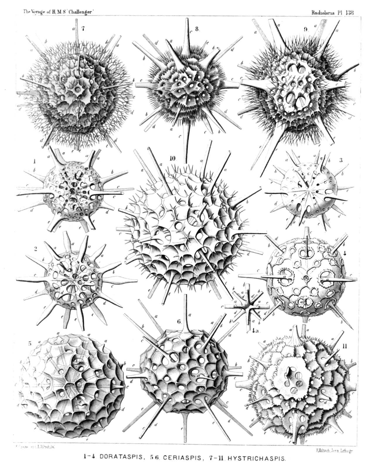 | |||||||||||||||||||||||||||||||||||||||||||||||||||||||||||||||||||||||||||||
| PLATE 138. | |||
| Dorataspida. | |||
| Diam. | Page. | ||
Fig. 1. Coscinaspis peripora (vel Dorataspis peripora), n. sp., | × | 300 | 826 |
| Fig. 2. Dorataspis fusigera, n. sp., | × | 400 | 813 |
| Fig. 3. Dorataspis micropora, n. sp., | × | 300 | 815 |
| Fig. 4. Dorataspis typica, n. sp., | × | 300 | 815 |
| Fig. 4a. Polar view of the central union of the twenty spines, | × | 300 | 815 |
| Fig. 5. Ceriaspis inermis, n. sp., | × | 400 | 821 |
| Fig. 6. Ceriaspis favosa, n. sp., | × | 400 | 821 |
| Fig. 7. Hystrichaspis fruticata, n. sp., | × | 300 | 825 |
| Fig. 8. Hystrichaspis pectinata, n. sp., | × | 300 | 822 |
| Fig. 9. Hystrichaspis furcata, n. sp., | × | 400 | 822 |
| Fig. 10. Hystrichaspis dorsata, n. sp., | × | 300 | 823 |
| Fig. 11. Hystrichaspis cristata (vel Siphonaspis cristata, n. sp.), | × | 400 | 823 |
PLATE 139.
Legion ACANTHARIA.
Order PRUNOPHRACTA.
Families Belonaspida et Hexalspida.
| 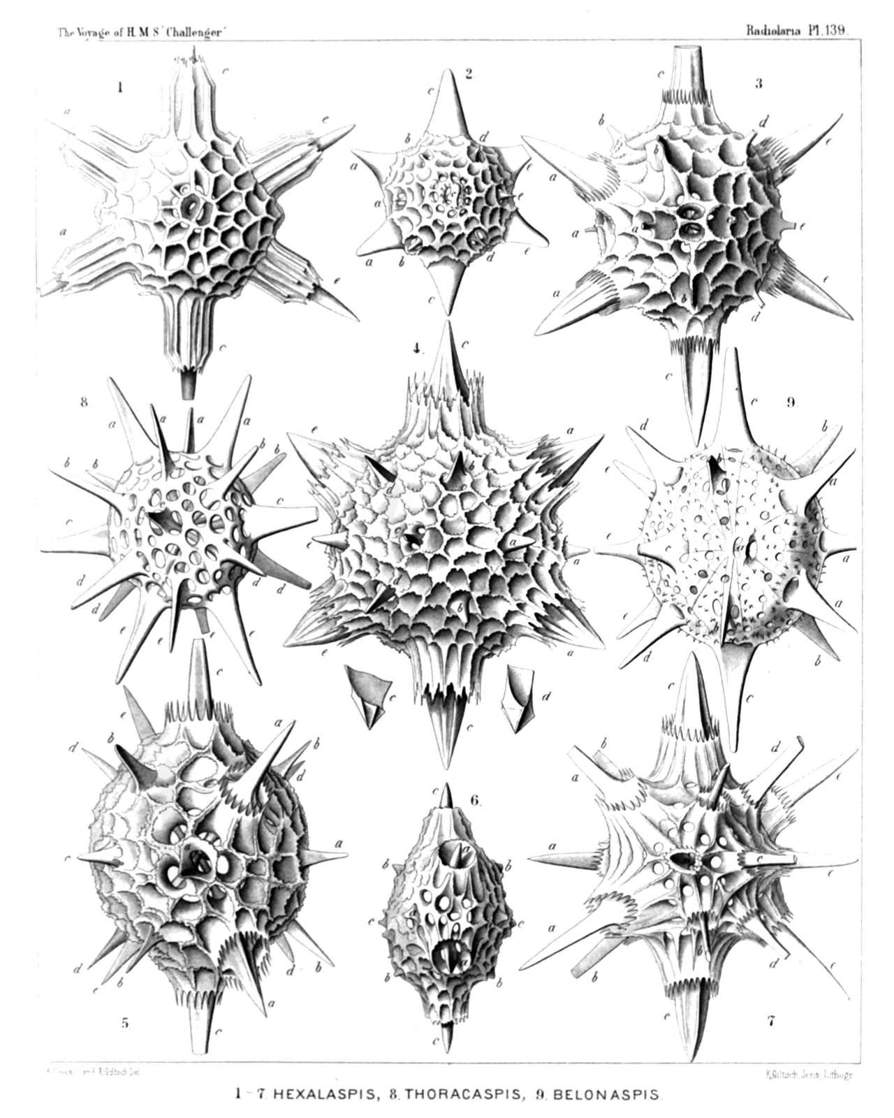 | |||||||||||||||||||||||||||||||||||||||||||||||||||||||||||||||||||
| PLATE 139. | |||
| Belonaspida et Hexalspida. | |||
| Diam. | Page. | ||
Fig. 1. Hexacolpus nivalis, n. sp., | × | 300 | 880 |
| Fig. 2. Hexalaspis heliodiscus, n. sp., | × | 300 | 875 |
| Fig. 3. Hexaconus ciliatus, n. sp., | × | 300 | 876 |
| Fig. 4. Hexaconus serratus, n. sp., | × | 300 | 877 |
| c, Central base of an equatorial spine; d, central base of a tropical spine. | |||
| Fig. 5. Hexaconus coronatus, n. sp., | × | 300 | 877 |
| Fig. 6. Hexaconus velatus, n. sp., | × | 300 | 877 |
| Marginal view of the shell. | |||
| Fig. 7. Hexaconus vaginatus, n. sp., | × | 300 | 877 |
| Fig. 8. Thoracaspis bipennis, n. sp., | × | 300 | 862 |
| Fig. 9. Belonaspis datura, n. sp., | × | 400 | 863 |
PLATE 140.
Legion ACANTHARIA.
Order PRUNOPHRACTA.
Families Belonaspida, Hexalaspida et Diploconida.
| 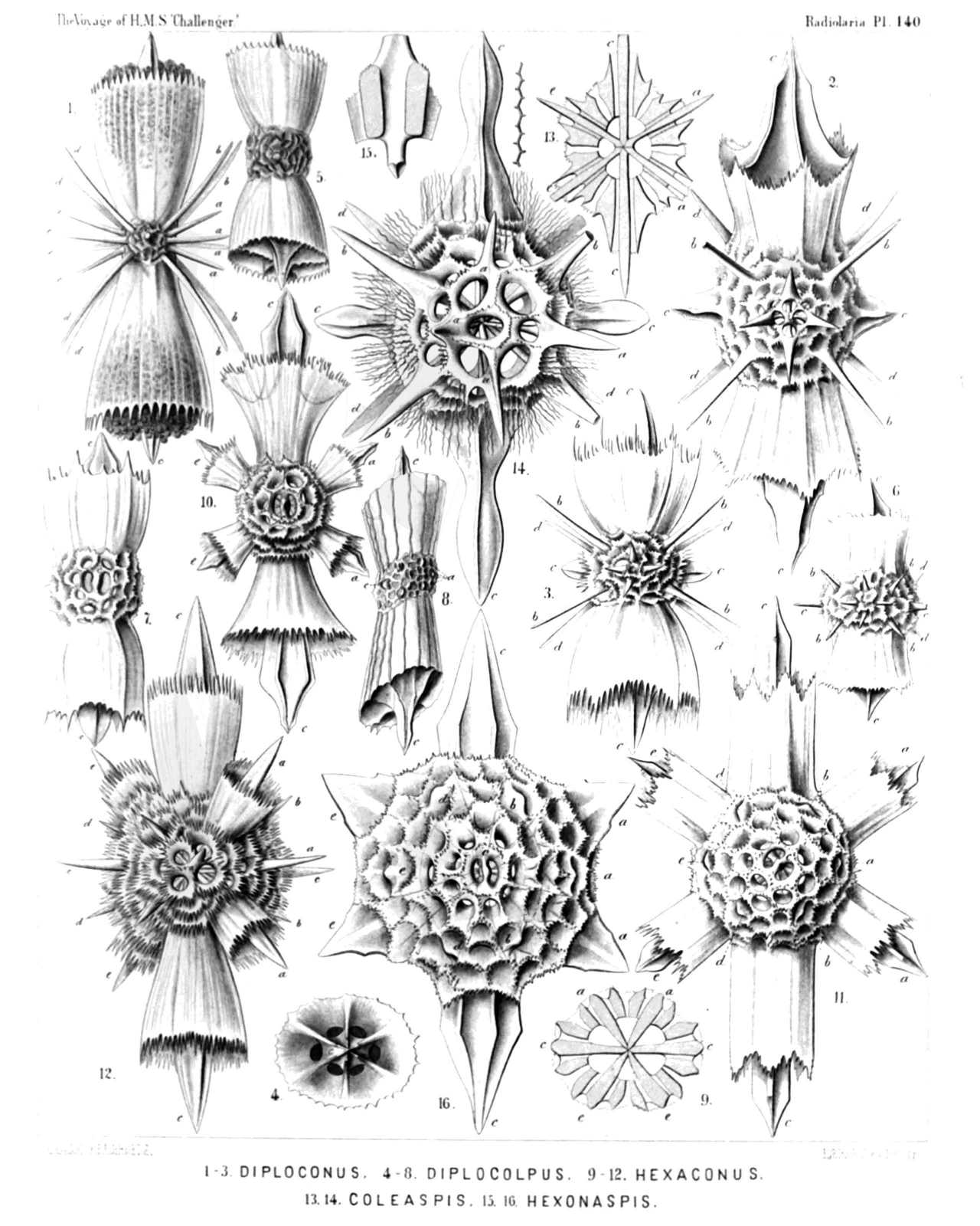 | |||||||||||||||||||||||||||||||||||||||||||||||||||||||||||||||||||||||||||||||||||||||||||||||||
| PLATE 140. | |||
| Belonaspida, Hexalaspida et Diploconida. | |||
| Diam. | Page. | ||
Fig. 1. Diploconus amalla, n. sp., | × | 300 | 885 |
| Fig. 2. Diploconus hexaphyllus, n. sp., | × | 300 | 886 |
| Fig. 3. Diploconus cyathiscus, n. sp., | × | 300 | 885 |
| Fig. 4. Diploconus cotyliscus, n. sp., | × | 400 | 886 |
| Polar view. | |||
| Fig. 5. Diplocolpus serratus, n. sp., | × | 300 | 888 |
| Fig. 6. Diplocolpus cristatus, n. sp., | × | 400 | 887 |
| Fig. 7. Diplocolpus costatus, n. sp., | × | 400 | 887 |
| Fig. 8. Diplocolpus sulcatus, n. sp., | × | 300 | 888 |
| Fig. 9. Diplocolpus dentatus, n. sp., | × | 300 | 888 |
| Meridional section through the centre of the shell. | |||
| Fig. 10. Hexacolpus infundibulum, n. sp., | × | 300 | 881 |
| Fig. 11. Hexacolpus trypanon, n. sp., | × | 300 | 881 |
| Fig. 12. Hexaconus echinatus, n. sp., | × | 300 | 878 |
| Fig. 13. Coleaspis vaginata, n. sp., | × | 300 | 866 |
| Meridional section through the shell. | |||
| Fig. 14. Coleaspis hydrotomica, n. sp., | × | 400 | 867 |
| Fig. 15. Hexonaspis hexapleura, n. sp., | × | 400 | 879 |
| A single spine with its thick apophyses. | |||
| Fig. 16. Hexonaspis hastata, n. sp., | × | 400 | 879 |